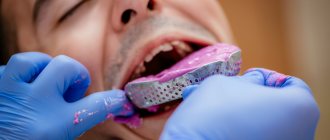
An impression is one of the important elements of dental work, serving as a means of communication between the doctor and the dental technician. A copy of the jaw row, which is a kind of “sketch,” is used to reflect elements that need correction, as well as to form a prototype of the future replacement structure. Understanding technical capabilities, taking into account nuances to eliminate further adjustments, as well as competent planning and forecasting are factors that determine the successful use of a dental impression.
General overview
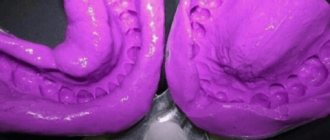
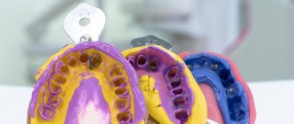
From a technical point of view, the impression is a negative, reflecting the position of the elements of the dentition and the surrounding tissues of the oral cavity. Based on the impression, a positive is created - a model that actually copies the anatomical structure of the jaw, which serves to form a replacement structure. The term "impression", which is quite common in dentistry, is usually used to describe a positive, relief prototype made of plaster or silicone. The formulation “taking an impression of the dentition” can be considered as a definition that combines two procedures: taking an impression and forming a model.
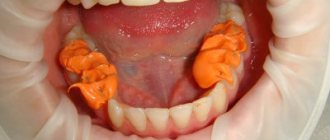
Making an individual plastic spoon in the laboratory.
In this case, an anatomical cast is taken with a standard spoon and a plaster model is cast from it. On the model, the dental technician draws the boundaries of the future individual tray . On the upper jaw, the border of the tray passes from the vestibular side along the transitional fold, not reaching the deepest point of its arch by 1-2 mm. On the distal side, it overlaps the maxillary tuberosities and runs along line “A” behind the palatine fossae by 1-2 mm. On the lower jaw, the border of the spoon passes from the vestibular side along the transitional fold, not reaching 1-2 mm to the deepest point of its arch, while bypassing the cords and frenulum of the lip. In the retromolar region, it is located behind the mucous tubercle, overlapping it by 1-2 mm. On the lingual side, the border of the spoon overlaps the area corresponding to the retroalveolar region (muscleless triangle), not reaching the deepest place of the sublingual space by 1-2 mm and going around the frenulum of the tongue. From the above it is clear that on both the upper and lower jaws the border of the individual tray is 2-3 mm less than the borders of the prosthesis. This is done so that there is space left for the impression material. The extruded impression material forms the edges of the impression. And, conversely, the distal boundaries of the tray must be larger than the boundaries of the prosthesis so that the anatomical formations, which are landmarks of the distal edge of the prosthesis, are well imprinted when taking an impression.
After drawing the boundaries, the dental technician coats the model with Izokol insulating varnish and begins making an individual tray from quick-hardening or base plastic. To make an individual spoon from quick-hardening plastic, the required amount of material is kneaded to a dough-like stage and a plate is made from it in the shape of the upper or lower jaw, which is pressed onto the model along the outlined boundaries. Then small pieces of plas are used to make a handle perpendicular to the surface of the spoon, rather than tilted forward. This position of the handle will not interfere with the design of the edges of the print. If the alveolar part of the lower jaw is significantly atrophied and the spoon turns out to be narrow, then the handle is made wider, almost up to the premolars: with such a handle, the doctor’s fingers will not deform the edges of the impression when they hold it on the jaw. After the plastic has hardened (10-15 minutes), the spoon removed from the model and processed with cutters and carborundum heads ( the individual spoon is not polished), making sure that the edges of the spoon correspond to the boundaries marked on the model. The thickness of the edge of the spoon should be at least 1.5 mm, because With a thinner edge, it is difficult to obtain the volume of the edge of the print. An individual spoon can be made from base plastic using the polymerization method. To do this, the heated wax plate is pressed tightly onto the model, giving it the shape of an impression tray, and excess wax is cut off with a spatula along the marked boundaries. The wax mold of the spoon is plastered into the cuvette in the reverse way and the wax is replaced with plastic. When making a spoon from AKR-P plastic, standard plates are softened in hot water and crimped according to the model. The excess is cut off with scissors after the corresponding area has been softened. The handle is made from scraps of material and glued to the spoon with a hot spatula (the heat melts and welds the plastic).
Individual spoons made of plastic are classified as rigid spoons. They can be used, as well as thermoplastic trays, to take compression impressions. Advantages and disadvantages of custom plastic impression trays . Plastic spoons are rigid and do not deform in the oral cavity, but, like any laboratory-made spoons (in two visits), they require subsequent correction in the oral cavity. In addition, spoons made in this way provide a modified display of soft tissues, because they are compressed and stretched during the taking of the anatomical impression.
Individual wax trays for the upper and lower jaws
Individual wax spoons can be made either in the laboratory or directly in the mouth. Wax trays using the CITO method are made in one visit directly on the jaw of the prosthetic patient. Such spoons are more accurate than individual ones made from an anatomical cast, because they display the soft tissues of the prosthetic bed at rest. The disadvantage of such trays is that the soft wax is deformed during fitting in the oral cavity and when taking an impression (it cannot withstand pressure), so the wax spoon can only be used to remove decompression impressions. Individual spoons , regardless of the method and material they were made from, must be placed in the oral cavity. A correctly fitted spoon sticks to the jaw and does not lag behind it when moving the lips and cheeks. , the technique of fitting individual spoons using Herbst functional tests has become widespread Functional Herbst tests are recommended when fitting individual trays and obtaining functional impressions .
Five tests are used on the lower jaw:
1) swallowing and wide opening of the mouth;
2) movement of the tongue to the sides along the red border of the upper and lower lips;
3) touching the cheeks with the tip of the tongue with the mouth half closed;
4) movement of the tip of the tongue forward beyond the lips towards the tip of the nose;
5) pulling the lips forward. Three tests are used on the upper jaw:
1) wide mouth opening;
2) cheek suction;
3) shifting the lips forward (pulling).
Classification
Differentiation adopted in dentistry involves dividing impressions into two categories:
- Working, or precision – precise impressions used to create functioning replacement structures installed on a permanent or removable basis;
- Auxiliary - as the name implies, this group combines all additional impressions that eliminate information gaps and make it possible to clarify the specifics of the occlusal relief to improve the quality of wearing the prosthesis.
Another category is occlusal recorders, whose task is to functionally complement and combine these types of impressions. If there are a sufficient number of surviving units, their use is not always advisable, but the desire to achieve optimal occlusion and shorten the adaptation period are good reasons for using recorders.
In terms of the number of jaws represented by the impression, there are one- and two-jaw options, with the second option being used much less frequently in dentistry. The effectiveness of using a separate impression for each row of teeth is determined by the quality of the resulting pattern.
Based on the technique of designing the marginal areas, anatomical and functional impressions are distinguished, however, it is quite difficult to establish a clear line of differentiation between them. Functional variation involves the use of clarifying movements of active and passive properties, allowing one to obtain additional information necessary for the formation of a prosthetic system. In turn, anatomical impressions are used for permanent replacement, when technical interest is primarily in the characteristics of the tissue structure of the prosthetic bed, while the marginal design loses its original priority.
Two-stage impressions
Two-stage impressions essentially differ from single-stage impressions not so much in that the impression tray is brought into the oral cavity twice, but in the rule that a larger thickness of the layer of polymerizing material has a greater degree of shrinkage than a smaller thickness of such a layer. Therefore, the two-stage technique offers a smaller layer of impression material, which gives less shrinkage, and the high fluidity of the corrective mass determines the high quality of the resulting impression. In this case, an impression using only the base impression material, the production of which is the first stage, is a kind of individual impression tray, which makes it possible to reduce the thickness of the simultaneously polymerizing layer of impression material, which will be the corrective impression mass.
The first stage and the principle of obtaining the base layer of the print are half similar to the method of obtaining a one-step print. Half because it is more important to correctly place the tray with the impression material on the tissue, while separating and removing it does not require such care to avoid deformation - only for the patient’s comfort. In addition, you should not wait for complete polymerization of the material, since, while maintaining plasticity, deformations will help create additional space for the corrective layer of impression material. Plus, at this stage you can further loosen the impression a little to create more space for the impression material.
| At the stage of obtaining the base layer of a two-stage impression, the correct location of the teeth relative to the sides of the tray is important, and not the accuracy of the display |
In the resulting impression, using a sharp instrument, the interproximal septa are removed and additional grooves are created for the outflow of excess corrective material. At this stage there is a danger of creating insufficient space for the flowable impression material. From a theoretical point of view, what complicates the situation is that different studies provide different conclusions: some of them indicate that creating grooves to remove excess material does not affect the quality of the impression, but in practice these same excesses can cause the inferiority of the impression. In addition, the need to create additional space is indicated by the fact that in practice it is extremely difficult to position the print exactly in the same place. The quality of the two-stage print is also indicated by the uniformity of the color of the correction layer, without pressing through the base layer, which will indicate a high probability of deformation. Therefore, the operator’s experience in creating both sufficient and not excessive space for the correction layer is important at this stage.
| There is no need to remove thick interdental septa in the base layer, since a thick layer of corrective mass will lead to large deformations when taking an impression and casting models |
At the second stage, it is important to apply the primary impression with the corrective mass added to it as accurately as possible. This is sometimes difficult, for which you should not spare any effort, but you must be careful not to injure the patient.
After complete polymerization of the material, separation and removal of the impression from the oral cavity are similar to those with a one-step technique.
| After extracting the print, it is necessary to assess its quality |
Materials and features of the procedure
To obtain high-quality dental impressions, the preferred option is to combine materials with different viscosity parameters. The high-viscosity formulation provides sufficient rigidity, while fine-grained correction materials improve imaging accuracy. Standard protocols also provide for the possibility of three-phase (the number of phases means the number of compounds) impression taking, with the addition of medium-viscosity material.
One of the elements provided by the technology for obtaining dental impressions is an impression tray. From a technical point of view, this is a design that is mass-produced or made individually, the structure of which repeats the anatomical structure of the jaw and looks like a horseshoe.
To obtain two-phase impressions, a one- or two-step approach is used. In the first case, an individual tray is used for simultaneous application of both compositions, while the corrective mass can also be applied to the surface of the tissue area of the prosthetic bed - followed by indentation of the element. The two-stage method differs in the number of placements of the spoon in the oral cavity - in the second case, the instrument is used twice, first with the base material and then with the corrective material. Practice shows that such an approach provides an accurate result, but leaves the possibility of making a technical error, which is practically impossible to identify before manufacturing a replacement structure.
Successful impression taking. The first time. Every time
CM. Drobyshevsky, STIdent brand manager for products for orthopedic dentistry and denture equipment
The modern doctor deals with a myriad of "complex parts", especially when preparing and placing indirect restorations and fixed prostheses. Materials for indirect restorations and materials intended for cementation have greatly improved in recent years. In order to be confident in the success of the entire complex of restorations, such as veneers, inlays, crowns, bridges and removable dentures, it is necessary to correctly observe and correctly perform a series of “simple things”. One of these critical “simple things” is the impression.
Although various CAD/CAM systems are already available in today's dentistry, most indirect restorations are fabricated by dental technicians. A good impression accurately depicts the details of the preparation and allows the technician to produce a functional, esthetic restoration with a good fit. Modern precision elastomeric impression materials have excellent physical properties. They have dimensional stability, precision in the transfer of parts, are resistant to tearing and an exceptionally good surface of reproducible parts. Moreover, they are usually mixed using automatic mixers, which ensures the correct base/catalyst ratio and a homogeneous consistency without air pockets. Materials are becoming less and less hydrophobic, which reduces the number of problems associated with oral moisture. Unfortunately, dental technicians report that a large percentage of impressions received by laboratories are unsatisfactory. These prints are replete with problems such as missing or unclear edges, pores and voids, drawbacks and other distortions. Bad prints are produced unintentionally. In fact, sometimes an “acceptable” print is all that can be achieved. Therefore, the prevalence of “weak” impressions suggests that impression materials are often misused and not examined with due attention to detail. Doctors may become frustrated by failures to take impressions and usually begin to look for a better (read different) material to solve their problems. However, material choice is just one variable in the print equation. Even if you have one of the best materials, if you use it incorrectly, you can get an unsatisfactory final result. Over the years, the quality of impression materials has improved significantly, making the choice of the correct impression-taking technique clearer. The choice of technique can make the difference between success or failure in a given print. Despite this fact, during primary or postgraduate training, impression materials are considered in passing, and therefore, due attention is usually not given to this important aspect of prosthetic dentistry. Most practicing doctors learn impression-taking techniques through trial and error. There is nothing wrong with learning by doing, but an informed practitioner can gain more knowledge with less effort. An informed clinician can use the basic principles developed by others to improve his or her impression-taking techniques. Today, orthopedic dentists use a wide range of methods for taking impressions. A full range of different materials with different properties is available to cover these user needs. And clinicians who are aware of these properties can select the most appropriate material for the specifics of each clinical case. The choice of material is influenced not only by its viscosity but also by other factors. For example, some materials may have an unpleasant odor or taste. Some of them require immediate casting of the model, while others, on the contrary, require you to wait a certain time before casting. Some materials are difficult to disinfect or require expensive equipment. From a practical point of view, impression materials can be classified according to just three main characteristics: • Viscosity. • Hydrophilicity. • Hardening time. These properties have a major influence on the choice of impression material depending on the indication, type of preparation and impression technique.
Viscosity.
Viscosity describes the flow characteristics of an uncured material. Materials with high viscosity have a weak degree of fluidity, while materials with low viscosity, on the contrary, are highly fluid. Materials currently available range from low to very high viscosity.
Low Viscosity - Type 3. Low viscosity materials may be known to us as light body or wash materials. They are usually able to flow very well around the surface of the tooth, flowing inward and displaying the smallest details of the tooth, oral mucosa and preparation area. They are rarely used as a single impression material. Typically, low-viscosity materials are used in combination with a second, more viscous material that forces them onto the tooth and holds them in place during the hardening process. Low viscosity materials are usually mixed in modern automatic mixing cartridges.
Medium Viscosity - Type 2 Medium Viscosity materials are very versatile and are sometimes known to us as monophasic. This is why medium viscosity materials can be used as one material when taking an impression. Their viscosity is sufficient to prime a syringe and capture fine detail accurately, but not so low that a second material is required to apply additional pressure and hold them in place during the curing process. Medium viscosity materials can be used in combination with wash materials that require support, pressure and fixation during the curing process. In addition, materials of medium viscosity can be used in combination with more viscous (type 1) or putty materials - in this case they are squeezed out of the syringe. Type 2 materials are conveniently used in cartridges for automatic mixing, squeezed into a spoon or directly through a cannula in the mouth. They can also be supplied in traditional tubes for manual mixing and syringe application.
High Viscosity - Type 1. High viscosity impression materials are known by the English term “heavy body” as impression base materials. Typically, these materials do not have sufficient fluidity to accommodate preparations, sulcus, and other highly detailed tissue surfaces. This means that these materials are primarily used as a print base in combination with “wash” or other less fluid materials. In this case, the high viscosity material provides the necessary pressure to push through the lighter, more fluid material, ensuring good contact with the surface on which the impression is being made. High viscosity materials are used exclusively for the base of the impression. High-viscosity materials are now available in special cartridges for automatic mixing in Heraeus Dynamix® type machines.
Very High Viscosity - Type 0. The most viscous materials are known to us as putty or moldable materials. Like high-viscosity print base materials, they are used in combination with “wash” or “light body” materials. “Putty” materials provide the necessary pressure to support the “wash” material, which imprints the finest details of the print. Putty materials are so viscous that they are only available in jars for manual mixing or in special cartridges for automatic mixing in Dynamix® type machines. Automatic mixing is not only much more convenient to use and saves time, but also improves the properties of the material for reproducing details, which in turn improves the quality and reliability of work when taking an impression.
Hydrophilicity.
Among other things, impression materials are characterized by the degree of their hydrophilicity, in other words, their ability to be moistened. Materials can be identified as: • Hydrophobic. • Hydrophilic. • Hydroactive.
Hydrophobic materials.
Hydrophobic materials (Figure 1) have low hydration capacity and tend to reject any moisture present on the tooth or other oral tissues. A good analogy for the reaction of hydrophobic materials to a humid environment is water droplets on a freshly waxed car body. Although no water absorption will occur, this repulsion may mask the surface to be printed, preventing uniform contact between the material and the surface, thereby reducing the extent to which desired details of the print are visible. Inaccurate details in the impression may result in further inaccurate fit of the final restoration. More moisture means worse results.
Hydrophobic materials: • Provide poor surface wetting. • Imprint surface details to a low degree. • In contact with moisture, they can mask the surface and prevent uniform contact. • Will not absorb moisture.
Hydrophilic materials.
Hydrophilic materials (Fig. 2) have a high ability to wet and are generally considered the most ideal option for taking impressions. Their ability to develop well in a humid environment means that they provide good surface wetting, allowing the material to capture the details of the printed surface to the highest degree. However, their ability to absorb moisture may lead to some dimensional instability or changes in the physical properties of the material in the event of excess moisture in the oral cavity.
Hydrophilic materials: • Provide good surface moisture. • Provide highly detailed surfaces. • May change and become inaccurate when exposed to excess moisture.
Hydroactive materials (artificially hydrophilic)
Since the 1980s, it has become possible to make some inherently hydrophobic materials more hydrophilic through changes at the chemical level. The material that has undergone this process may belong to the class of hydroactive or artificially hydrophilic (Fig. 3). They are usually made hydrophilic by adding surfactants called surfactants (special molecules with a hydrophilic group at one end of the molecule and a hydrophobic group at the other). Hydroactive materials have excellent wetting ability, but unlike truly hydrophilic types of materials, they do not absorb moisture. When an artificially hydrophilic material is placed in contact with moisture, surfactants react and help provide the material with maximum contact with the tooth and oral tissues, allowing for an excellent impression in the smallest detail. Thus, moisture tends to be displaced, but does not enter the impression material. This means that the material properties and its dimensional stability remain unchanged.
Hydroactive materials: • Provide excellent surface hydration. • Provides a high degree of detail display. • Provide dimensional stability. • Displaces moisture from the tooth and tissue surface without absorbing it.
Hardening time.
In reality, setting time is the total time required from the start of mixing until the impression is completely set and can be removed from the mouth without deformation. Within the total curing time there is a period known to us as working time. Working time is measured from the start of mixing until the point where the material can no longer be manipulated without risking distortion of the print. Therefore, the material must be mixed and placed in place before the end of working hours. After which it stabilizes until it hardens completely. Curing times for elastomeric materials range from about 1 minute for fast-curing alginates to about 10 minutes for polysulfides. Based on the hardening time, we distinguish materials as slowly hardening (low set), normally hardening (regular set) and quickly hardening (fast set). As a rule, the hardening time is related to the working time. Thus, materials with a slow curing time have a prolonged working time. In turn, quickly hardening materials have a short working time. Hardening times and working times set by manufacturers are characterized by values that can affect them. Such as: ambient temperature, air humidity and mixing technique applied to the material. The only material with variable hardening time on the Russian market is Variotime® A-silicone material (Fig. 4), produced by the German company Heraeus. Variable hardening time was achieved by increasing the sensitivity of the material to oral temperature. This means that when working with Variotime material, its total curing time, as well as working time, are under the control of the user. Once mixed, the correction material has a working time of 1 to 2:30 minutes. The final hardening time is activated by the temperature of the oral cavity, is 2:30 minutes and always remains unchanged. Thus, the total curing time of the material is controlled by the user.
A-silicones (Addition-curing silicones). Today, A-silicones are used by most qualified orthopedic dentists. These materials are also known as vinyl polysiloxanes or VPS (vinyl polysiloxanes). A-silicones have a more pleasant odor and taste than polyesters or polysulfides. They have a lower percentage of distortion (permanent deformation) than any other existing impression material, making A-silicones a good choice for undercut impressions. For the most part, A-silicones are not as hard as polyesters. Their tensile strength varies and mainly depends on viscosity. Low viscosity A-silicones tend to have lower strength than C-grade silicones, while high viscosity A-silicones have much higher tensile strength than "C" grade silicones. A-silicones have a different hardening mechanism compared to C-silicones and do not release accompanying compounds during polymerization. This means that A-silicones exhibit good dimensional stability, allowing impressions to be cast many weeks later. One of the specific qualities of A-silicones is their sensitivity to sulfur compounds. Because sulfur is used in the manufacture of latex gloves and can also be found in some hemostatic agents, the clinician must be careful to avoid contamination of the material, which could inhibit its hardening process. Automatic mixing is one of the well-tested methods that allows you to fill a stock or syringe without touching the material. Review of techniques for taking impressions.
The simplest way to describe methods for obtaining impressions is by the number of working steps and material requirements. As a rule, each technique uses several types of materials or their combinations. The main group of techniques can be classified as follows:
• One material - one stage. • Two materials - one stage. • Two materials - two stages.
Impression techniques vary in the number of materials used and work steps.
Let's consider the applied impression-taking techniques (Table 1) using the example of the innovative A-silicone material Variotime®, produced by the famous German company Heraeus. With Variotime®, the doctor has the ability to adjust the impression taking process to suit his or her personal requirements. As noted above, Variotime® has flexible working hours and a short hardening time in the mouth, which is very comfortable for the patient and reduces the time of the entire procedure.
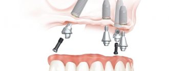
The material is supplied in 6 different viscosity options and three packaging options, thereby giving the doctor the opportunity to choose the material “for himself.” The Variotime® material system is multifunctional and has a wide range of indications: • Crowns and bridges. • Inlays and overlays. • Transfers and implants. • Functional impressions.
One material, one stage.
This technique seems to be the simplest, since it uses one material and one stage of obtaining an impression. The base material acts in the same way as the second correction layer, which is why this technique is known to us as monophasic (Fig. 5). Working steps of the monophase technique (using Variotime Monophase): 1. The impression tray is coated with adhesive and dried according to the manufacturer's instructions. 2. The spoon is filled with Variotime Monophase material. 3. Variotime Monophase is injected from a syringe into the gingival sulcus around the preparation margins. 4. The impression tray with the material is slowly inserted into the oral cavity and gently seated in place. 5. Next, the impression is held in place in a calm position, without excessive pressure, until the material has completely hardened (about 2:30 minutes). 6. The impression is then removed from the patient's mouth. 7. Evaluation of the finished print.
Two materials, one stage (first layer and correction):
This method, by definition, requires two different materials that are mixed individually and then used to create the impression in one step. In most clinical cases, a low viscosity material is pressed out around the preparation and a more viscous base or putty material is immediately inserted on top, before both materials begin to harden. When used correctly, both materials are joined monolithically, which allows you to obtain a highly accurate impression. The more viscous base material provides support and hydraulic pressure to the correction material, allowing this less viscous material to imprint fine details on the preparation and surrounding tissue. To obtain an impression with two different materials in one step, there are two options: “double mixing” and “sandwich” method. Double mixing uses a medium to high viscosity material as the base material. The sandwich method uses the highest viscosity putty material as the base layer, combined with rigid, non-perforated spoons that will not deform the putty material under pressure. It is very important to understand the difference between the “sandwich” method and the two-step (“putty/wash”) method, which will be discussed briefly below.
Steps for the Double Mix Method/Sandwich Technique (using Variotime Heavy Tray or Putty in combination with Variotime Medium Flow or Light Flow): 1. A rigid non-perforated tray is coated with adhesive and dried according to the manufacturer's instructions. 2. Variotime Heavy Tray or Variotime Putty is mixed and placed in a spoon. 3. To create space for the teeth, a groove is pressed into the material. 4. A thin layer of correction material Variotime Medium Flow (or Light Flow) is applied to the resulting groove. 5. Variotime Medium Flow (or Light Flow) is squeezed into the gingival sulcus along the preparation margin. 6. The impression tray is placed in the oral cavity. 7. The spoon should be inserted slowly, ensuring smooth pressure. 8. Next, the impression is held in place in a calm position, without excessive pressure, until the material has completely hardened. 9. The impression is then removed from the patient’s mouth. 10. Evaluation of the finished print. The double mixing method is extremely flexible and versatile because it can handle materials of varying viscosities. This method is also suitable for most clinical situations. However, not all materials can be used by this method, since one of its conditions is the simultaneous hardening of two materials in the impression. This is why the most suitable materials for this situation are A-silicones.
Two materials, two stages (putty/wash method):
This method (Fig. 7) also uses two different materials, but they are applied in two separate stages. The first step is to obtain an initial impression using the highest viscosity material (putty). The second step is to make the final impression using a low-viscosity correction material, using the primary impression as an individual tray. Before taking the final impression, the initial impression must be carefully trimmed to remove undercuts and create a uniform space for correction material. To guarantee adhesion of the main layer of the impression to the correction material, it is necessary to clean the primary impression from any contamination (blood, saliva, etc.).
Steps of the Putty/wash method (using Variotime Heavy Tray or Putty in combination with Variotime Extra Light Flow): 1. A rigid non-perforated tray is coated with adhesive and dried according to the manufacturer's instructions. 2. Variotime Heavy Tray or Variotime Putty is mixed and placed in a spoon. 3. To create space for the teeth, a groove is pressed into the material. 4. The impression tray is placed in the oral cavity, pressed lightly and held until the material hardens (about 2:30 minutes). 5. After final hardening, the impression is removed from the oral cavity, rinsed with water, trimmed along the periphery, interdental spaces are removed, and drainage channels are cut out in the area of the preparation boundaries. 6. A mixing tip is placed on the Variotime Extra Light Flow material cartridge and the material is extruded onto the initial impression. 7. The intraoral cannula is placed on the mixing tip of the Variotime Extra Light Flow cartridge and the material is applied into the gingival sulcus around the preparation margins. 8. The spoon is inserted into the oral cavity with short pressure to ensure that Variotime Extra Light Flow penetrates into the required areas. Then the impression is held in a calm position without unnecessary pressure until the material is completely hardened (about 2:30 minutes). 9. The final impression is taken from the mouth and evaluated.
Below we provide a summary table of the individual characteristics of the Variotime family of materials and methods of their application.
Variotime Putty • Soft mixing consistency. • High final hardness for applying pressure to the correction material and low impression deformation. • Easy to trim in a two-step technique. • Available in both manual mixing and Dynamix cartridges for automatic mixing. • For two-stage and sandwich techniques.
Variotime Heavy Tray • High final hardness for precision and dimensional stability. • Exceptional properties for transfer impressions and implant impressions. • Available in Dynamix auto-mix cartridges and manual dispenser cartridges. • Suitable for two-step and double-mix techniques.
Variotime Monophase • Balanced final hardness for consistent impression accuracy and ease of removal. • Pronounced thixotropy. • Can be used as a base material or applied from a syringe around preparation margins. • Available in Dynamix auto-mix cartridges and manual dispenser cartridges. • Suitable for monophasic impressions.
Variotime Medium Flow • Medium viscosity correction material. • Stable hydrophilicity for the periodontal sulcus. • High elasticity and strength. • Ideal for double mixing and sandwich techniques as it is more viscous than Variotime Light Flow, complementing the high viscosity of the impression base materials in these techniques. • Available in cartridges for hand dispensers.
Variotime Light Flow • Correction material with light consistency. • Hydrophilic properties for high fluidity. • Elasticity and tensile strength. • Ideal for double mixing and sandwich techniques as it is more viscous than Variotime Extra Light Flow, complementing the high viscosity of the impression base materials in these techniques. • Suitable for double mixing, sandwich prints.
Variotime Extra Light Flow • Correction material with extra light consistency. • Highest hydrophilicity at the level of polyester for the transfer of details of the periodontal sulcus. • High elasticity and strength. • Especially recommended for two-stage impressions, since the low viscosity of Variotime Extra Light Flow is very suitable for obtaining a thin layer of correction material. • Available in cartridges for hand dispensers. • Suitable for two-stage impressions.
Standard spoons
There is a wide range of standard impression trays on the market today. The first factor to take into account when choosing is the working jaw. For example, spoons for a moving jaw are equipped with a projection for the tongue and are shaped like a horseshoe, while products for a fixed jaw are similar to a regular spoon.
When it comes to choosing a partial impression, it is important to take into account the working side of the jaw.
Segmental designs
We are talking about spoons designed for simultaneous removal of models of both jaws. Instead of a bed of correctional mass, they are equipped with a thin layer of nylon or gauze. Like standard modifications, these spoons have sides.
The model removing compound is applied on both sides of the intermediate layer. Simultaneous taking of impressions from the lower and upper jaws allows you to reduce the duration of the procedure due to the absence of the need to additionally register the bite.
However, the practicality of using such a technique is controversial. In some cases it is quite difficult to obtain a universal impression. It is worth keeping in mind that insufficient rigidity of the spoon can negatively affect the quality of the finished model.
Another important aspect is the condition of the dentition. Spoons are made for patients with partial or complete edentia.
Material of manufacture
Spoons can be made of polystyrene, aluminum, steel and other materials, depending on the intended purpose. For example, metal products are used to obtain the most rigid impressions, and plastic structures are widely used in the field of implantology. In a number of clinical cases, plastic and metal products are interchangeable.
Disinfectants for disinfection of dental impressions, denture blanks, articulators
| Home | Information | Disinfectants for disinfection of dental impressions, denture blanks, articulators | Disinfectants | Antiseptics |
Disinfection of medical products in dentistry requires careful selection of the method and means of disinfection.
Let us dwell on the issue of disinfection of dental impressions, denture blanks, suction systems, etc. First of all, the choice of disinfectant should be determined by the type of material from which the objects are made, and the products should also be easily washed off with water.
We present to your attention several disinfectants developed and produced by the Federal State Unitary Enterprise “SSC “NIOPIK” for disinfection in dentistry.
ADS-521 is a disinfectant specially designed for the disinfection of dental impressions made of alginate, silicone materials, denture blanks made of metals, ceramics, plastics and other materials, impression trays, corrosion-resistant articulators. It has good cleaning properties and is easily washed off with water.
Aquaminol Forte is a powerful disinfectant with excellent cleaning properties based on triamine and HAC. The product has a wide range of applications, including recommended for use in dentistry for the disinfection of impressions, denture blanks, articulators and impression trays, suction systems of dental units and spittoons, and dental instruments.
Alaminol Plus is a highly effective, economical universal disinfectant based on aldehydes and QAS. In dentistry, it is used to disinfect denture blanks, articulators and impression trays, suction systems of dental units and spittoons, and dental instruments.
Disinfection modes
| Disinfectant | Concentration of working solution, % (by preparation) | Holding time, min | Application | Microbiology |
ADS-521The product can be used multiple times throughout the day | ||||
| Active ingredients: glutaraldehyde - 1%, alkyldimethylbenzylammonium chloride - 0.5% pH of the product - 3.7-4.7 | Without dilution | 10 | disinfection of impressions made of alginate, silicone materials, denture blanks made of metals, ceramics, plastics and other materials, corrosion-resistant articulators | For infections of bacterial (including tuberculosis), fungal (candidiasis) and viral etiology |
Aquaminol Forte | ||||
| Active ingredients: N,N-bis(3-aminopropyl) dodecylamine (amine) – 8.0%, alkyldimethylbenzylammonium chloride (QAC) – 8.0%; pH of 1.0% product solution – 11.0-13.0 | 3,0 | 15 | Dental impressions (silicone, alginate, etc.) and denture blanks made of ceramics, metals, plastics | For viral, bacterial (including tuberculosis) infections, candidiasis, dermatophytosis |
| 3,0 | 30 | Dental suction systems | ||
| 2,0 3,0 | 60 30 | Saliva ejectors, spittoons | ||
Alaminol Plus | ||||
| Active ingredients: alkyldimethylbenzylammonium chloride (QAC) - 30%, glutaraldehyde (GA) - 0.6%, glyoxal - 6.0% pH of 1% solution of the product - 3.0 - 6.0 | 1,5 2,0 | 30 15 | Dental impressions from various materials | For viral, fungal (candidiasis, dermatophytosis) and bacterial (including tuberculosis) infections |
| 1,0 1,5 2,0 | 60 30 15 | Denture blanks, articulators and impression trays, suction systems of dental units and spittoons | ||
Disinfection procedure
Disinfection of dental impressions is carried out after their preliminary washing with water in compliance with anti-epidemic protection measures. Disinfection of impressions is carried out by immersion in a disinfectant solution. After disinfection, the impressions are washed with water to remove any remaining disinfectant.
Dental impressions, blanks and dentures are disinfected after use on patients and after receipt from the dental laboratory immediately before use.
Disinfection of dental suction systems is carried out after completion of work, for which a solution of a disinfectant recommended for these purposes is pumped through the system; The system filled with solution is left for the time specified in the instructions for use of the product. After the end of the disinfection period, the solution is drained from the system, then washed with running water.
Disinfection of the surfaces of objects located in the treatment area (instrument table, control buttons, keyboard, air gun, lamp, spittoon, headrest and armrests of the dental chair) is carried out after each patient. For these purposes, disinfectants are used that are approved for use in the presence of patients and have a wide spectrum of antimicrobial (virucidal, bactericidal, fungicidal - (with activity against fungi of the genus Candida) action. The choice of disinfection modes is carried out according to the most resistant microorganisms - between viruses or fungi of the genus Candida (in tuberculosis medical organizations - for Mycobacterium tuberculosis).