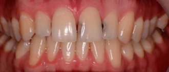
Periodontium is the connective tissue adjacent to the root system of the tooth. Accordingly, periodontitis is considered an inflammation or change in the structure of tissues in this area. The periodontium consists of blood vessels, nerve connections and lymphatic formations. It performs the function of feeding the organ and distributing the load from the tooth to the bone.
Periodontal inflammation is a fairly common disease, since this diagnosis occurs as a complication of advanced dental caries. The destruction of dental tissue opens access to the internal cavity of the organ. The next stage usually becomes pulpitis - inflammation of the nerve bundle of the tooth, and if this process is not stopped, then the infection through the root canal goes deeper into the root and, as a result, leads to periodontal infection.
Of the many types of periodontitis described below, the leading position is occupied by periodontal inflammation of infectious origin. Hemolytic streptococci, entering the pulp tissue, slowly corrode it, destroying the entire tooth structure.
Symptoms and manifestations
If pulpitis is diagnosed by acute pain upon contact with cold or hot food, then the characteristic symptoms of periodontitis are painful tapping or pressing on the tooth.
This diagnosis may be indicated by:
- swelling of the cheek or gum near the tooth;
- a feeling of fullness in the tissues of the organ;
- the occurrence of a fistula with the outflow of pus into the oral cavity;
- increased pain when eating or pressing on the causative tooth;
- the feeling of an “overgrown” tooth (due to compaction of the tissues under the root, the organ begins to bulge);
- pulsation in the root area;
- increased temperature in the area of the causative tooth.
The chronic type of this disease can be asymptomatic and detected only with the help of an X-ray examination. Therefore, the insistent recommendations of the dentists of the LeaderStom clinic to undergo a dental examination by a doctor once every six months are so important for the early detection of such diseases.
My gums hurt and are swollen, what should I do?
If you start to feel pain in your gums, you shouldn’t hope that maybe it will go away on its own; hurry up and see a doctor. It is he who will understand the reason that caused the development of inflammation. Determines the degree of the inflammatory process. Once diagnosed, treatment is very effective and you will soon feel healthy again.
For prevention purposes, we recommend choosing the right brush and toothpaste according to the condition of your teeth and gums. For example, sensitive gums are further damaged by hard bristles, so it is better to choose a brush with soft bristles. It is necessary to practice oral hygiene at least 2 times a day – in the evening and in the morning.
If you follow the recommendations described above, both your teeth and gums will not cause you any discomfort.
Types and differential diagnosis
Like most diseases, periodontitis can occur in acute or chronic form. Sluggish periodontal inflammation is divided into three types:
Granulating
Depending on the type of infection that affects the tooth tissue, and on the specific immune response of the body, various kinds of altered tissues are formed in the periodontal cavity. With chronic periodontitis of this type, granulating tissue appears. It looks like a loose red formation near the top of the tooth root, which quickly grows and thereby destroys the bone tissue of the jaw. Of all three types of chronic inflammation, granulating periodontitis is considered the most active and pronounced. Its characteristic manifestation is the formation of fistulas, which can keep the tooth in a latent form of the disease for a long time. Connective tissue can not only affect the plate of the alveolar process (jaw), but also spread even to the skin. Therefore, advanced cases of periodontitis are much more difficult to treat and can affect large areas of tissue, including adjacent teeth. An X-ray image allows differentiation of granulating periodontitis from other types of periodontal disease. It clearly shows darkening in the area of the apex of the tooth root with circular outlines of tissue or in the form of flames. At the same time, the image does not show sharp boundaries, as in some other classifications of periodontitis. These dark spots indicate that the bone tissue has dissolved as a result of inflammation and has been replaced by connective tissue.
Fibrous
This form of the disease is quite rare and is accompanied by an asymptomatic chronic course. As a result of sluggish periodontitis, connective fibrous tissue forms around the tooth root. It is denser and is visible on an x-ray as a periodontal tooth gap, more widened than usual. The result of the appearance of fibrous tissue can be:
- suffering from acute periodontitis, which was successfully treated;
- poor-quality filling of tooth root canals;
- lack of treatment for granulating periodontal inflammation.
If the disease has entered the stage of remission (quiescence), and the patient does not complain about the causative tooth, then fibrous periodontitis cannot be treated. However, in cases of exacerbation, therapy in the form of refilling the root canals of the tooth is possible.
Granulomatous
If granulating periodontitis does not have clear outlines on an x-ray, then granulomatous periodontitis is characterized by the presence of a capsule with purulent contents. It occurs at the apex of the tooth root and gradually grows, affecting the bone tissue.
There are three degrees of development of periodontal abscess:
- Granuloma. The size of the capsule with this diagnosis reaches 0.5 cm in diameter.
- Cystogranuloma. It is diagnosed when the dense membrane reaches 1 cm.
- Cyst. More than one centimeter in diameter suggests the cystic development of periodontitis.
An increase in granuloma indicates that the inflammatory process does not find a way out and continues to dissolve the bone tissue near the tooth. In this case, a whole chain of pathological processes occurs in the human body. Sometimes particularly advanced cases showed a cyst of up to 4-5 cm in the tissue, which significantly impaired the integrity of the lower jaw, increasing the risk of its fracture.
On the other hand, the accumulation of serous fluid in the tooth area regularly causes additional intoxication to the entire body, which can be accompanied by periods of weakness, fatigue, low-grade fever and other symptoms of periodontitis.
Among all forms of chronic
Granulomatous periodontitis is considered to be the most sluggish. The fibrous tissue of the capsule serves as a protective barrier that prevents pathogenic bacteria from entering the blood. Therefore, the liquid in the capsule can remain in the tooth tissue for years without showing itself. Depending on their location, granulomas can be located either immediately under the root apex or in the tissues of the lateral cavity.
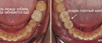
Why does food end up between teeth?
In dentistry, there is the concept of a contact point. This is the name given to the point of contact between the lateral surfaces of two adjacent teeth. The function of the contact point is to protect the gingival papilla from injury from pieces of chewed food. The papilla itself protects the interdental space from food entering there. Therefore, when the contact point is destroyed, problems begin.
This problem can arise for various reasons:
- due to poor-quality treatment of caries and incorrectly placed fillings - for example, when the anatomical relief of the tooth being restored is not accurately reproduced;
- in case of poor-quality dental prosthetics - with the formation of an overhanging edge of the crown, with its contour being too wide, due to cement remaining on the surface after fixing the crown, or due to an incorrectly designed hanging part of the bridge.
But the anomaly is not always caused by dental errors. Very often, food gets stuck in people with disturbances in the natural structure of the dentition: with crooked, tilted and crowded teeth, with a diastema - a gap between the upper or lower incisors, or with trema - gaps between the lateral teeth.
In addition, food remains in carious cavities and in interdental spaces formed after injuries and professional tooth extraction. Tartar formed above and below the gum also contributes to the sticking of pieces, which provokes the development of periodontal diseases.
Causes of development of chronic periodontitis
As already mentioned, one of the main causes of periodontal inflammation is untreated caries. However, the disease can develop for other reasons.
- Low quality filling materials. Some types of root fillings (especially those based on clove oil) can dissolve over time, forming empty cavities in the tooth tissue, which become a favorable breeding ground for anaerobic microbes. Other filling materials may shrink or crack, which can lead to the same result with bacteria and periodontitis. Partial filling of the root canal. Very often, when removing the pulp and cleaning the canals of the tooth, a small area remains at the apex of the root. If the canal is not completely sealed, then an infection develops in these tissues, which ultimately leads to periodontitis.
- Perforation of channels. When installing pins or cleaning channels, they sometimes break through. If a small hole in the root of a tooth is not disinfected and sealed hermetically, then inflammation will develop on the part of the periodontal tissue. This disease is characterized by the occurrence of periodontitis on the puncture side.
- Inflammation of the gums or periodontitis can also cause periodontal disease. There is a high probability of infection spreading if the patient has chronically inflamed gums with deep pockets of soft and then bone tissue. Particularly advanced cases can even lead to periodontitis of the apex of the roots.
- Tooth injuries. There is a category of people who do not consider it dangerous to crack a nut or open a beer bottle with their teeth. This bravado lasts until the tooth becomes inflamed or breaks off. Even such a “harmless” action as the habit of chewing a pen can turn into chronic periodontitis.
In addition to the obvious provocateurs of this disease, there are also secondary factors that indirectly cause or contribute to the development of incipient periodontitis in the dental tissues.
- Failure of metabolic processes in the body.
- Pathogenic microflora in the oral cavity.
- Malocclusion, which leads to injury to dental crowns.
- Chronic diseases of internal organs.
- Reduced immunity.
- Frequent infectious and viral diseases.
- Diabetes.
- Disturbances in the endocrine system.
A separate category of such factors should include poor nutrition, which leads to multiple imbalances in the body, and poor oral hygiene.
Treatment methods
Treatment of inflamed interdental papillae is aimed at eliminating increased sensitivity and pain. This may include:
- rinsing the mouth with antiseptic solutions and chamomile decoction,
- applications of anti-inflammatory gels and ointments,
- taking antibacterial drugs,
- professional teeth cleaning,
- coagulation of overgrown tissues.
The treatment regimen is chosen by the doctor taking into account the diagnosis, clinical picture, and individual characteristics of the patient’s oral cavity.
Acute periodontitis
The acute form of this disease is characterized by similar symptoms as in the chronic course of the disease, only in a more pronounced form. Sharp pain in a tooth can begin spontaneously, often at night. In this case, the slightest touch to the causative tooth leads to a “lumbago” effect. The periodontal tissues swell significantly, which causes the tooth to rise, as if to grow. This fact further injures the diseased tissue, since the enlarged tooth, when chewing, receives more pressure from the closing tooth from the other jaw.
The active phase of the disease is divided into two types:
- serous periodontitis;
- purulent periodontitis.
The first two to three days pass through a serous period, when fluid with a large number of leukocytes accumulates at the site of infection. After this time, the serous tissue becomes purulent and all the symptoms of periodontitis intensify. A so-called throbbing pain in the tooth appears, which practically does not subside. If a person has not yet consulted a dentist, then before the visit, acute pain can be eased by taking painkillers. It is simply impossible to delay going to the doctor at this stage, since the symptoms will not allow the person to eat, sleep, or live normally. The accumulation of purulent contents at the root of the tooth corrodes hard tissues, which leads to the organ’s staggering. The areas of the gums, cheeks and lips are also painful and have swelling of varying degrees. Acute purulent periodontitis is a rather dangerous disease, since in the absence of effective antibacterial treatment it can acquire various stages of complications.
- Periodontal stage. Pus accumulates in the area of the periodontal gap, causing the feeling of an overgrown tooth. This is the initial stage of dental tissue abscess, which must be stopped immediately.
- Endosseous stage. The infection, along with pus, enters the bone tissue. The process of damage to the alveolar plate begins.
- Subperiosteal stage. The amount of pus under the root of the tooth increases, and it accumulates under the periosteum. This formation is called flux. In parallel with the increase in flux, the swelling of the cheek intensifies. Acute and aching periods of pain worsen.
- Submucosal stage. It is characterized by a breakthrough of the periosteum and the release of exudate into the soft tissue. Opening the fistula relieves acute symptoms of pain in the tooth, transforming periodontitis into a chronic form. However, as soon as the outflow is disrupted, periodontitis immediately reactivates.
Periodontitis and implantation
With periodontitis, a paradoxical situation often arises: the disease leads to tooth loss, but it also prevents their restoration. It is impossible to attach a denture to mobile and weakened teeth. Classical implantation in case of periodontitis is often impossible, since the disease is accompanied by a rapid decrease in the volume of bone tissue necessary for reliable fastening of the implant.
Today, technologies have emerged that make it possible to restore missing teeth even with bone atrophy and inflammatory processes of the gums. Modern implants of a special design can also be installed for periodontitis. In certain clinical situations, the “all on 4/6” technology is actively used. The implants are placed at certain points of the jawbone that are least susceptible to changes, and the prosthesis is installed on such reliable supports. In some cases, bone tissue augmentation is also performed.
The service life of modern implants is 10 years or more. Survival rate is at least 95%. You can get more detailed information on the topic by signing up for a free consultation with Viodent specialists.
Periodontitis by location
There are two main divisions in the classification of periodontitis based on the location of the disease: apical and marginal. The first option (apical) is also apical, based near the apex of the tooth root. This type is the most common, since periodontal infection in most cases occurs through a descending channel: from caries to inflammation of the pulp, and then through the root canal the infection descends into the periodontal tissue. This process most often becomes chronic, since the protective mechanisms of the periodontium are much more powerful than those of the pulp. Therefore, the infection can enter the deep tissues of the tooth for years without causing signs of disease.
Marginal chronic periodontitis is located on the lateral walls of the tooth root and has the etiology of microtraumas.
Periodontitis, the treatment stages of which are a complex, complex process, requires immediate attention to a dental clinic. It is safe to say that such a diagnosis requires highly qualified specialists with extensive experience in solving such problems. Dentists of the LeaderStom network of clinics are rightfully considered the best in this area of dental therapy. The latest technologies, progressive techniques and extensive practice allow them to cope with the most complex, advanced cases.
What causes food getting stuck in teeth?
As a rule, those patients who become annoyed by the need to constantly remove food debris turn to a dentist for advice. They can be understood: over time, this procedure begins to take more and more time. Many people also note the appearance of bad breath. But these are only the very first signs of an unresolved and therefore progressive problem.
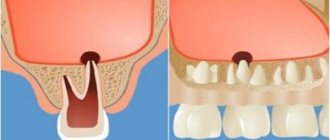
- By injuring the gingival tissue, food debris over time causes chronic inflammation, which is invisible to the patient due to its sluggish nature.
- Constant use of toothpicks and floss aggravates inflammation, transforming it into gingivitis and periodontitis. In the absence of treatment, in addition to the unpleasant odor, the patient will sooner or later be faced first with exposure of the tooth roots, and over time with loosening and premature loss of the teeth themselves. This is explained by the gradual resorption of the bone interdental septa adjacent to the source of inflammation.
- Poorly cleaned dental spaces become a breeding ground for pathogenic microflora that affects the contact surfaces of the teeth. Caries in these places is difficult to recognize at the initial stage and often manifests itself as severe pain already at the stage of development of pulpitis.
How to detect caries in interdental spaces?
One of the specific features of interdental caries is that it is difficult to diagnose. Since enamel damage is located at the junctions of teeth, it can be quite difficult to detect it yourself. Therefore, caries between teeth is rarely diagnosed at an early stage, when it is possible to manage with conservative methods and not prepare the tooth. Much more often it is detected already in the presence of symptoms in the form of pain or a reaction to cold/hot.
The dentist diagnoses interdental caries using several basic methods:
- External visual inspection using a mirror and probe. Only an experienced dentist can detect areas affected by caries and dark spots on the enamel in hard-to-reach areas.
- If the teeth are very closely spaced, a diagnostic examination may be difficult, and then the dentist will order an x-ray, which will show the presence of a carious cavity in the tooth.
- Sometimes, as an alternative to x-rays, a modern sound diagnostic method is used.
After making a diagnosis, the question arises of how caries between teeth is treated.
Interdental caries of the anterior teeth: what should be done?
The most unpleasant and “ugly” caries occurs between the front teeth. If the disease is not detected at the spot stage, then treatment will be carried out approximately according to the following scheme:
- a drug with an anesthetic effect is applied to the affected area;
- clean areas affected by caries from dental plaque and other deposits to prevent the spread of pathology to neighboring areas;
- isolate the affected teeth using a latex napkin (rubber dam) to prevent saliva from getting into them or being injured during dental treatment;
- carious cavities are prepared, the affected tissue is removed on the contact surfaces between the two front teeth using a drill;
- treat the interdental areas with antiseptic solutions (for example, chlorhexidine), and then with adhesive compounds, which improves the fixation of the filling to the tooth walls;
- teeth are filled, and then polished and ground in such a way as to restore their natural anatomical shape and the contours of the interdental spaces as much as possible;
- in case of severe damage, when it is not possible to restore the aesthetics of the front teeth with a filling, they are restored using crowns or veneers.
Both upper and lower teeth are treated in this way.
How is caries between teeth treated when molars and premolars are affected?
The algorithm is like this:
- First, anesthesia is performed. If the lesion is shallow, sometimes topical anesthesia is sufficient; for deeper stages, an anesthetic injection is given with a drug to which the patient is not allergic.
- The affected areas are cleaned of plaque and tartar, and a rubber dam is applied.
- Carious cavities are prepared; in most cases, not only the affected tissue is removed from the chewing teeth, but also some of the healthy tissue to provide the necessary access;
- The cavities are treated with disinfectants and treated with adhesives.
- The tooth is filled with modern photocomposite materials, which are illuminated with special lamps. To restore the side walls of the chewing teeth and ensure tight contact between them, the dentist uses special wedges and matrices.
- At the final stage, the restored surfaces are polished.