Inflammation of the nerve of the tooth is characterized by:
- Pain from temperature stimuli;
- Very severe pain that radiates to the ear, temple or larynx;
- Night pain.
In the first stages, pain appears rarely and quickly subside, but as pulpitis develops, pain appears more often and more severely. The pain becomes more severe and throbbing when pulpitis becomes purulent.
When the first symptoms appear, you should immediately consult a doctor, because pulpitis is dangerous due to its complications. Treatment of pulpitis consists of removing the dental pulp (nerve) under anesthesia or using arsenic paste, mechanical and medicinal treatment of the canals, filling them and placing a filling, if necessary, using pins.
To perform high-quality work in root canals, qualified doctors at our center use modern treatment methods. The work is carried out with “four hands” (doctor-assistant), which improves the quality of the doctor’s work and the patient’s comfort.
Treatment of tooth pulpitis from 6,500 rubles.
Prices
| Name of service | Price, rub |
| Initial consultation with a dentist | 450 |
| Initial consultation with a dentist for treatment/restoration | 650 |
| Application anesthesia | 100 |
| Infiltration anesthesia | 350 |
| Conduction anesthesia | 400 |
| Removing the seal | 150 |
| Removing an anchor pin | 500 |
| Application of arsenic paste | 200 |
| Installing an anchor pin | 600 |
| Electroodontodiagnosis (EDD) 1 tooth | 200 |
Make an appointment by phone from 9:00 to 20:00 +7, 20-88-20
What tools and equipment are used in our clinic?
First and most important
when treating tooth canals and treating pulpitis, this is an opportunity to quickly and effectively conduct an X-ray examination (radiovisiography). It is necessary for: making a correct diagnosis, determining the length and location of the canals, and monitoring the quality of their filling.
The advantage of a radiovisiograph is that the radiation exposure to the patient is 10–20 times less than when using a conventional X-ray machine.
Second
, the presence of an apex locator allows the doctor to know exactly in which part of the canal the instrument is located.
Third
, the presence of an endodontic motor allows you to work with advanced nickel-titanium instruments to expand and taper the canal. This allows you to efficiently clean and seal the canals.
Fourth
, the presence of optics allows the doctor to see the hidden mouths of the canals.
Fifth
, our clinic uses the latest advances in dentistry in canal treatment - three-dimensional obturation of root canals with hot thermoplasticized gutta-percha. During heating, gutta-percha becomes very plastic, due to which the tooth canal system is tightly sealed. This material is introduced on a special carrier made of a denser, modified gutta-percha - Gutta Core, preheated in a special oven. The tightness of the material virtually eliminates the risk of developing an infection in the tooth. Gutta-percha does not dissolve over time and does not irritate the tissue surrounding the tooth.
This method is very effective, it involves sealing and filling the entire system of lateral tubules, but it requires a highly qualified doctor and considerable financial and time costs.
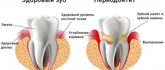
Periodontitis
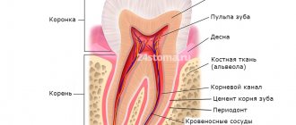
Enamel
Dentine
Pulp
Periodontitis (inflammation at the root apex)
Periodontitis is a process of inflammation that occurs in the bone tissue in the area of the tooth root. Infection from a carious cavity penetrates into the root canal, and from there enters the tissues surrounding the tooth root, destroying them. Periodontitis can have serous and purulent forms. If periodontitis occurs without pain, then the bone around the root of the tooth is reabsorbed, a granuloma appears (a sac at the apex of the root), which over time can turn into a cyst.
Dental granuloma is an inflammation of the periodontium, which is a small round-shaped formation located in the area of the tooth root. This disease, like a dental cyst, is characterized by a long asymptomatic course. Granuloma is aggravated under the influence of certain factors, which, as a rule, do not differ from the factors that cause exacerbation of dental cysts.
During therapeutic treatment, canals require filling not only to preserve the tooth, but also to prevent infection of the body as a result of the decay of dead tissue.
Acute periodontitis is manifested by sharp and severe pain in the tooth area, aggravated by touching it. There may be swelling of the lips, cheeks, bad breath, and sometimes fistulas on the gums. The tooth becomes mobile.
Chronic periodontitis develops slowly and does not cause characteristic pain. In the chronic form of periodontitis, the symptoms are less pronounced, but swelling and redness of the gums and increased temperature may occur. In some cases, a fistulous tract forms on the gum.
Diagnostics
To diagnose periodontitis, it is enough for an experienced dentist to examine the patient’s oral cavity. The presence of fistulous openings, inflammation of the gums around the tooth and the presence of symptoms typical of periodontitis will help to recognize the disease. To differentiate pathology from pulpitis and periodontal disease, use:
- X-ray of the tooth - the images will show rarefaction of the periodontium, the presence of a purulent focus, granuloma or cystic neoplasm at the root apex;
- CT is a more informative method of radiation diagnostics, which allows you to obtain a three-dimensional detailed image of the tooth and its surrounding tissues, on which the boundaries of periodontal inflammation will be visible, and its sources will also be determined.
Treatment of periodontitis
In order to save a tooth with periodontitis, complex and lengthy professional treatment is necessary. In case of chronic periodontitis, the canal of the affected tooth is first treated: infected tissue is removed and the canal is disinfected. Then anti-inflammatory and antimicrobial medications are injected into the canal, and physiotherapeutic procedures are prescribed if necessary. To assess the success of treatment of such a tooth, a control radiovisiographic study is prescribed after 6 months. In very advanced forms of periodontitis, it is sometimes necessary to remove a tooth.
In case of acute periodontitis, the tooth is first left open for the outflow of inflammatory fluid and antibiotics are prescribed, and when the process subsides, treatment is continued according to the same scheme described above. Under X-ray control, root canals are filled using modern filling materials.
Depending on the degree of destruction, the tooth can be restored in different ways:
1. made of composite filling material directly in the mouth. With this type of tooth restoration, titanium or fiberglass pins are used to better fix the filling;
2. restore the missing part of the tooth using a post and a crown.
Treatment of periodontitis from 8,000 rubles.
How to check the quality of canal filling?
Quality control of fillings is an integral part of the treatment process and is performed at every stage. X-ray control is especially important after filling. It allows you to identify areas of insufficiently dense obturation, detect pins or sealer residues protruding beyond the root apex, identify fragments of files and other defects. All this can lead to severe pain after treatment, as well as to the development of complications.
The x-ray image should clearly show the canal cavities, densely filled with filling material. There should be no enlightenment. The filling material should reach the very top of the canal.
Following simple rules of prevention also helps to avoid complications. Pain after treatment can last from several days to a month and does not always indicate ineffectiveness or low quality of treatment. However, if they appear, you should consult your dentist. Also, after treatment, it is recommended to abstain for some time from physical activity, drinking alcohol, and eating hot and spicy foods.
To prevent pulpitis, carefully monitor your oral hygiene. Use only professional products. If during the day you do not have the opportunity to brush your teeth after eating, use Asepta Fresh mouthwash. It disinfects the oral cavity, effectively fights caries, and normalizes acidity.
Treatment of dental cyst
A dental cyst is the last stage in the development of chronic periodontitis. It is a cavity of various sizes, which is formed in the thickness of the jaw and is connected to the root of the tooth. This cavity is lined with a membrane and filled with liquid. When microbes enter, the contents of the cyst suppurate. Usually the cyst grows for a long time without any subjective sensations or clinical manifestations. The first symptom that attracts attention is the deformation of the jaw in the form of a protrusion at the site of the growing cyst.
Tooth cyst
If the cyst does not suppurate, then it can reach a significant size without causing any reaction from the general condition of the patient and without causing him pain.
Types of cysts according to their causes:
- Eruption cyst – most often occurs in children aged 7 – 10 years.
- Paradental (retromolar) cyst - appears when there is difficulty in the eruption of a wisdom tooth and its chronic inflammation.
- A follicular (tooth-containing) cyst is formed due to infection of a tooth germ or an unerupted or supernumerary tooth.
- Primary cyst - is formed when the development of a tooth is disrupted from the remains of dental tissue.
- Radicular cyst - a cyst that forms on the root of a tooth and usually develops due to chronic periodontitis
- A residual cyst appears in the bone after tooth extraction.
Treatment of pulpitis under a microscope with our experienced endodontists:
- extended warranty
- saving 98% of teeth with cysts and periodontitis - no removal
- treatment using modern equipment under magnification and a microscope (fillings last 10 years or longer)
- use only certified materials from Germany, USA, Japan, Israel
- thousands of positive reviews
- high-quality diagnostics , and not treatment “by eye”
- detailed treatment plan with no hidden fees
- quality treatment at prices in the residential area of Yasenevo
Signs and symptoms of a dental cyst
Often the development of a cyst is completely asymptomatic or with barely noticeable symptoms: rare slight pain when biting on a tooth or mild pain when pressing on the gum. In this case, the cyst is detected completely by accident - on x-rays during the treatment of other teeth.
The main signs of a cyst begin to appear already at a late stage of development. Main symptoms of a cyst:
- Aching or nagging pain that only gets worse all the time. It is difficult to get rid of it with the help of simple analgesics and folk remedies. Initially, pain may occur when chewing on the causative tooth.
- The appearance of edema. When a cyst occurs, the gums around the diseased tooth become red and swollen.
- High temperature due to associated infection.
- Suppuration, flux and fistulous tract are external manifestations of an already formed cyst.
- X-rays and tomography are necessary to clarify the diagnosis.
Why is a dental cyst dangerous? Consequences of the disease
A cyst that is not detected in time grows, which leads to the destruction of bone tissue and its replacement with connective tissue formations. As a rule, at this stage complications appear that lead to tooth loss. Possible complications with a dental cyst:
- Purulent inflammation of the cyst.
- Melting of the jaw bone due to cyst growth.
- Inflammation of the lymph nodes.
- The appearance of chronic sinusitis due to the growth of a cyst into the maxillary sinus.
- The appearance of osteomyelitis or periostitis.
- Formation of an abscess on the gum or cheek.
- Formation of phlegmon of the neck due to prolonged purulent inflammation.
- The development of sepsis - blood poisoning.
- Spontaneous fracture of the jaw, which appears due to the growth of the cyst and thinning of the bone.
What is pulpitis
Pulpitis is a dental disease that is accompanied by inflammation of the pulp. Contains a large number of nerve endings, blood vessels, located in the deep tissues of the tooth under the enamel and dentin. The disease is often bacterial in nature and develops against the background of caries and dental trauma. In the International Classification of Diseases (ICD - 10), it is coded K04 - which means “Diseases of the pulp and periapical tissues”.
Therapeutic treatment of cysts
If the size of the cyst does not exceed 8 millimeters, drug treatment is necessary. The entire treatment process takes place in several stages:
Opening of the causative tooth, instrumental and thorough medicinal treatment of the canals. The tooth canals are filled with medicinal paste, which has antimicrobial and anti-inflammatory effects. Periodically, the medicinal paste must be changed, so the tooth is opened again, and the cavity is filled with a new portion of medicinal paste. The number of repetitions depends on how successful the treatment is. The doctor monitors the condition of the cyst using x-rays.
- If the treatment is successful, as evidenced by a decrease in the size of the cyst, the dentist decides to permanently fill the canals and takes measures to restore the tooth.
- The process of further reduction of the cyst and regeneration of bone tissue is monitored for two years. To do this, it is necessary to take a photo every few months, since if the treatment is insufficiently effective, timely measures must be taken.
The only drawback of conservative therapy is its duration. During treatment, regular visits to the dentist are necessary, and further observation by the attending physician is necessary.
Advantages of contacting PROFI-Dent
The main objective of the PROFI-Dent clinic is to provide a full range of dental services at the highest level of quality.
Our advantages:
- modern equipment that allows you to avoid diagnostic errors and act precisely on the affected area;
- a full range of services, including tooth-preserving operations;
- absence of pain and discomfort even during the most invasive procedures;
- use of the latest materials and new generation drugs;
- attractive price.
Treating tooth pulpitis in PROFI-Dent dentistry means solving the problem quickly and as efficiently as possible!
All our regular clients can undergo a free preventative dental examination once every 6 months!