Atheroma on the face is not just a cosmetic defect. It occurs due to blockage of the sebaceous glands. If infection penetrates inside, purulent inflammation may develop. Inflammation of atheroma on the face is very dangerous, especially if the process is located above the line of the mouth. This is due to the peculiarities of the blood supply to the head. The veins of the face drain into the venous sinuses of the skull, which leads to the rapid progression of the disease and the development of abscesses in the brain. The degree of danger of atheroma depends on other factors, including the size of the formation and the presence of underlying chronic diseases, such as diabetes. Therefore, when facial atheroma appears, treatment should only be carried out by a doctor.
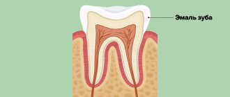
Rice. 1 Facial atheroma occurs due to blockage of the sebaceous glands
Diagnosis of atheroma on the face
Atheroma looks like a small subcutaneous ball. The tumor itself does not resolve and gradually increases. With long-term existence, atheroma on the face can reach 7 cm in diameter. On its surface you can always see a black dot that blocks the sebaceous duct. Atheroma has clear boundaries and a spherical shape. The patient does not feel pain upon palpation. Visually, atheroma is similar to lipoma, hygroma, fibroma, hemangioma, so a doctor must make an accurate diagnosis. If there are difficulties in diagnosis, an MRI may be prescribed to determine the nature of the tumor. After removal of any formation, including atheroma, a histological examination is carried out.
Rice. 2 Atheroma looks like a small ball under the skin of the face
Precancerous neoplasms (precancrosis)
Precancerous neoplasms are those that, under the influence of congenital or current causes, have acquired a tendency to malignant degeneration. As a rule, these are chronic conditions that are observed in a person for a long time.
Thus, precancerous tumors are dangerous neoplasms on the skin that can lead to the development of oncological processes. These include:
- Actinic keratoma is a keratosis in which dry crusts and scales appear on the skin of older people. When they peel off, slight bleeding may occur.
- Xeroderma pigmentosum is a hereditary tumor that develops due to increased sensitivity of the skin to ultraviolet radiation. Rarely encountered, these are pigmented spots that become warty growths.
- The cutaneous horn is a cone-shaped tumor that looks like a horn. Has a yellow or brown color. occurs in exposed areas of the body that are regularly subjected to friction or pressure. Typical for older people
- Bowen's disease is an intraepidermal cancer. Without treatment, it can transform into invasive skin cancer. In the early stage, Bowen's disease appears as a small reddish-brown spot measuring 2-50 mm. has a flaky surface and raised, uneven edges. After removing the scales, a weeping but not bleeding surface remains.
The danger of atheroma on the face
Sebum and keratin masses accumulated in the atheroma capsule have a cheesy consistency. This is a favorable environment for the development of bacteria. An attempt to squeeze out the atheroma can lead to infection getting inside the capsule and causing suppuration. The main symptoms of its onset are:
- redness of the skin around the atheroma;
- pain in the affected area;
- Migraine-like headaches;
- temperature increase.
Inflamed atheroma can lead to phlegmon - inflammation of adjacent tissues without precise boundaries.
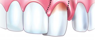
Rice. 3 Self-squeezing of atheroma usually leads to inflammation of facial tissues
Why does my cheek swell?
Periostitis of the jaw
It is the most common dental cause of the symptom. Develops against the background of dental diseases: periodontitis, pulpitis, alveolitis, periodontitis, suppurating jaw cyst. It is provoked by open fractures of the jaws, infected facial wounds, operations, and tooth extraction. In some cases, it becomes a consequence of hematogenous or lymphogenous spread of infection from distant foci. Depending on the form, it is accompanied by the following manifestations:
- Acute serous periostitis.
Moderate swelling of the soft tissues of the cheek, redness of the mucous membrane, and enlargement of regional lymph nodes are detected. The general condition suffers slightly, sometimes there is an increase in body temperature to subfebrile levels. - Acute purulent periostitis (flux
)
.
The contours of the face are sharply changed, the predominant localization of edema is determined by the location of the inflammatory focus in the periosteum. Patients complain of sharp pain radiating to the temple, eye, ear, and neck. General hyperthermia, chills, weakness, headache, and regional lymphadenitis are observed. When examining the oral cavity, an area of swelling with a fluctuation in the center is detected - a subperiosteal abscess. - Chronic periostitis.
Slight swelling of the cheek, thickening of the jaw, and enlargement of the submandibular lymph nodes are typical. The patient complains of periodic moderate pain. The mucous membrane of the affected area is swollen, hyperemic, with a bluish tint.
Other dental diseases
Other possible dental causes of cheek swelling include the following:
- Perimaxillary abscess.
It occurs as a result of infection of the soft tissues of the perimaxillary area due to boils, tonsillitis, wounds, abrasions, and some dental diseases. Swelling is preceded by toothache, which gets worse when biting. Then dense swelling appears, the temperature rises, and appetite disappears. After spontaneous opening of the abscess, the condition improves, but subsequently the pathology may recur. - Vincent's stomatitis.
Develops when immunity decreases due to diseases, injuries, and stressful situations. The leading symptom is the formation of multiple ulcers on the mucous membrane. Swelling of the cheek is detected in severe cases of the pathology. - Noma.
An area of ulcerative-necrotic lesions of the lip or oral mucosa appears. Necrosis covers the gums, tongue, cheek tissue, and facial bones. The tissues around the area of necrosis are swollen; in severe cases, swelling from the cheek spreads to the chin and infraorbital area. - Salivary gland adenoma.
The parotid gland is most often affected. Local swelling with clear boundaries forms on the outer surface of the cheek, in the parotid zone, and spreads to the angle of the jaw, the area under the earlobe. Characterized by slow growth, painlessness at the initial stage, progression of discomfort and dry mouth as the formation increases. - Tumors of the salivary glands.
Along with adenomas, benign connective tissue neoplasia, intermediate and malignant neoplasms can form in the area of the salivary glands. The area of swelling is located in the same place as with adenomas. The swelling slowly increases in benign tumors and spreads quickly in malignant ones. - Purulent parotitis.
Swelling occurs in the parotid area, which spreads to the adjacent part of the cheek. There is severe pain, difficulty when trying to open the mouth, severe intoxication, and severe hyperthermia.
Cheek swelling
Traumatic injuries
All facial injuries are accompanied by rapidly increasing soft tissue swelling, spreading to adjacent anatomical structures. Swelling of the cheek of traumatic origin can occur with the following injuries:
- Injury.
Swelling without clear boundaries. Along with edema, pain, hyperemia, and sometimes hemorrhage are detected. The pain intensifies when opening the mouth and active facial movements. Speech and the ability to eat were preserved. - Hematoma.
Formed against the background of a bruise. A compaction appears in the area of diffuse swelling, which, as a rule, resolves on its own within 1-2 weeks. - Fracture of the upper jaw.
The most pronounced swelling is observed in Le Fort type 1 fractures, combined with neurological symptoms, hemorrhages in the conjunctiva and periorbital area. Type 2 is manifested by edema, hemorrhages in the periorbital zone, and changes in facial parameters. In type 1 fractures, swelling is more noticeable in the area of the upper lip and medial cheek. - Fracture of the lower jaw.
Injuries to the ramus, lateral and angular fractures of the bone body are characterized by swelling of the lower or outer edge of the cheek. Facial asymmetry, hematomas, bruises, articulation disorders, and stepped dentition are observed. - Fracture of the zygomatic bone.
Swelling appears in the cheekbone area, quickly spreads down the cheek, up the infraorbital region. Bruises and hemorrhages form in the conjunctiva. Along with pain, victims are sometimes bothered by nosebleeds and double vision.
Allergic reactions
Swelling of both cheeks is observed with angioedema, combined with swelling of the eyelids and lips, and breathing problems. The condition develops acutely, within a few minutes, less often – hours. It is provoked by contact with an allergen, insect bites. Along with the listed symptoms, angioedema in children may be accompanied by abdominal syndrome, and sometimes by neurological symptoms.
The cause of swelling of the cheek from the oral cavity may be an allergy to prosthetic materials. The pathological condition occurs several months or years after the installation of prostheses and is characterized by a burning sensation in the area where the prosthesis is fixed, tongue, cheeks, soft palate, changes in taste sensitivity, thirst, and dry mouth. When using metal products, a metallic taste may appear.
Ophthalmic diseases
In patients with acute dacryocystitis, swelling of the lacrimal sac area is complemented by swelling of the cheek, eyelid, and dorsum of the nose. In the chronic form of the pathology, swelling is noticeable along the upper inner edge of the cheek and covers the inner edge of the lower eyelid. Cellulitis of the lacrimal sac is characterized by sharp pain and swelling along the inner edge of the eyelid, combined with fever, weakness, weakness, headache, swelling of the cheek and paranasal area.
Neurological pathologies
Local symptoms of cavernous sinus thrombosis are exophthalmos, blurred vision, swelling and pain in the eyeball, swelling of the temple, part of the forehead, cheek, upper lip, and mastoid process. The clinical picture also includes headache, nausea, vomiting, and in case of infectious genesis of the pathology - hyperthermia, intoxication syndrome.
Swelling of the cheeks can be detected with one of the types of angioneurosis - rosacea. There is constant redness of the cheeks, nose, forehead, chin, and the formation of spider veins. The cause of swelling is persistent dilation of blood vessels, which, with a long course of pathology, leads to skin changes.
Skin lesions
Minor swelling of the cheek may result from simple contact dermatitis. The severity of the symptom increases against the background of prolonged contact with the irritant and secondary infection. With allergic dermatitis, the swelling is more noticeable and is combined with itching of the skin. Atopic dermatitis is characterized by mild swelling combined with the formation of vesicles.
Due to the abundant blood supply to the face and the structural features of the soft tissues, a boil on the cheek is accompanied by significant swelling. In the center of the swelling area there is a limited round or cone-shaped formation with a black rod in the center. After the boil has matured, yellowish pus appears around the shaft. Increasing twitching pains are noted.
In young children, swelling of the cheeks is often provoked by superficial pyoderma and occurs against the background of pustular rashes. A severe form of facial skin lesions in adults and children is erysipelas. The disease manifests itself with itching, bloating, and a burning sensation. Subsequently, the cheek swells, and a focus of clearly defined hyperemia with uneven edges, reminiscent of a geographical map, forms on it. Fever and intoxication syndrome are observed.
ENT diseases
Slight swelling of the cheeks is possible with the development of acute sinusitis or exacerbation of chronic inflammation of the maxillary sinuses. Other symptoms include pain, impaired nasal breathing, nasal discharge, weakness, fever, and signs of intoxication. Patients with large odontogenic cysts of the paranasal sinuses are bothered by a feeling of tension and heaviness. Objectively, diffuse swelling of the cheek on the affected side and protrusion of the bottom of the nasal cavity are detected.
Other reasons
Swelling of one or two cheeks is observed with the following pathologies:
- Myxedema.
The swelling is bilateral, uniform, covering the forehead and chin, making the face look puffy. In severe cases, swelling spreads to the entire body. Symptoms of hypothyroidism are observed. - Parotitis.
Due to inflammation of the salivary glands, the perimaxillary area, the outer part of the cheeks, swells. The deformation is bilateral, often uneven. The disease manifests itself acutely, accompanied by fever, chills, and signs of general intoxication. - Melkersson-Rosenthal syndrome.
The leading manifestation is periodic swelling of the lips. Possible swelling of the tongue, cheeks, eyelids. Neuritis of the facial nerve is often detected. - Phlebolith.
Stones in the veins of the cheek are often asymptomatic, but can manifest as pain and swelling, and sometimes inflammation of the affected area.
Treatment of facial atheroma
Getting rid of atheroma using traditional methods of treatment, drugs of different release forms, or self-dissection is ineffective and dangerous . Atheroma can only be removed surgically along with the capsule. The smaller the formation, the easier the procedure will be and the greater the likelihood of being left without a scar on your face. Several methods of operating on atheroma on the face are practiced.
- Laser destruction of atheroma is the most effective and painless treatment method, after which there is no scar. Used to remove formations with a diameter of up to 5 mm.
- Laser excision of atheroma is an innovative treatment method that does not require a recovery period.
- The classic operation with a scalpel is the removal of atheroma. It is performed under local anesthesia.
During the inflammatory process, the doctor cleans out the contents and disinfects the atheroma cavity, then performs an operation to remove the capsule and, if necessary, the affected tissue. Even if the atheroma does not bother you and “does not spoil the beauty,” it still needs to be removed. Pressure on nearby tissues causes them to become deformed, and the possibility of inflammation puts your health at risk.