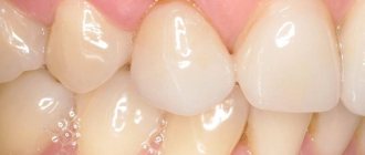
Causes of receding gums
If the gums have receded onto the tooth, this may indicate a dental disease that has caused thinning and reduction in the volume of soft tissues.
In most cases, the causes of gum recession are:
- neglect of hygiene - with irregular cleaning, tartar forms in the oral cavity, causing inflammation of the gums and their decline;
- mechanical trauma to the gingival tissue (for example, during brushing your teeth or while eating);
- gum atrophy caused by natural aging of the body;
- injuries during treatment, implantation or teeth whitening, as a result of which an inflammatory process developed;
- advanced or severe periodontal disease, accumulation of pus in periodontal pockets;
- incorrectly or poorly made dentures that rub soft tissues, errors in orthodontic treatment;
- anomalies and defects of bite;
- pathologies of the structure of the mucous membrane, in which the gums are initially thin and weakened;
- excessively thin cortical bone, causing circulatory problems;
- general diseases of the body that impair blood circulation and the functioning of the immune system.
Only a dentist can determine the exact reason why the gums on the teeth recede, so at the first signs of recession you should contact the clinic.
Apical periodontitis: pathogenesis
Why does inflammation appear in the peri-root zone? Pathogenic bacteria in tissues are the most likely cause of apical periodontitis. It is called an infectious form of periodontitis, it is a logical continuation of untreated caries, which subsequently develops into pulpitis. If the patient continues to ignore the symptoms and does not seek treatment, the pulp dies, allowing pathogenic bacteria to enter the periodontium through the apical foramen.
Another cause of the disease can be mechanical damage - a blow, bruise, displacement or fracture of a tooth. Then the patient is diagnosed with traumatic periodontitis.
The third reason is the penetration of caustic, potent substances into the peri-root tissue, for example, medications. Almost always this is a consequence of a medical error, most often - inaccurate treatment of pulpitis. Such cases are called drug-induced apical periodontitis.
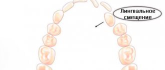
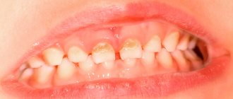
Stages of gum tissue recession
Treatment of the pathology is prescribed based on its stage and form. The following symptoms will help identify them (according to the Miller classification accepted in dentistry):
- tissue loss is hardly noticeable and is visible only at the junction of the dental crown to the gum - the initial stage of subsidence, easily treatable;
- at the point of contact between the gum tissue and the neck of the tooth, the gum level is below normal by 1-2 mm, while the defect does not appear between the teeth;
- receding gums by 3 mm or more, at this stage the necks of the teeth are exposed, soft tissues may bleed, the bone level remains at the same level;
- receding gums by 5 mm or more, accompanied by exposure of tooth roots and mobility of dental units, the most severe stage.
At advanced stages, the gums on the front teeth most often recede - the defect is clearly visible without additional diagnostics.
At the beginning of the disease, it can be determined by the following symptoms:
- the presence of inflammation and bleeding;
- swelling of the mucous membrane;
- formation of periodontal pockets;
- increased sensitivity of enamel;
- pain when brushing teeth;
- whitish tint of gums.
It is worth adding that the disease can affect both the lower and upper jaws, or both at once. In addition, recession can be localized - within 1-2 dental units, and generalized, when the deformation of the gingival contour spreads to the entire jaw.
Regardless of whether the gums have receded on the lower teeth or on the upper ones, you need to seek help from a specialist.
Apical periodontitis of the tooth - what is it?
The diagnosis of periodontitis is made when the patient's periodontal tissues near the apex of the tooth root become inflamed, which is why it is called apical. It affects deep gingival and bone structures, so it is not easy to treat - unlike, for example, caries. Nevertheless, this disease requires immediate intervention, because if the patient is not in a hurry to treat apical periodontitis, then he risks encountering unpleasant consequences of the disease: the appearance of granulomas and cysts, destruction of the alveolar bone, and in the most severe cases, even sepsis.
Potential Complications
Recession of gingival tissue is an independent disease that requires competent therapy. If left untreated, it can lead to much more serious consequences than a violation of the aesthetics of a smile.
First, food gets clogged into the gaps between the teeth, causing bad breath. Next, inflammation develops, which can be accompanied by severe pain, discharge of pus and loosening of the teeth.
Increased sensitivity and soreness develop in the area of the gum margin, which makes eating difficult.
Teeth and roots are affected by caries. In advanced cases, lack of treatment can lead to the loss of one or several dental units at once.
The easiest way to notice when the gums on the front teeth recede, what to do in this case is well known - sign up for a consultation with dentistry to choose a treatment method.
Anatomical structure of the tooth
Anatomy distinguishes three elements of tooth structure:
Tooth root
- This is an invisible “part” that is hidden in the jaw. A tooth can have from one to three roots, depending on its functions. However, there are known cases when one tooth had up to 5 roots. The root is attached to the alveolus (tooth socket), tightly surrounded by connective tissue.
Tooth neck
- This is the transitional part of the tooth from the root to the crown. It is also covered by the mucous membrane of the gums and connected to the bone substance of the alveoli.
Crown of the tooth
– this is the visible part, in fact, what we call a tooth.
The shape of teeth depends on the functions they perform. Nature has provided for all stages of chewing here.
A man takes a bite of food. The front teeth come into play. They feature a thin edge and cut off pieces of food. Such teeth are called incisors. Then the pieces are sent to the pointed outer teeth. The fangs tear them into smaller pieces. Premolars and molars - large lateral teeth - complete the process - chewing food, grinding it, so that the food ground into porridge is sent into the esophagus.
Treatment for gum recession
If the gums on a tooth have receded, the dentist decides how to treat and restore the atrophied tissue after examination and diagnosis.
The method of treating recession depends on the form of the pathology, the number of affected teeth and other factors.
In the initial stages of the disease, therapeutic measures can be used:
- professional cleaning, removal of tartar and plaque;
- cleaning periodontal pockets using closed curettage;
- laser therapy, electrophoresis, current therapy, UV irradiation;
- therapeutic injections based on biogels;
- taking medications - antibiotics, vitamins;
- hygienic rinses with lotions and herbal decoctions.
In the mild stage, it is important to stop the inflammatory process and restore blood circulation in the tissues, as well as eliminate the cause of the pathology.
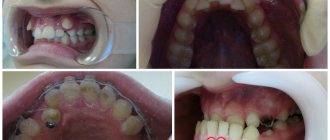
In advanced cases, surgery cannot be avoided. It is carried out in several ways.
Flap reconstruction or plastic surgery. The gum tissue is cut and the root canals are cleaned. The missing tissue is transplanted using tissue from a nearby unaffected area or from the patient's palate. The technique is traumatic, but allows you to quickly and completely restore the gums.
Regeneration or regrowth of damaged tissue. At the first stage, the cause of the recession is eliminated, after which a membrane is applied to the gum, which stimulates the growth of bone and soft tissue. Subsequently, the membrane is removed or resorbed using resorbable membranes.
If there is atrophy of the gingival contour on the upper jaw or if the gums on the lower teeth have receded, what to do and what treatment to resort to depends on the clinical picture.
And timely diagnosis and regular examination by a specialist will help avoid the development of pathology and unnecessary problems.
Hyperesthesia (increased tooth sensitivity) - symptoms and treatment
Treatment of hyperesthesia is aimed at reducing the speed of fluid flow in the dentinal tubules. This can be achieved in the following ways:
- blockage of enamel micropores with the help of desensitizers - drugs that reduce tooth sensitivity;
- reducing the size of micropores using mineralizing agents, for example, gels with a high content of fluoride and calcium. These include gels ROCS Medical Minerals, GC Tooth Mousse;
- closing or narrowing of dentinal tubules using a laser [14];
- preparation and filling of teeth for carious lesions;
- surgery on the gum when it recedes as a result of periodontal disease;
- correction of the bite.
In 1935, dentist L. Grossman identified the main properties of an ideal remedy for combating hyperesthesia [12]. In his opinion, it should act quickly and be effective for a long time, not irritate the pulp, not provoke pain and not change the color of the teeth. There are quite a lot of such drugs now. Desensitizing agents (reducing tooth sensitivity) are produced in the form of gel, toothpaste, rinse, varnish, etc. Toothpastes are most often used.
For mild hyperesthesia , as a rule, applications of various solutions are sufficient.
The drug Remodent in the form of a 3% aqueous solution for applications is applied to areas of hyperesthesia for 15-20 minutes. For the course of treatment to be effective, it is necessary to make 8-28 such applications (one 2 times a week) until a positive result is achieved. Sometimes a 3% solution of remodent is used as a rinse (4 times a week). The full course includes about 40 procedures.
Strontium chloride is used in the form of a 25% aqueous solution and 75% paste. When rubbing the paste, stable compounds of strontium appear with the hard tissue of the tooth. This remedy is a good prevention of hyperesthesia after tooth preparation and removal of dental plaque.
To eliminate hyperesthesia, a 30% aqueous solution of zinc chloride is also used. After this, an application is carried out with a 10% solution of potassium ferrocyanide. It seals the tubules with the resulting sediment. The duration of the applications is one minute. The use of a paste containing alkalis is also indicated: sodium bicarbonate, sodium carbonate, potassium, magnesium. It attracts dentinal fluid, which is contained in the micropores of the enamel, and reduces tooth sensitivity.
A modern treatment for hyperesthesia is bifluoride-12, a special colorless varnish containing fluoride, and fluocal, a dental gel containing sodium fluoride. They are carefully applied to the teeth with a brush or foam swab. Both products create a coating that saturates the enamel and dentin with fluoride ions, which leads to a decrease in sensitivity. Fluocal can also be used in the form of an application solution. Typically, a course of treatment with such applications includes only 2-3 procedures. Varnish is usually used as an auxiliary method that enhances the effect of the main drug [15].
Treatment of hyperesthesia with laser has been shown to be the preferred treatment method. It is aimed at narrowing the dentin tubules by combining the proteins of the dentin fluid, partially melting the hard tissues and relaxing the internal nerve receptors. Laser therapy has no side effects [15].
Treatment of generalized hyperesthesia is aimed at eliminating the cause of the disease, so it not only relieves symptoms, but also affects the course of the disease. Treatment must be comprehensive. In addition to treating the main cause of hyperesthesia, it is necessary to restore the mineral composition of enamel and dentin, and normalize the content of phosphorus and calcium in the body. For this purpose, calcium glycerophosphate, clamine or phytolon, as well as multivitamin preparations are prescribed. Taking phosphorus-calcium drugs throughout the course of treatment provides a good prognosis. Otherwise, restoration of normal sensitivity will take a long time.
To eliminate pain at the beginning of treatment, in addition to using toothpaste with phosphate, electrophoresis with a 2.5% solution of calcium glycerophosphate is required. The course of such physiotherapy is 10 sessions. In case of hyperesthesia against the background of impaired neurological status, the use of a galvanic collar according to Shcherbak is indicated (also at least 10 sessions).
Studies have shown that the effect of complex treatment of hyperesthesia occurs quickly and lasts for quite a long time. The content of phosphate and calcium in the dental tissues increases, the level of calcium and phosphate in the blood normalizes.
During the treatment of hyperesthesia of any degree it is necessary:
- select personal hygiene products;
- control bad habits (do not bite nails, pencils);
- adjust your diet (exclude soda, sticky sweets, consume vegetables, fruits and dairy products more often);
- wear a mouth guard at night (for bruxism) [14].
Treatment methods for apical periodontitis of the tooth
How to treat chronic apical periodontitis and how does the treatment method for acute apical periodontitis differ? In both cases, the patient will need several visits to the doctor, since the therapy is carried out in several stages.
- The first stage:
opening the tooth, thoroughly cleaning the dental canals from the remains of necrotic pulp and areas affected by caries. Expansion of channels. - Second stage:
relief of inflammation. It is produced by filling the channels with antiseptic and anti-inflammatory materials. Additionally, the doctor may prescribe medicinal rinses, and in advanced cases, a course of antibiotics. - Third stage:
filling the canals and monitoring the results of treatment using radiography.
In particularly difficult cases, surgical intervention may be required to gain access to the affected root through an opening in the alveolar bone. If none of the treatment methods brings results, the tooth is removed.
Exacerbation of periodontitis: how does it manifest?
The chronic form of the disease may not have any symptoms for a long time, but sometimes exacerbation phases may occur.
At such moments, the disease begins to resemble the acute stage of development: the gums swell, severe pain occurs, and pus appears. When there is no more pus left in the lesion, the pain goes away and the symptoms disappear, but this does not mean that periodontitis has been cured: it has simply entered the chronic stage again. After some time, an exacerbation will occur again, accompanied by the same symptoms.
Factors such as hypothermia, various diseases, or simply a weakened immune system can provoke an exacerbation of the disease.
Classification of pulpitis
In dentistry, pulpitis is divided into varieties, each with its own characteristics. Each one can be identified with the help of serious diagnostics. Types of pulpitis:
- Spicy:
- focal;
- diffuse.
- Chronic:
- fibrous;
- gangrenous;
- hypertrophic or proliferative;
- chronic with exacerbation.
Exacerbations of chronic pulpitis are difficult to diagnose. They are often mistakenly interpreted as one of the forms of acute pulpitis. In such cases, the history of the disease (history) plays an important role.
Complications after treatment of apical periodontitis
Treatment of acute apical periodontitis (as well as chronic) is a rather complex process that requires accuracy and concentration from the doctor. Like any intervention, it can lead to complications under unfavorable circumstances. What symptoms should you pay attention to after treatment of apical periodontitis?
- A sharp pain that occurs immediately after the end of the anesthesia may indicate that when filling the roots, the filling mass has gone beyond the boundaries of the tooth and is pressing on the gums. There is also a possibility of root perforation and breakage of the thin end of the endoscopic instrument in the root canal.
- After putting the medicine into the canals, the tooth hurts when pressed, and the gums are slightly swollen. These symptoms indicate that the doctor has used a strong medication that irritates periodontal tissue. It could also be a sign of an allergic reaction.
However, in most cases, timely treatment of apical periodontitis ends successfully and does not cause complications.
Diagnosis of apical periodontitis
It should be remembered that the symptoms of apical periodontitis are similar to the symptoms of some other oral diseases: pulpitis, sinusitis, hilar cyst and others. To make an accurate diagnosis, the following studies are necessary:
- electroodontometry
- helps to assess the degree of pulp necrosis; - radiography
- allows you to see tissue changes in the apical region; for chronic apical periodontitis, x-ray is the best diagnostic method, but it may not detect acute inflammation at an early stage; - A blood test
is an additional diagnostic method. With apical periodontitis, an increase in the level of leukocytes and ESR is observed.