Description and causes of the disease
Both children and adults are susceptible to pathology. Depending on the etiological reasons, acquired and congenital anomalies of dental development are distinguished. The first appear throughout life as a result of various external and internal factors affecting the dental system. Congenital are formed due to the presence of unfavorable heredity, or a violation of the embryonic development of the fetus.
Main reasons:
- genetic predisposition;
- teratogenic effects of various substances on the embryo during the formation of organs and systems;
- hormonal imbalance (hypothyroidism, hypocortisolism), vitamin deficiency during pregnancy;
- infections that cause genetic mutations in the fetus (herpes, rubella, HIV, syphilis, toxoplasmosis, cytomegalovirus, etc.);
- fetal hypoxia;
- toxicosis of a pregnant woman;
- bad habits of childhood: prolonged use of a pacifier, bottle feeding, finger sucking, etc.;
- difficult childbirth, birth injuries of the jaw;
- infectious diseases, rickets, hypovitaminosis in the first three years of life, leading to impaired development of segments.
Various factors can provoke abnormalities in the development of teeth in children: alcohol, drugs used by the expectant mother before and during pregnancy, unbalanced nutrition, sudden climate change, radiation, lack of fluoride, calcium, etc.
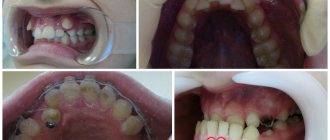
Features of diagnosing dental anomalies
The diagnosis is made even if the tooth is rotated only a few degrees around its axis. Cases of turning at an angle of up to 45° are classified as minor, and all others are classified as significant. The maximum rotation angle of the dental crown is 180°.
To diagnose and develop a pathology correction program, the patient may be prescribed:
- X-ray of the problem area;
- orthopantomogram, which allows assessing the quality of tissue;
- anthropometric analysis, during which, using specially prepared models of the patient’s jaws, the parameters of the components of his dental system are determined and their relationship is analyzed.
Diagnostics makes it possible to identify the cause of tortocclusion and develop a treatment program that corresponds to the degree of its severity and the condition of the dental tissues. When the teeth are rotated by 10–20°, they resort to straightening the teeth with braces, a trainer or a plate. When the angle is 20–40°, special orthodontic devices based on a spring arch and elastic rings are used. In even more complex cases, orthodontic treatment is combined with surgical treatment.
Types of dental anomalies
| View | Characteristic |
| Size pathology | Macrodentia. The crown is significantly enlarged. The developmental anomaly is associated with the fusion of several rudiments into one due to hormonal imbalance. Defective units are more often located in the upper row of the smile zone. Microdentia. Excessively small units. Pathogenesis is associated with genetic predisposition. The violation affects the incisors, mainly the upper ones. |
| Anomalies of dental structure | Hyperplasia. “Pearls” are formed on the crown - formations of enamel of various shapes, 1.5 - 3.5 mm in diameter. Droplets are most often localized near the neck of the segment, or in the bifurcation zone of the root. Dysplasia. The surface of the unit is heterogeneous, thinned in places, and gray spots are present. There are many serrations on the incisors. Chipping and increased sensitivity may occur. Hypoplasia. The enamel layer is sharply thinned, sometimes absent (aplasia). Leads to premature development of inflammatory diseases of hard tissues (caries, pulpitis). Dentinogenesis imperfecta. Units are amber in color, opalescent. Enamel and dentin are fragile, susceptible to abrasion and destruction. Insufficient amelogenesis. It manifests itself as thinning of the enamel, the appearance of brown spots, and hyperesthesia to stress factors. |
| Form defect | Spine-shaped (in the form of a thorn or awl). The segments are wide at the base and sharply taper towards the periphery. Possible combination with microdentia. The surface is uneven and spotty. The pathology is characteristic of segments of the smile zone. Barrel-shaped (Hutchinson's teeth). The neck is thickened, the cutting surface has a recess in the center of which there is no enamel layer. Molar segments with a shortened surface and enamel hypoplasia, etc. |
| Growth disorder | Anomalies in tooth growth are associated with their abnormal spatial arrangement: vestibular displacement (outward); oral (inside); high or low localization; trema, diastema (gaps between units); tortoanomaly (rotation of a segment around an axis); crowding; transposition (change of place). |
| Pathology of numbers | Supernumerary (hyperdentia). Extra units. Hypodentia (insufficient quantity). Adentia (complete absence of units in the mouth). |
| Impaired eruption | Retention (retention of a unit in the jaw or alveolar process). Late eruption of incisors. Impaired eruption of paired segments. Premature eruption. |
| Abnormalities of tooth roots | Characterized by a change in the number of roots (downwards or upwards), their curvature |
Common symptoms that occur with most developmental anomalies are: discomfort in the mouth (sometimes soreness), problems with chewing food, and speech defects. Such pathologies are often accompanied by other genetic defects (cleft palate, lips, etc.).
Hyperdontia or supernumerary groups
The mechanism of the appearance of supernumerary teeth is not completely clear. Some authors are of the opinion that atavism is to blame, while others believe that this occurs as a result of splitting of the tooth germ. Another group of scientists thinks that such teeth grow under the influence of both reasons. Supporters of the first hypothesis believe that gradually such an anomaly will cease to manifest itself, since atavism will become obsolete. In addition, this theory explains the appearance of only teeth near the canines or incisors. As for the remaining teeth, many still adhere to the second point of view.
As a result, we can conclude that such teeth are a genetic anomaly, which is represented by the growth of teeth, most often of an irregular anatomical shape: cone-shaped, oval or multifaceted. It is extremely rare that the form is correct. As a rule, treatment involves only surgical intervention.
Types and classification of malocclusions
Bite is the position of the segments at the moment of tight closure of the jaw bones. Occlusion is assessed in 3 planes:
- by crown height (vertical);
- lateral arrangement of segments relative to the jaws (transvesial);
- ratio of the length of the row and the jaw (sagittal plane).
Malocclusion can be of 2 types: skeletal and dental. In the first case, the cause of pathological closure of the elements is the non-standard size of the jaws or their position in the mouth. Dental abnormal bite is formed due to dental defects (number, size, shape, etc.).
Classification of dental malocclusions:
- Open: the segments of the top and bottom rows do not meet each other vertically. The mouth remains slightly open. Due to the defect, speech, breathing, and chewing are impaired.
- Deep. The lower crowns are significantly (more than ½ covered by the upper ones). The face takes on a flattened shape. Often leads to injuries to the mucous membrane, rapid abrasion of teeth, and defects of the temporomandibular joint.
- Distal (prognathic). Occurs due to disproportionately developed jaws (overdevelopment of the upper or underdevelopment of the lower).
- Mesial: the lower row overlaps the upper segments.
- Crossed: on one side of the jaw, the teeth are located without pathology, and on the other, the lower crowns protrude above the upper ones.
Why does thorotoanomaly occur?
Most often, the tooth is forced to take a pathological position due to lack of space in the jawbone due to macrodentia (large teeth in relation to the bone) or an excessively narrow jaw. In this case, it tends to turn around so as to take up as little space as possible, and the minimum will be achieved when turning at an angle of 45°. But pathology arises not only due to the primary lack of free space.
- The crown can rotate in the socket due to constant pressure exerted on it from the side or below, either by a supernumerary tooth, or by complete “neighbors” that erupt not entirely physiologically - for example, with a delay.
- Quite often, erupting permanent teeth unfold due to the persistence of the milk teeth that precede them. If during the period when baby teeth fall out, the “old” one is in no hurry to fall out, then the “new” molar will have to bypass the obstacle, deviating to the side or turning around the vertical. Pathology can be avoided by timely removal of worn-out milk teeth.
- Sometimes the cause of non-physiological tooth eruption is a dental follicle that is incorrectly formed during pregnancy or in early infancy.
- Often a permanent tooth rotates as a result of a mechanical injury received by a child at the stage of changing the primary bite to a permanent one.
Possible consequences
Improper development of teeth and abnormal bite cause uneven distribution of chewing load. This leads to the development of various complications:
- rapid abrasion of closing teeth;
- caries, pulpitis;
- gingivitis, periodontitis;
- bleeding of the mucous membrane;
- displacement of the articular head of the temporomandibular joint.
When bite deviations occur, problems arise with oral hygiene and microcirculation in the periodontal tissues is disrupted. Ultimately, cellular nutrition, gas exchange, and metabolism are disrupted. A complex of pathological processes leads to early destruction of dentin, periodontal ligaments and tooth loss. Timely consultation with a doctor will help prevent adverse consequences. In Moscow, jaw bite correction is carried out by the Center for Aesthetic Dentistry near the Otradnoye metro station.
The effect of malocclusion on teeth
Initially, nature built a high margin of safety into all systems of the body, provided that they work in a given rhythm and pace. Incisors with fangs seem to be the strongest, along with bones, and at the same time they are very vulnerable.
A change in the natural position of the jaws provokes damage to the enamel, the appearance of cracks and chips, through which pathogenic bacteria quickly reach the inner layer, penetrating deeper, causing inflammation and tissue destruction. The entire row loses support, subject to gradual deformation. In addition to the aesthetic effect, this arrangement will certainly affect the elasticity and resilience of the ligaments.
Knowing the dangers of malocclusion in adults and what consequences are possible, consult a doctor in time to correct the situation and maintain a beautiful smile.
Methods for correction and treatment of anomalies
Treatment depends on the clinical situation and is determined by the doctor after a comprehensive dental examination. Correction of dental abnormalities may include:
- surgical removal of wisdom teeth or supernumerary units;
- prosthetics: for adentia, hypodentia;
- orthopedic correction with a crown, veneer, lumineer: for size anomalies;
- restoration with crowns, composite materials, veneers: for non-standard shapes;
- if the structure of enamel and dentin is damaged, complex therapeutic, orthodontic, orthopedic treatment, enamel mineralization, and drug therapy are prescribed;
- eruption disorders: physiotherapy, massage. Sometimes orthopedic treatment (plates, aligners), removal of a baby tooth (if a permanent tooth has formed underneath it), surgical care (for impacted segments), or orthodontic preparation (system braces) may be required in order to create conditions for stretching the segment retained in the gum.
Malocclusions are most often corrected with fixed orthodontic structures (braces, aligners) in adults. Children are mostly fitted with removable devices.
Phenomena that occur after teething
- plaque of various origins, tooth pigmentation;
- increased abrasion of hard tissues;
- defects called wedge-shaped;
- erosion;
- traumatic lesions;
- hyperesthesia.
Changes in tooth color and the appearance of pigment spots on it may depend on several factors:
- taking special medications and food dyes;
- resorcinol-formalin method for treating pulpitis;
- application of silvering of root canals;
- poor quality filling;
- oxidation of instruments left during treatment;
- hemorrhages into the pulp (the enamel turns pink);
- jaundice (yellow color);
- pulp necrosis (dull enamel). Treatment depends on what caused the tooth discoloration.
Increased abrasion of hard tissues
Increased tooth abrasion is a loss of hard dental tissues, which can be caused by both internal (genetic predisposition, diseases of the endocrine system, etc.) and external factors (functional load on the teeth in the absence of some of them, malocclusion pathologies, unreasonable prosthetics). This pathology is accompanied by both functional changes and aesthetic defects.
This disease is quite common and affects about 12% of middle-aged people. Men are more susceptible to it than women.
The first sign of the disease is increased tooth sensitivity, which may decrease as the pathology progresses due to the formation of replacement dentin. Abrasion can occur right up to the neck of the tooth, and causes a decrease in the height of the lower part of the face and changes in the bite, which in turn provokes a change in the ratio of the components of the temporomandibular joint and disruption of its function.
Treatment in this case requires orthopedic completion in most cases. First, the diseases and causes that caused the pathology are eliminated. If other diseases, such as fluorosis, contribute to erasure, treatment is carried out for them as well. The sharp edges of the teeth are ground to avoid injury to the oral mucosa. The crown part of the tooth is restored using inlays or metal-ceramic crowns.
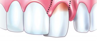
Wedge-shaped tooth defects
If the form of abrasion is localized, the doctor makes special caps with molded chewing surfaces soldered on them. When the height of the lower part of the face is reduced, the installation of prostheses, both removable and non-removable, is used. Wedge-shaped dental defects are often provoked by endocrine diseases, as well as certain pathologies of the central nervous system and gastrointestinal tract.
In this case, the defects are localized on the vestibular surfaces in the area of the crowns of the same teeth from different sides. At first it looks like the appearance of a gap or a kind of crack, but as the pathology develops, such gaps widen and take the shape of a wedge, hence the name of the pathology. Such a wedge has smooth edges, walls without roughness and a hard bottom. The formation of so-called secondary dentin avoids opening the tooth cavity. Further, as the pathology progresses, retraction of the gingival margin is formed, then the necks of the teeth are exposed and increased sensitivity of the tissues to the influence of the irritant occurs.
Treatment of a wedge-shaped defect can be carried out in different ways, and it most often consists of applying medications, filling formed cavities, making crowns from various materials, but it is easier to prevent the occurrence of pathology with the help of orthopedic treatment - timely correction of the bite by installing braces, crowns and grinding of teeth.
Erosion of hard dental tissues
Erosion of hard dental tissue is essentially a progressive loss of hard tissue, and the reasons for this are not fully understood. The disease begins with the formation of an oval-shaped or rounded enamel defect with a hard, shiny bottom, without roughness, formed on the most prominent area of the vestibular surface of the dental crown. Further, the erosion deepens and expands, this is accompanied by a change in the color of the enamel, often also by abrasion of hard tissues.
Treatment of erosion includes a list of measures to remove pigments, remineralizing therapy, filling with composite and glass ionomer materials, and deep fluoridation of teeth is recommended for prevention. Hyperesthesia is an increased sensitivity of dentin, which characterizes pain when the tooth comes into contact with irritants. The main treatment consists of closing enamel micropores and dentinal tubules with special preparations, and remineralization therapy of teeth, as well as recommendations for further dental care for prevention, the main one being the daily use of special toothpastes.
Retention period
Tortoanomalies corrected with orthodontic appliances require a long period of retention as the periodontal and interdental ligaments strive to return the tooth to its previous abnormal position.
Retention methods are divided into orthodontic, surgical and physiotherapeutic:
- Orthodontic retention primarily involves the use of lingual retainers bonded to the inner surface of the teeth. The device used to correct the anomaly can also be used as a retainer.
- The surgical method of retention involves dissection of the interdental papillae with ligaments that tend to return the displaced unit to its previous position.
- The physiotherapeutic method consists of vibration stimulation of the rotated tooth with a frequency of up to 50 Hz. Vibrations are accompanied by repair of the bone tissue of the alveolar sockets, which helps maintain the tooth in a rotated position. Vibration stimulation sessions are carried out several times a week.
The retention time by orthodontic methods depends on the severity of the tortoanomaly and the correction method used, and can be several months.
Characteristic manifestations
The main symptom of the disease is the presence of twisted elements of the dentition. The pathology does not have pronounced symptoms and is detected at an early age as the defective unit develops. In some cases, side effects are observed, such as:
- Regular damage to mucous tissues due to incorrect position of the cutting edge of the problem tooth;
- Difficulty biting food;
- Periodic resumption of stomatitis;
- Formation of interdental gaps – diastemas and trema.
Before starting the correction, it is necessary to conduct an examination to identify the root cause of the development of the pathology.
Preventive measures
Specific methods for the prevention of tortoanomalies have not yet been developed. This is quite understandable, given their polyetiological nature and the impossibility of accurately predicting how teeth will behave during the process of eruption and growth.
You can help reduce the risk and consequences of an anomaly only by regular preventive visits to a doctor , which allow you to identify the development of disorders at the initial stage and take timely measures to prevent them.
The recommended frequency of visiting a doctor depends on the age of the child. In the period from the second to the fifth year (the period of growth and development of baby teeth), it is recommended to visit the dentist 3 times a year; in the future, you can reduce visits to 2 times a year.
It is also necessary to take into account the possibility of a hereditary predisposition to tortoanomaly; it should be the reason for genetic analysis.