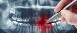
Computed tomography (CT) allows you to most accurately diagnose the condition of the patient’s oral cavity. That is why this procedure must be done before implantation. Only based on the results of such diagnostics can the dentist make a conclusion whether implantation is indicated or, conversely, contraindicated for a particular patient.
CT allows you to obtain a three-dimensional image of the jaw, and in great detail. This is a kind of successful alternative to an x-ray. In dentistry, spiral or multispiral dental tomographs specially created for this purpose are used. They scan tissues and transmit data to a monitor.
Advantages of computed tomography
The results of computed tomography make it possible to detect any pathologies in the oral cavity at the earliest stage of their development. Thanks to this, treatment can be prescribed on time and, accordingly, proceed faster than with an advanced problem. For successful dental implantation, three-dimensional photographs are simply necessary for the dentist; this is the only way to choose an error-free option for installing implants. Statistics show that in 65% of cases of implant failure, dentists did not have three-dimensional photographs of the patients’ jaw bones. On the other hand, if such images are available, successful implantation is observed in 98% of cases.
Indications for this type of examination
3D tomography has a lot of possibilities, for which it is valued by doctors. Thus, it will allow you to assess the condition of not only the jaw, but also the gums, see the quality of previously installed fillings, and reflect all pathologies and anomalies. Indications for this may include:
- abnormalities in the structure of the jaw and the shape of the teeth;
- suspicions of pathological changes in dental tissues;
- identification of the rudiments of permanent teeth in children, assessment of the condition of milk teeth;
- the presence of cysts, inflammation, cracks in the roots of the tooth;
- suspicion of traumatic injuries to the jaw;
- preparation for implant installation;
- the presence of tumors in the jaw area;
- preparation for plastic surgery;
- preparation for operations in the jaw area.
Thus, a detailed three-dimensional image is necessary in all complex cases that require detail and a thorough assessment of the patient's current condition. It is especially important in assessing abnormally growing teeth, as well as in case of complex fractures and dental pathologies.
On a note! A CT scan is also often performed during preparation for implant placement. Moreover, its efficiency and effectiveness are much higher than that of orthopantomography. It is often necessary when performing a so-called sinus lift.
Table. Areas of application of CT.
| Sphere | Explanation |
| Orthodontics | The ability to assess how distorted the bite is. |
| Surgery | Assessment of the possibility of certain manipulations by the surgeon, as well as the possibility of installing teeth. |
| Maxillofacial Surgery | Assessment of the condition of the jaw and surrounding areas of the skull. |
| Therapy | The ability to detect deep-lying caries and choose the right treatment option. |
Benefits of CT
- Minimum X-ray exposure. CT scan is indicated even for young children.
- Diagnostics takes from 8 seconds to 5 minutes.
- There is no need to decipher the diagnostic result.
- The highest quality and resolution of images.
- Adjustable image contrast allows you to reveal the smallest details.
- The ability to study the problem area in three planes.
- Ability to change the scale of the image or part of it.
- The ability to see the location of nerves and thereby optimally select places for bone drilling.
- The ability to quickly make copies of a photo.
- The ability to detect complications invisible to the eye.
Computed tomography is a completely painless procedure. It does not even cause the slightest discomfort in the patient. Depending on the type of tomograph, diagnostics can be performed while the patient is sitting, lying, or standing. No preliminary preparation is required to carry it out. A computed tomography scan can be done even when the patient comes only for a consultation. Pictures can be received instantly. Computed tomography is important for both the patient and the dentist; there is no more accurate diagnosis.
Interpretation of results
After completing the scanning procedure, the radiologist deciphers and interprets the data obtained. The main task of the radiologist when interpreting tomograms is to describe the nature and density of detected tumors, tissue lesions and abnormalities in the development of the jaw. However, the diagnostician does not have the right to make a definitive diagnosis, recommend treatment or give advice on the use of medications. Therefore, after a CT scan of both jaws, the patient should always make an appointment with the attending physician. Based on the issued x-ray report, CT images of the jaw, examination and other studies, the specialist makes a diagnosis and makes decisions on the course of further treatment of the patient.
Study Details
- Doctor's referral: Required for CT scans for children
- Preparation: Yes in case of CT angiography, CT OMT, CT OBP
- Application of contrast: As prescribed by a doctor and with CT scan of blood vessels
- Time: 5-7 minutes
- Contraindications and restrictions: Pregnancy, allergy to iodine on CT with contrast, renal failure
- Results delivery time: 30-40 minutes
| List of studies | Price | Stock |
| CT scan of a large joint (knee, elbow, shoulder, hip, ankle) | from 2500 rub. | At night |
| CT scan of small joints (wrist joint, foot and toe joints, hand joints and fingers) | from 2500 rub. | At night |
| CT scan of the jaw joint and jaw | from 1300 rub. | At night |
| CT scan of bones (one zone) | from 2000 rub. | At night |
| CT scan of the skull | from 2000 rub. | At night |
Disadvantages of Computed Tomography
Alas, this seemingly completely harmless procedure has its drawbacks. They consist in the fact that CT has a number of contraindications. These include the following reasons:
- an allergic reaction to a contrast agent that is used to pre-treat the patient’s oral cavity;
- pregnancy at any stage;
- breastfeeding a baby;
- the impossibility of diagnosing excessively active young children;
- panic fear of closed spaces;
- diabetes;
- kidney diseases;
- thyroid diseases.
The introduction of a contrast agent into the oral cavity allows you to obtain the highest quality image of the oral cavity. However, as a last resort, you can do without this, which experienced dentists sometimes do.
A CT scan of the oral cavity can be done no more than twice a year. Moreover, over the next year it is necessary to refrain from this procedure.
Description of the diagnostic procedure
To understand how computed tomography is done, it is worth considering a step-by-step description of the process:
- immediately before the procedure, the specialist will ask you to remove all metal jewelry to avoid equipment malfunctions,
- then the patient puts on a special protective vest to reduce the degree of radiation exposure to the body as a whole,
- the patient stands or sits with his back to the device, and his chin is fixed using a special stand - this is necessary to eliminate unnecessary movements and get the most accurate image possible,
- after turning on the device, a scanner with an emitting tube begins to rotate around the patient’s head - it is this that transmits the three-dimensional image to the computer.
The photo shows a tomography being performed.
The procedure lasts less than a minute. The diagnosis is completely painless and does not require any serious preparation from the patient.
How is a tomographic image taken?
A three-dimensional dental tomograph consists of a scanner and a computer. Scanning of the oral cavity is performed using very weak x-rays.
Currently, the industry produces two types of tomographs for dentistry. Tomographs of the first type have a scanning device in the form of a cylinder, through which a table with the patient moves. Tomographs of the second type are equipped with a head stand. The stand is installed on the rotating part of the device. If the first type of tomograph is universal, then the second is designed specifically for the needs of dentistry.
Both types of tomographs work on the same principle. The scanner takes from two hundred to six hundred images of the patient’s oral cavity within one hundred seconds. All of them are sent to the computer in electronic format. The program installed on it analyzes the images and produces an accurate image of the area under study. A three-dimensional image is formed by superimposing layers of different thicknesses on top of each other. Each such layer is separately saved as a DICOM file and can be examined separately.
The tomographic diagnostic procedure itself is not at all burdensome and is done in the following sequence.
- The patient is freed from all metal objects that he has with him.
- The patient places his chin on a support.
- The patient remains completely motionless for several seconds.
That's the whole procedure. If the diagnosis requires the administration of contrast, the patient must not eat or drink anything for 4 hours before the diagnosis.
Preparatory stage
CT scan of the upper and lower jaw according to the basic (native) protocol does not require special preparation. If a CT scan of the jaw with contrast is prescribed, the diagnosis is usually carried out with a break in food for 2 hours. If the patient suffers from renal failure, tests to determine the level of creatinine in the blood are preliminarily prescribed before contrast tomography. This way, the doctor can assess the possible risks of developing nephropathy after administration of a contrast agent. Before entering the CT room, it is better to change into comfortable clothes and remove all jewelry.
What exactly does CT help with successful dental implantation?
Computed tomography is not a mandatory procedure when preparing a patient for dental implantation. You can do without it by trusting the experience of the dentist. However, in this case, there is a high probability of unsuccessful implantation. Simply put, there is no complete guarantee that the implant will grow into the bone safely.
Computed tomography allows the dentist to obtain:
- comprehensive information on jaw bone density;
- a complete picture of existing foci of inflammation and defects in the oral cavity;
- an accurate picture of the location of the maxillary sinuses, nerves and blood vessels;
- the ability to accurately calculate the size of implants and the optimal depth of their immersion into the bone;
- the ability to accurately determine the optimal angle of inclination of the implant in relation to the dentition;
- the ability to calculate the maximum permissible mechanical load on the implant.
Agree, each of the listed points is extremely important for successful implantation. Under no circumstances should any of these points be neglected.
Individual application
Dental implantation is not some cookie-cutter procedure. On the contrary, it requires an exclusively individual approach, since no two identical jaw bones exist in nature.
The key to successful implantation is the complete surrounding of the implant surface with bone tissue. The implantation should be completed without the slightest gap between the bone and the artificial root. This process occurs differently for each person.
The dentist’s task, among other things, is to choose the right place to drill a hole for the implant. In this place, the density of bone tissue should be maximum, but it is far from being the same throughout the entire volume of the jaw.
No less important is the choice of the angle of inclination of the implant. If there is an error, the crown will not fit into the overall shape of the remaining dentition.
The drilling depth must also be calculated as accurately as possible to avoid damaging the nerve.
Without computed tomography data, it is extremely difficult to do all this.
Risks and dangers of dental implantation
The most risky is dental implantation in the lateral parts of the upper jaw. This is due to the presence of the maxillary sinuses in this area. The trouble is that after the loss of teeth, these sinuses tend to increase in size. It is almost impossible to find out their size without a computed tomography scan. Problems in the maxillary sinuses can significantly complicate dental implantation in the upper jaw, and therefore diagnosis of the sinuses is done as carefully as possible.
If there is chronic inflammation, tumors, polyps or cysts, dental implantation is not performed until all these problems are eliminated. If this is not done, then the implants in the bone tissue simply will not take root and will be rejected by the body. An opinion on the readiness of the upper jaw for implantation is given by two specialists - a dentist and an otolaryngologist. This approach minimizes the likelihood of implant failure.
Read also
What is dental restoration
If the aesthetic properties of teeth are lost, a person experiences certain discomfort.
What to do if your tooth aches
Sometimes aching pain in a tooth can appear for no apparent reason at first glance.
What are the consequences of not having a three-dimensional image?
Refusal to conduct a computed tomography scan can lead to very sad consequences. The fact is that without a three-dimensional image, the dentist makes a decision almost intuitively, and this, of course, is a risk. The consequences may be as follows:
- nerve damage;
- inflammation of the gums due to uneven load on the implants;
- premature wear of the structure;
- the likelihood of resorting to treatment for problems with the oral cavity;
- risk of re-installation of the implant.
CT scan after implant placement
Many patients have questions about whether a CT scan can be performed after implants, crowns or pins have been installed. This question is asked for a reason. Many people know that when performing an MRI, artificial materials present in the oral cavity create a “phonic” sound and thereby confuse dentists. Nothing like this happens with a CT scan. Moreover, it is advisable to do a computed tomography scan before and after dental implantation. This will allow, firstly, to verify the success of the operation, and secondly, to notice in time possible problems with implantation.
However, if there are metal crowns in the oral cavity, it is better to refuse computed tomography. The fact is that metal interferes with the normal operation of the scanner. As a result, the picture turns out to be of poor quality. The same applies to metal pins.