X-ray examinations in dentistry are divided into two types: traditional two-dimensional radiography and 3D computed tomography. The advantage of the latter is that it allows you to examine each tooth from literally all sides - in all conceivable sections and sections. As a result, the doctor receives the most complete and reliable information about the condition of the patient’s dental system.
What is dental computed tomography?
In modern clinics, 3D photographs of teeth are taken using cone beam tomographs. The design of such devices is based on a C-shaped arc, on one side of which there is an X-ray source, on the other - its detector. During the examination, the arc makes a full revolution around the patient's head, scanning his jaw from different angles. During this time, the detector manages to take about 200 two-dimensional images, which are then converted by software into a three-dimensional model of the jaw.
On a computer, the doctor can study the created 3D model in the way that is convenient for him: rotate, enlarge individual fragments or make sections of potentially suspicious teeth, take bone measurements necessary for implantation. Digital images are high resolution and clear. Therefore, with their help, it is possible to identify features of the dental system that cannot be seen on a regular x-ray.
Advantages of the MRI method
Magnetic tomography is a safe, painless and most informative type of diagnostics for a wide range of diseases:
- blood vessels;
- brain;
- thyroid and mammary glands;
- joints;
- internal organs;
- soft tissues;
- spine;
- tumor processes.
MRI eliminates radiation exposure to the body, does not require special preparation, and has only the presence of metal implants in the body as contraindications. This type of study can be carried out during pregnancy and at a very early age. There are no restrictions on the frequency of tomography repetitions. It is also used to evaluate the effectiveness of treatment.
Indications for computed tomography
- Upcoming orthodontic or orthopedic treatment
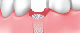
- Planning for dental implantation (in this case, photographs are taken with markings of missing teeth)
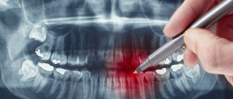
- Diagnosis of hidden dental pathologies
- Diagnosis of cysts, tumors of the dental system, diseases of the temporomandibular joint
Contraindications
- Pregnancy
- Acute respiratory infections
How is the preparation carried out?
Computed tomography of teeth does not require special preparation. Before the examination, the radiologist will ask you to remove items that may interfere with the operation of the tomograph: earrings, hair clips, piercings, glasses, hearing aids. If you have removable dentures, you will need to remove them from your mouth. Before the procedure, the patient must wear a protective lead apron.
What can dental tomography show?
Dental computed tomography allows:
- determine the position of the teeth;
- identify damage under fillings and fixed dentures;
- assess the condition of the dental canals;
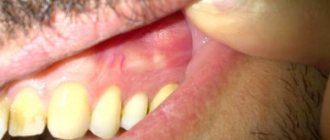
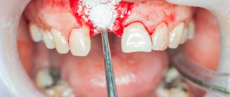
- identify tumors, cysts, fistulas, foci of inflammation in the gums;
- detect the consequences of injuries (fractures of varying complexity, etc.)
How is the research going?
The examination is carried out in 1-3 minutes:
- The doctor takes the patient to the CT scanner and helps fix the head in the desired position. Depending on the design of the tomograph, the examination takes place while sitting or standing.
- A small plate covered with a disposable cover is placed in the mouth, which fixes the position of the jaws.
- The scanning arc of the tomograph makes one revolution around the patient's head. The exposure time to X-ray radiation does not exceed 20 seconds.
The specialist writes the obtained data onto a CD and, if necessary, prints it onto film.
Carrying out dental tomography
No preparation is required for a dental CT scan. The study is not carried out during pregnancy.
The research procedure itself does not cause any difficulties for patients. Dental computed tomography is performed in a standing position. It is necessary to place the chin on a special stand. After which the protection is put on. The picture is taken by the rotating part of the device in one revolution. Do not swallow during the examination.
You can get a dental computed tomography scan in Moscow at the Dental Center of Polyclinic No. 5 of JSC “Family Doctor”.
Sign up for diagnostics Do not self-medicate. Contact our specialists who will correctly diagnose and prescribe treatment.
How much does it cost to take a dental photograph in Moscow?
The cost of a 3D dental photo in Moscow depends on a number of factors: the clinic, the type of equipment and the size of the area being examined. The price of the service usually already includes recording images onto a CD. Printing of photographs is paid separately.
We compared the cost of dental CT scans in two well-known Moscow dental chains: “All Yours”, “Novadent” and in the clinic of the Open Association of Dentists ROOTT.
Dental network “All Yours”
- Cost of initial examination at the clinic: Free.
- Cost of CT scan: The clinic only takes a picture of both jaws for orthodontic or periodontal treatment. The cost of 3D tomography for new clients is 3,700 rubles, for regular clients - 2,700 rubles. The clinic’s website does not indicate the cost of a CT scan with image markings (for patients planning implantation).
- The cost of deciphering the results: 1000 rubles.
- Cost of printing photos: No information on the website. More information about the clinic.
Dental network "Novadent"
- Cost of initial examination at the clinic: Free.
- Cost of CT: Only the lower price threshold is indicated - from 4050 rubles. The minimum shooting area is 10X10 cm (i.e., both jaws are scanned at once).
- Cost of interpretation of results: Included in the cost of CT.
- Cost of printing photos: No information on the website. More information about the clinic.
Clinic of the Open Dental Association ROOTT
- Cost of initial examination at the clinic: Free.
- Cost of CT: In the area of 3 teeth - 1300 rubles, for two jaws - 4000 rubles, for two jaws + sinuses + temporomandibular joint - 5000 rubles. Marking for the implantation of 3 teeth with printing on film costs 1000 rubles, for implantation of one or both jaws with printing on film - 2500-4500 rubles. When receiving treatment at the ROOTT clinic, image marking is performed free of charge.
- Cost of decoding results: Included in the cost of the service.
- Cost of printing photos: Included in the cost of the service. More about the clinic
In our opinion, performing computed tomography in ROOTT clinics is most beneficial for patients, especially for those planning implantation. In addition, this is one of the few clinics in Moscow where you can take three-dimensional photographs of not only both jaws, but also individual segments of the oral cavity, jaw joints, maxillary sinuses and lacrimal canals. Such images are often required by therapists (when treating root canals) or surgeons (when planning an operation). Computed tomography of teeth allows patients to further save on treatment and reduce radiation exposure.
Equipment of centers and cost of services
We use modern PHILIPS ACHIEVA tomographs, expert-class equipment with a power of 1.5 Tesla. This makes it possible to make scanning as accurate and effective for diagnosis as possible. Our prices are truly affordable - at least 25% lower than the average market price for MRI in Moscow.
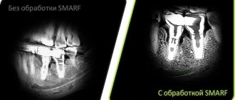
Approximate image quality at 0.3-0.5 Tesla.
Image quality is 1.5 tesla. Higher quality means lower probability of error.
Prescribing a CT scan of the upper and lower jaw before installing implants
Diagnosis of teeth and jaw structures
3D X-rays make it possible to examine areas of the maxillofacial system - from one tooth to a complete set of cross-sectional sections of the jaw and maxillary joint. Tomography before implantation allows:
- identify hidden carious cavities of neighboring teeth, curvature, length of canals, number of broken tooth roots to draw up a treatment plan before implantation;
- detect impacted, supernumerary teeth that may interfere with the installation of implants;
- evaluate bone septa, areas of pathological reorganization of bone tissue;
- visualize inflammation, cysts, granulomas, dental abscesses near the planned implantation area, which must be treated before surgery;
- identify inflammation in the maxillary sinuses and lacrimal ducts, which can become a temporary obstacle to implantation;
- evaluate bone tissue parameters - volume, density, degree of resorption, alveolar process inclination, thickness of cortical plates in order to correctly select the size and shape of the implant;
- clarify the anatomical structure of the maxillary sinuses, mandibular canal and other bone structures in order to plan the angle of inclination when installing an artificial rod;
- identify anomalies of the dentofacial system, pathologies of the temporomandibular joint for the correct modeling of the orthopedic structure for the implant;
- control the quality of implant and crown installation;
- assess the bone density around the installed implant;
- clarify the severity of the injuries received if the dentition is being restored after an injury
The information received in digital format is recorded on a medium (disk, flash drive) and sent by e-mail.
3D modeling of the operation CT results allow you to simulate the installation of implants of the required size, determine the angles of implementation bypassing the anatomical structures and simulate the final result of implantation.
The tomograph data is loaded into a computer program, and a 3D model of the jaw is created. The implantologist creates a virtual operation plan - selects the shape and size of artificial roots, their number, determines the installation location and angle of inclination. This allows you to take into account the nuances in advance.
A customized surgical template is printed using a 3D printer. This is an overlay with guides for future implants, which accurately determine the location and angle of installation of artificial roots. During the operation, the template is tightly applied to the gums, and the implants are installed with maximum precision.
Which is better - multislice or cone-beam CT?
Multislice MSCT
The device scans the area under study in a spiral, the examination is carried out in a horizontal position. Most often used in maxillofacial surgery. Layer-by-layer visualization occurs, soft and bone tissues are examined. The results are indispensable for TMJ dystrophy and jaw injuries. But for traditional implantological treatment, MSCT has some disadvantages:
- results may be inaccurate
due to the horizontal position of the patient - distortion of jaw closure occurs; - high degree of radiation
up to 1000 μSv - such radiation exposure is impractical, since after the operation more than one control examination is carried out in the first year.
Cone beam CBCT
This is a newer method, used in our clinic before implantation. The device rotates around the patient's head, the procedure is performed in a vertical position. Scanning of the area under study occurs in different planes. CBCT is important when planning dental implantation and has advantages over MSCT:
- radiation exposure is less - 25–50 μSv, can be carried out several times a year;
- fast diagnostics;
- wide range;
- cheaper than MSCT.
Contraindications for 3D dental imaging
The radiation exposure of CT scans ranges from 0.045 to 0.06 mSv. This is relatively little, given the recommendations of SanPiN, which indicate the upper possible threshold of radiation exposure for research purposes equal to 1 mSv per year. However, if you compare a CT scan with a dental x-ray, where radiation occurs in the range of 0.002 - 0.003 mSv, a 3D photograph of teeth no longer seems so harmless. In this regard, there are a number of contraindications to CT scanning. If we are talking about simple tomography, then, as a rule, it is not done during pregnancy, especially in the first trimester. The doctor will always consider the need for radiation exposure for a pregnant patient based on the balance of benefit to the mother and risk to the fetus. If tomography with contrast is necessary (for better visualization of soft tissues and blood vessels, it is rarely used for CT of the jaw), then the number of contraindications expands. It should not be given to pregnant women, nursing mothers, or to persons suffering from thyroid disease, severe diabetes mellitus, renal failure, or an allergy to iodine. If you have any allergies or have ever experienced drug intolerance, be sure to tell your doctor.
What is a 3D dental image?
If you visit an orthodontist, maxillofacial surgeon, implantologist, or even a plastic surgeon for a consultation, most likely one of his first appointments will be to do a computed tomography (CT) scan of the jaw, or a 3D dental scan. This is a modern and very accurate diagnostic technique that allows the doctor to view an image of the patient’s jaw from any angle and in any projection, even when the patient has already left his office. CT, unlike an orthopantomogram, provides a three-dimensional image of the jaw without distortion and allows you to look into any layer of tissue, making a kind of virtual section, without the need to perform unnecessary traumatic procedures on the patient live. The efficiency and safety of diagnosis and treatment are thus significantly increased.
CT scan of the jaw: what it shows, how it is done, radiation doses
Dental computed tomography is an important part of diagnostic procedures in the dental field. This is a very informative method that allows you to see the structural features of the jaw, dentition, and also identify pathological processes. Previously, X-rays were used for this, but CT is a more advanced diagnostic method that allows you to get the most accurate picture.
3D dental diagnostics are carried out using a tomograph. In one procedure, you can obtain detailed information regarding the condition of the dentition. The main feature of this method is a three-dimensional image that allows you to see the object under study in full. This is extremely important for making an accurate diagnosis and drawing up a detailed treatment plan.
Content
- The main advantages of dental tomography
- Indications for CT scanning
- Main contraindications
- Is it harmful to have a dental CT scan?
- How often can a dental CT scan be done without harm to health?
- Basic rules of preparation
- How are dental CT scans done in specialized clinics?
- What does this procedure show?
- Computed tomography after implant installation
- Which is better - CT or X-ray?
- Computed tomography of the jaw in St. Petersburg
The main advantages of dental tomography
Compared to other diagnostic methods, computed tomography has the following advantages:
- The entire procedure lasts no longer than 1 minute.
- High level of information content.
- CT is safe for the patient’s health, so it can be performed repeatedly if necessary.
- The high quality of the image in the image allows you to clearly see the structural features of bone tissue.
- The radiation dose received is minimal.
At the end of the procedure, the resulting 3D image is recorded on a CD with universal software, which is installed automatically on the attending physician’s computer for the purpose of further diagnosis.
Indications for CT scanning
The information obtained after a CT scan is necessary to solve a number of problems in the field of dentistry.
The procedure is carried out in the following cases:
- Damage to the jaw after minor injuries. It is with the help of CT that dislocations and fractures can be determined.
- Detection of pathologies in the structure of the dentition. This procedure is commonly used before any type of orthodontic correction.
- Drawing up a computer model of the jaw for the manufacture of an individual brace system.
- As a preparatory procedure before the upcoming operation.
- Presence of neoplasms in the jaw.
- Diagnosis of hidden caries.
- Complications in the dental canals.
- Monitoring the success of previous treatment.
A CT scan is performed before dental implantation, because based on a 3D image, a suitable implant is selected, its installation is carried out and the quality of the procedure is checked.
Main contraindications
During the procedure, the body is exposed to a certain dose of radiation, so computed tomography is not performed during pregnancy. As for women during lactation, the procedure is possible, but after its completion it is forbidden to breastfeed the baby for 24-48 hours.
It is prohibited to perform dental CT scans on preschool children. In this case, alternative diagnostic methods are used. The same applies to patients with a pacemaker.
Is it harmful to have a dental CT scan?
Computed tomography is based on the principle of passing x-rays, with the help of which a series of layer-by-layer images are produced. Accordingly, a certain radiation dose is present during a CT scan of the jaw. It depends on the number of images and the total area of study.
When examining the chest and abdominal cavity, this figure is 11-14 mSv. The critical level for one procedure is 50 mSv (the maximum permissible annual dose is 150 mSv). If this figure is exceeded, there is a high risk of developing cancer.
So is it harmful to have a CT scan of your teeth? The study of this area is considered the most gentle, because the radioactive dose during the procedure is only 0.1-0.3 mSv. Therefore, computed tomography is absolutely safe for the health of patients (except in cases of contraindications).
How often can a dental CT scan be done without harm to health?
Despite the fact that a dental CT scan exposes the body to a minimal dose of radiation, there is no need to overuse this procedure. The frequency of the procedure depends on the degree of need for this, but it must be taken into account that radiation can accumulate in the human body. Therefore, the standard frequency of procedures is no more than 2 times a year.
But there are times when it is necessary to exceed the permissible limit. Based on the permissible radiation exposure, CT scans of the face and jaw should be performed no more than once every 2-3 months.
Basic rules of preparation
There are no special requirements that must be strictly observed before tomography of the dental jaw. It is better not to eat for three hours before the procedure.
The remaining requirements are standard, as in the case of x-rays. You will need to remove all jewelry and objects containing metal. This is necessary to obtain the most accurate research results.
How are dental CT scans done in specialized clinics?
Compared to the old X-ray machine, the tomograph is much more compact. A CT scan can be performed lying down, standing, or sitting. The choice depends on the physiological characteristics of the person, his age, as well as the type of pathology itself.
In cone beam computed tomography, the patient's head is placed between two scanners. To ensure maximum immobility, clamps are provided for the jaw, chin and temples. To understand how a dental CT scan is done, we will describe the procedure in stages:
- The procedure is carried out standing, the platform for fixing the jaw, chin and temples is set in accordance with the patient’s height, and a protective lead vest is put on before the procedure. The head is placed on a special tomograph stand and pressed against the equipment rack. And in this position it is fixed by means of stabilization.
- After preparing the patient, the specialist turns on the tomograph. The moving part of the device rotates around the patient's head. During the entire procedure, about 200-300 images are obtained, which are immediately displayed on the monitor.
- During the procedure, the patient may hear noise - this is quite normal and there is no need to be afraid of it. When it subsides, you do not need to immediately remove your head from the platform or make other movements; you must wait until the x-ray technician reports the end of the study and asks you to leave the scanning area.
During the procedure, the patient does not experience pain or any discomfort. The only discomfort is the forced need to remain immobile. Also, for 10-20 seconds during the image, the patient can feel the heat emanating from the moving scanners.
CT scan of the jaw: what does this procedure show?
The resulting CT image of the teeth is a three-dimensional image of the jaw, which is obtained using hardware positioning and the use of a three-point fixation system.
Computed tomography of teeth allows you to clearly see all pathological processes and disorders in the structure of bone tissue. With this study you can see:
- Crowns and fillings.
- Condition of the paranasal sinuses.
- Location of canals, roots and unerupted molars.
- TMJ condition.
- Various pathologies of the jaw.
The high accuracy of the results obtained allows us to accurately determine the nature of the pathology. If a CT scan is performed for dental implantation, then layer-by-layer images allow you to get the clearest picture of the condition of the maxillofacial area in different projections. The specialist sees the maxillary sinuses, canals, TMJ and blood vessels.
This type of research is the most accurate source of information about the dental system. Only with the help of CT can you correctly plan the upcoming implantation procedure. If you do not use CT, there is a high risk of installing a smaller implant, which will lead to unnecessary stress on the bone tissue.
Computed tomography after implant installation
Quite often the question arises: is it possible to do a CT scan with dental implants? Unlike magnetic resonance imaging, when foreign bodies can produce a “phonic effect,” there is no such problem with computed tomography. Moreover, it is not only allowed, but recommended after implantation.
CT is necessary in the following cases:
- Identification of the reasons for the instability of the installed structure.
- If you need to determine whether the artificial root has taken root or not.
- Determination of the quality of implantation performed. This is especially necessary if computer modeling of the jaw was not carried out at the preliminary stage.
- Identification of complications hidden during visual examination.
If the patient has metal crowns installed, then preliminary consultation with a specialist is necessary. The fact is that metal elements can create unwanted optical effects in images, which will complicate diagnosis and diagnosis.
Which is better - CT or X-ray?
Computed tomography is considered one of the most informative diagnostic methods used in dentistry. Using this method, it is possible to obtain a multilayer image of the area under study. A 3D image is much more informative compared to a conventional X-ray, which only provides a planar image.
From the point of view of affordability, x-rays are preferable, but they do not always provide an accurate picture of the condition of the dental-maxillary system. As practice shows, if a CT scan is performed, then in 99% of cases it is possible to accurately determine the cause of toothache and other symptoms.
Computed tomography of the jaw in St. Petersburg
If the doctor has prescribed a CT scan for you, but you don’t want to wait and sit in lines, then X-ray will offer you to undergo this study on their newest tomographs. The procedure takes no more than a minute, is absolutely painless and effective.
Today we told you how a CT scan of the jaw and teeth is done without negative consequences for your health. You can also record the examination results on a CD, send them by e-mail and make an online appointment. You can find out more detailed information by phone.
Sign up for a study by phone
+7 (812) 332-52-54