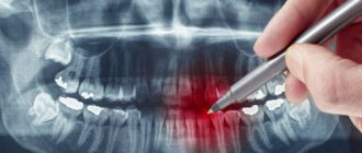
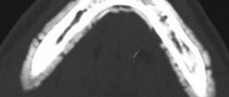
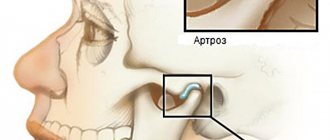
CT scan of the upper or lower jaw allows you to create a three-dimensional model of the structure under study and thereby plan the course of surgical intervention. The main indication for this type of examination is implantation. Thanks to digital images and computer planning of the placement, shape and size of implants, the safety and predictability of the dental restoration procedure is significantly increased.
CT of the jaw is an innovative method of visualizing the maxillofacial area, with greater diagnostic value than panoramic X-rays, allowing for highly accurate three-dimensional anatomical images of the teeth. This is a non-invasive examination of bone density and tooth structure from the inside.
The examination does not require special preparation. Before performing it, you only need to remove jewelry, glasses, removable dentures and orthodontic devices from your mouth.
Make an appointment with a specialist, without queues, at a convenient time
+7
Sign up
Is special training needed?
The preparatory stage includes an initial consultation with a dentist, who conducts a survey to identify possible contraindications. Further preparation depends on what type of research needs to be carried out:
- Normal (without contrast injection). Such a CT scan will not require additional preparation, except for the general requirements: it is recommended to remove all metal objects from the head - earrings, hairpins, etc. if present, remove the hearing aid and removable dentures; warn the radiologist if there are non-removable orthopedic structures in the mouth (this will allow the specialist to adjust the device in a special way).
The lack of serious preparation before taking a CT scan of two jaws or teeth allows diagnostic studies to be carried out immediately after receiving an order from a doctor.
Contraindications and restrictions
The patient’s weight – heavy weight may not allow the examination to be carried out since CT scanners have weight restrictions.
CT scanning is not advisable for persons under 14 years of age, since at this age the body is actively growing and radiation exposure can be risky.
The presence of kidney disease, decompensated heart disease, and pregnancy are contraindications for CT scanning.
The presence of an allergy to iodine-containing products often does not allow CT scanning with contrast, since contrast agents have iodine atoms in their structure, and this sharply increases the risk of dangerous allergic conditions such as anaphylactic shock.
How is a CT scan of teeth and two jaws done?
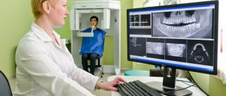
After the preparatory stage described above, the procedure itself begins with a short instruction - the radiologist explains how and why it is necessary to remain still while the device is operating. After this, the CT scan proceeds as follows:
- if necessary, a contrast agent is introduced at this stage;
- The patient wears a vest with a lead layer, which will provide additional protection to the chest area from radiation;
- depending on the type of scanning equipment used, the patient is asked to stand or sit near the tomograph;
- the radiologist fixes the patient’s head with special supports provided in the tomograph (fixation is carried out in the frontal, temporal and chin parts), and the hands are asked to be placed comfortably on the handrails;
- the specialist starts the equipment and the sensor, which takes and transmits 3D images, begins to rotate around the head (in one revolution, the sensor takes 150-200 high-resolution 2D images, after which it converts them into a three-dimensional picture);
- the sensors take readings for no more than 30 seconds and to obtain the highest quality results and accurate images, it is recommended to remain motionless during this time;
- The radiologist releases the patient's head from fixation on the supports and this is where the procedure ends.
Next, it will take a few minutes for the device to process the scanned data and write it to disk. You may be interested and useful to know - in the article “CT of the jaw for dental implantation” we talk in detail about the features of tomographic studies before implanting artificial roots.
You might be interested in:
Dental diagnostics
Computed tomography of teeth and jaws
X-ray of teeth
3D image of teeth
CT scan of the jaw: what it shows, how it is done, radiation doses
Dental computed tomography is an important part of diagnostic procedures in the dental field. This is a very informative method that allows you to see the structural features of the jaw, dentition, and also identify pathological processes. Previously, X-rays were used for this, but CT is a more advanced diagnostic method that allows you to get the most accurate picture.
3D dental diagnostics are carried out using a tomograph. In one procedure, you can obtain detailed information regarding the condition of the dentition. The main feature of this method is a three-dimensional image that allows you to see the object under study in full. This is extremely important for making an accurate diagnosis and drawing up a detailed treatment plan.
Content
- The main advantages of dental tomography
- Indications for CT scanning
- Main contraindications
- Is it harmful to have a dental CT scan?
- How often can a dental CT scan be done without harm to health?
- Basic rules of preparation
- How are dental CT scans done in specialized clinics?
- What does this procedure show?
- Computed tomography after implant installation
- Which is better - CT or X-ray?
- Computed tomography of the jaw in St. Petersburg
The main advantages of dental tomography
Compared to other diagnostic methods, computed tomography has the following advantages:
- The entire procedure lasts no longer than 1 minute.
- High level of information content.
- CT is safe for the patient’s health, so it can be performed repeatedly if necessary.
- The high quality of the image in the image allows you to clearly see the structural features of bone tissue.
- The radiation dose received is minimal.
At the end of the procedure, the resulting 3D image is recorded on a CD with universal software, which is installed automatically on the attending physician’s computer for the purpose of further diagnosis.
Indications for CT scanning
The information obtained after a CT scan is necessary to solve a number of problems in the field of dentistry.
The procedure is carried out in the following cases:
- Damage to the jaw after minor injuries. It is with the help of CT that dislocations and fractures can be determined.
- Detection of pathologies in the structure of the dentition. This procedure is commonly used before any type of orthodontic correction.
- Drawing up a computer model of the jaw for the manufacture of an individual brace system.
- As a preparatory procedure before the upcoming operation.
- Presence of neoplasms in the jaw.
- Diagnosis of hidden caries.
- Complications in the dental canals.
- Monitoring the success of previous treatment.
A CT scan is performed before dental implantation, because based on a 3D image, a suitable implant is selected, its installation is carried out and the quality of the procedure is checked.
Main contraindications
During the procedure, the body is exposed to a certain dose of radiation, so computed tomography is not performed during pregnancy. As for women during lactation, the procedure is possible, but after its completion it is forbidden to breastfeed the baby for 24-48 hours.
It is prohibited to perform dental CT scans on preschool children. In this case, alternative diagnostic methods are used. The same applies to patients with a pacemaker.
Is it harmful to have a dental CT scan?
Computed tomography is based on the principle of passing x-rays, with the help of which a series of layer-by-layer images are produced. Accordingly, a certain radiation dose is present during a CT scan of the jaw. It depends on the number of images and the total area of study.
When examining the chest and abdominal cavity, this figure is 11-14 mSv. The critical level for one procedure is 50 mSv (the maximum permissible annual dose is 150 mSv). If this figure is exceeded, there is a high risk of developing cancer.
So is it harmful to have a CT scan of your teeth? The study of this area is considered the most gentle, because the radioactive dose during the procedure is only 0.1-0.3 mSv. Therefore, computed tomography is absolutely safe for the health of patients (except in cases of contraindications).
How often can a dental CT scan be done without harm to health?
Despite the fact that a dental CT scan exposes the body to a minimal dose of radiation, there is no need to overuse this procedure. The frequency of the procedure depends on the degree of need for this, but it must be taken into account that radiation can accumulate in the human body. Therefore, the standard frequency of procedures is no more than 2 times a year.
But there are times when it is necessary to exceed the permissible limit. Based on the permissible radiation exposure, CT scans of the face and jaw should be performed no more than once every 2-3 months.
Basic rules of preparation
There are no special requirements that must be strictly observed before tomography of the dental jaw. It is better not to eat for three hours before the procedure.
The remaining requirements are standard, as in the case of x-rays. You will need to remove all jewelry and objects containing metal. This is necessary to obtain the most accurate research results.
How are dental CT scans done in specialized clinics?
Compared to the old X-ray machine, the tomograph is much more compact. A CT scan can be performed lying down, standing, or sitting. The choice depends on the physiological characteristics of the person, his age, as well as the type of pathology itself.
In cone beam computed tomography, the patient's head is placed between two scanners. To ensure maximum immobility, clamps are provided for the jaw, chin and temples. To understand how a dental CT scan is done, we will describe the procedure in stages:
- The procedure is carried out standing, the platform for fixing the jaw, chin and temples is set in accordance with the patient’s height, and a protective lead vest is put on before the procedure. The head is placed on a special tomograph stand and pressed against the equipment rack. And in this position it is fixed by means of stabilization.
- After preparing the patient, the specialist turns on the tomograph. The moving part of the device rotates around the patient's head. During the entire procedure, about 200-300 images are obtained, which are immediately displayed on the monitor.
- During the procedure, the patient may hear noise - this is quite normal and there is no need to be afraid of it. When it subsides, you do not need to immediately remove your head from the platform or make other movements; you must wait until the x-ray technician reports the end of the study and asks you to leave the scanning area.
During the procedure, the patient does not experience pain or any discomfort. The only discomfort is the forced need to remain immobile. Also, for 10-20 seconds during the image, the patient can feel the heat emanating from the moving scanners.
CT scan of the jaw: what does this procedure show?
The resulting CT image of the teeth is a three-dimensional image of the jaw, which is obtained using hardware positioning and the use of a three-point fixation system.
Computed tomography of teeth allows you to clearly see all pathological processes and disorders in the structure of bone tissue. With this study you can see:
- Crowns and fillings.
- Condition of the paranasal sinuses.
- Location of canals, roots and unerupted molars.
- TMJ condition.
- Various pathologies of the jaw.
The high accuracy of the results obtained allows us to accurately determine the nature of the pathology. If a CT scan is performed for dental implantation, then layer-by-layer images allow you to get the clearest picture of the condition of the maxillofacial area in different projections. The specialist sees the maxillary sinuses, canals, TMJ and blood vessels.
This type of research is the most accurate source of information about the dental system. Only with the help of CT can you correctly plan the upcoming implantation procedure. If you do not use CT, there is a high risk of installing a smaller implant, which will lead to unnecessary stress on the bone tissue.
Computed tomography after implant installation
Quite often the question arises: is it possible to do a CT scan with dental implants? Unlike magnetic resonance imaging, when foreign bodies can produce a “phonic effect,” there is no such problem with computed tomography. Moreover, it is not only allowed, but recommended after implantation.
CT is necessary in the following cases:
- Identification of the reasons for the instability of the installed structure.
- If you need to determine whether the artificial root has taken root or not.
- Determination of the quality of implantation performed. This is especially necessary if computer modeling of the jaw was not carried out at the preliminary stage.
- Identification of complications hidden during visual examination.
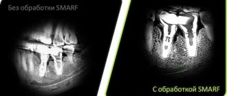
If the patient has metal crowns installed, then preliminary consultation with a specialist is necessary. The fact is that metal elements can create unwanted optical effects in images, which will complicate diagnosis and diagnosis.
Which is better - CT or X-ray?
Computed tomography is considered one of the most informative diagnostic methods used in dentistry. Using this method, it is possible to obtain a multilayer image of the area under study. A 3D image is much more informative compared to a conventional X-ray, which only provides a planar image.
From the point of view of affordability, x-rays are preferable, but they do not always provide an accurate picture of the condition of the dental-maxillary system. As practice shows, if a CT scan is performed, then in 99% of cases it is possible to accurately determine the cause of toothache and other symptoms.
Computed tomography of the jaw in St. Petersburg
If the doctor has prescribed a CT scan for you, but you don’t want to wait and sit in lines, then X-ray will offer you to undergo this study on their newest tomographs. The procedure takes no more than a minute, is absolutely painless and effective.
Today we told you how a CT scan of the jaw and teeth is done without negative consequences for your health. You can also record the examination results on a CD, send them by e-mail and make an online appointment. You can find out more detailed information by phone.
Sign up for a study by phone
+7 (812) 332-52-54
How long does it take to do a CT scan?
The duration of tomography depends on the type of diagnostic equipment used. Before doing a CT scan of the jaw and teeth, it is better to ask what kind of tomograph is used in the clinic. Modern devices allow you to carry out the necessary manipulations as quickly as possible with minimal radiation. If you are offered a procedure that will last more than 15 minutes, then you should refuse. Only innovative equipment allows you to carry out the required research in just a few minutes, guaranteeing highly detailed images.
On average, everything is done in no more than 10 minutes, taking into account the instruction time and the preparatory stage. Please note that 3Shape is also used for 3D diagnostics, but this intraoral scanner is not suitable for all types of diagnostic studies. The attending physician will be able to determine which scan is required for you after collecting a general clinical picture and based on the reason why you came to the dentist.
Preparation
Before performing a CT scan of the jaw, no special preparation is required, with the exception of studies with contrast. Before conducting a contrast study, it is recommended to refrain from eating 6-8 hours before the procedure.
During the procedure, the patient lies on the scanner table. The head can be fixed to achieve immobility during the examination. The duration of the procedure is on average 20 minutes. If contrast is used, which is administered intravenously before tomography, the duration of the study can increase to 30 minutes.
How is a CT scan done?
For a CT scan to be completely completed, it is not enough just to have the scan results. It is necessary to obtain a conclusion, that is, a decoding of what the tomography gave. Typically, a radiologist is responsible for this step. Carefully studying the resulting 3D model, the specialist conducts an analysis and issues a conclusion, assessing the condition of the teeth, gums, root canals, and also identifies the presence of inflammatory processes and neoplasms, and assesses the volume and density of the bone tissue of the jaws.
As a result, a conclusion is given about whether all parameters correspond to the norm. Special attention is paid to the assessment of that part of the jaw system that is described in the direction of the CT scan. All the work of the radiologist at this stage takes no more than 10-15 minutes. After this, the patient receives the scan results in digital format (on disk or as a file via email), a printout (if required) and the described conclusion. Based on these data, the attending physician can make an accurate diagnosis, prescribe effective treatment, or state the results of previously performed dental procedures (if a CT scan was done for control). Now, knowing how a CT scan of teeth and two jaws is done, you can go for a tomography without fear to obtain the necessary diagnostic data.
The service of computed tomography of teeth and jaw is available in all clinics of the Smile Factor network. You can find out more about the procedure and current prices ON THIS PAGE of the website.
Indications
CT scanning of the maxilla and mandible is indicated for various conditions where visual information is needed to confirm the diagnosis. CT is indicated in the following cases:
- Implantation planning
- Diagnosis of pathological changes in the jaws
- Abnormal arrangement of teeth requiring extraction
- 3D reconstruction and measurement of deformities and developmental anomalies in the lower and upper jaw
- Planning of surgical intervention for jaw tumors (lower and upper)
- Injuries of the maxillofacial area
- Sinus infections (sinusitis)
- Tumors
- Facial nerve damage
- Pathology of the mandibular joint
- Pathology of the salivary glands
Read also
What is dental restoration
If the aesthetic properties of teeth are lost, a person experiences certain discomfort.
What to do if your tooth aches
Sometimes aching pain in a tooth can appear for no apparent reason at first glance.
Contraindications to CT scan of the jaw
There are a number of contraindications for which the procedure is not recommended.
These include:
- weight more than 150 kg or girth more than 150 cm (the device is designed for a load of up to 150 kg);
- pregnancy (the risk of developing defects in the fetus increases);
- lactation period (during the procedure, rays can directly affect the composition of a nursing woman’s milk);
- severe mental illness (during the procedure the person must be at rest);
- the presence of metal objects in the area of interest, as they can reduce the information content of the results;
- children under 5 years old;
Contraindications
A CT scan of the jaw can be performed on an adult patient if there is a referral from a doctor or on personal initiative. For children, due to the presence of radiation exposure to the body, computed tomography is performed strictly on the direction of a doctor and exclusively for strict medical reasons.
When scheduling a CT scan, patients should consider the following contraindications:
- the patient is pregnant at any stage;
- The age of the subject is up to 3 years.
Computed tomography of the jaw: price in Moscow
The cost of a dental CT scan in Moscow will primarily depend on the size of the area being examined, the type of device used, and also on whether you need to print the image on film (this will be more expensive) or whether recording the image on a CD is sufficient. For a dental CT scan, the price indicated below already includes recording the image on a CD, as well as a written analysis of the image by a radiologist.
Cone beam computed tomography of teeth: price
- CT scan area 6*6 cm (3 teeth) – RUB 1,700.
- CT scan area 6*10 cm (1 jaw on one side) – 2900 rub.
- CT scan area 7.5*10.0 cm (2 jaws on one side) – RUB 3,700.
- CT scan area 7.5*14.5 cm (both jaws) – RUB 4,200.
- CT scan of the paranasal sinuses – RUB 3,900.
- CT scan 13.00*14.5 cm (both jaws, paranasal sinuses, TMJ) – 4500 rub.
- CT scan of both temporomandibular joints - 6,000 rubles (but if such an image is taken not using cone-beam tomography, but using a cheaper linear tomograph, then its price will be about 1,500 rubles).
Important: if, in addition to a CD, you need a printout of a photograph on film + its markings, then you will have to pay extra for this. The cost of marking the image will depend on the number of missing teeth in the area of which implantation is planned. You can then send such a photograph with markings by e-mail to any clinic, and your presence will not even be required for the initial consultation.
