In 1913, Nikolai Ivanovich Taratynov (1887–1919), a young doctor and employee of the Department of Pathological Anatomy at Kazan University, received material for research and clarification of the diagnosis. It was a fragment of tissue taken from a patient with a contusion of the cranial vault - some time after the injury, a granulomatous formation formed in this place.
Figure 1. 3D reconstruction of skull bones with solitary eosinophilic granuloma https://radiopaedia.org/cases/eosinophilic-granuloma-skull
It was assumed that this was bone tuberculosis, but instead of the classic tuberculous tubercle, Taratynov saw accumulations of mononuclear cells (tissue macrophages or histiocytes) and eosinophils, as well as Charcot-Leiden crystals, which had previously been found in the sputum of patients with bronchial asthma. The morphological picture indicated “the existence of granulomas, clinically and macroscopically completely similar to tuberculous ones, recognizable only microscopically and consisting almost exclusively of eosinophils.”
Figure 2. Eosinophilic granuloma of bone: lesion of the hip in an 11-year-old girl (x-ray). Swelling and pain were noted for 3 months. https://radiopaedia.org/cases/langerhans-cell-histiocytosis-skeletal-manifestations
The previously unknown disease was named Taratynov's disease. Currently, this name is mainly used in Russian-language sources and has rather a historical meaning.
In the 1940s. similar cases were presented in articles by US doctors Sadao Othani, Joseph Ehrlich, Louis Lichtenstein and Henry Jaffe under the names “solitary granuloma of bone” and “eosinophilic granuloma of bone”.
Similar histiocytic infiltrates were described by other authors - Alfred Hand Jr, USA in 1893, Arthur Schüller (Austria) in 1915, Henry Cristian (USA) in 1920, Erich Letterer , Germany) in 1924, Sture Siwe (Sweden) in 1933, in connection with which the terms “Hand-Schüller-Christian disease” and “Abt-Letterer-Siwe disease” were introduced. However, later it was concluded that all of these are forms of the same disease, different in severity and localization of foci, called “histiocytosis X”
in 1953.
Figure 3 . Eosinophilic granuloma of the skull (X-ray) https://radiopaedia.org/cases/eosinophilic-granuloma-1
The answer to the question of what the histiocytes that form granulomas are was given in 1973 by pediatrician and morphologist Christian Nezelof (France), who identified them as Langerhans cells (a type of antigen-presenting cells localized in the epidermis). In this regard, since 1987, the name “histiocytosis X” was replaced by “Langerhans cell histiocytosis” ( LCH ).
The head of the Department of Pathology at St. Petersburg State University, Leonid Churilov, believes that Doctor Taratynov became the prototype of Doctor Zhivago. This bold assumption is supported by the coincidences in the life of Nikolai Taratynov and his family with the story of Yuri Zhivago: both victims of the revolution, military doctors, whose daughters were orphaned early and were raised by their father’s relatives. Nikolai Ivanovich Taratynov was shot and killed by a sailor, the brother of a patient who died in the hospital, in 1919 at the age of 32. You can learn more about Taratynov’s biography here.
Etiology
The question of the reasons for the development of granulomas remains open. One theory considers Langerhans' histiocytosis to be a neoplastic process. This is supported by such features as monoclonality of pathological cells (origin from a single pathological cell), increased expression of proliferation activators and factors inhibiting apoptosis. At the same time there are no genomic defects in the cells , and spontaneous remission of the disease is possible. At the site of granuloma development, activation of T-lymphocytes and T-regulatory cells with suppressor activity has been described. These signs indicate that other immunopathological processes may underlie the development of the disease.
Figure 4. Eosinophilic granuloma: pathological changes. In the last image, the S-100 protein expressed by Langerhans cells is colored. https://radiopaedia.org/cases/eosinophylic-granuloma-histology-1
Causes of granuloma
The reasons for the occurrence of this type of formation can be different, the main ones are:
- damage to the body by infectious diseases (syphilis, typhoid fever, typhus, tuberculosis);
- collagen diseases (rheumatism, scleroderma);
- drug effects;
- diseases caused by dust (silicosis, talcosis, asbestosis);
- dental diseases.
The mechanism of granuloma formation can be divided into 4 stages:
- A collection of young cells prone to phagocytosis (a process by which cells take up and digest particulate matter).
- Active proliferation of formed phagocytes.
- Transformation of phagocytes into epithelial cells.
- A collection of epithelial tissue that forms a granuloma.
Clinical manifestations
Eosinophilic granuloma (EG), also known as Taratynov's disease, is a relatively benign variant of Langerhans' histiocytosis with the appearance of single foci in flat or tubular bones. The appearance of two or three or more lesions is much less common. Typically, Langerhans cell histiocytosis occurs in children and adolescents (up to 15–20 years), more often in boys (approximately 1.5:1). Frequency of occurrence: less than 1 person. per 100,000 population, which is 60–80% of all cases of LCH. In adults, eosinophilic granuloma, like hysteocytosis X in general, is much less common.
The most commonly affected bones are the skull bones, femurs, and less commonly, the pelvic bones, ribs, and vertebrae. There are also cases of the appearance of pathological foci in the thymus, skin, bladder, parathyroid glands, hypothalamus, lungs and gastrointestinal tract.
The clinical picture is determined by the location of the granuloma. An intraosseous formation may not cause any symptoms, but usually leads to the development of swelling, pain, and sometimes pathological fractures. When lesions appear in the jaw, tooth loss, damage to the mastoid process or temporal bone may occur with the development of otitis media. When the walls of the orbit are involved in the process, exophthalmos may develop. Sometimes polymorphic rashes are observed on the head, back, armpits, perianal and genital areas in the form of spots or plaques, small nodules or nodes with ulcerations. Common manifestations of Taratynov’s disease are common: increased fatigue, weakness.
Figure 5. Photo of the manifestations of eosinophilic granuloma on the skin - typical elements of purpura, must be differentiated from seborrheic dermatitis
Symptoms of granuloma
Since most granulomas are not an independent disease, symptoms can be varied and depend on the background of the pathology they appeared against. Sometimes the first signs may go unnoticed.
Inflamed nodules first look like bright red spots. Next, a pink papule appears. In most cases, granuloma is not accompanied by discomfort. Often it can disappear and reappear on its own.
Nodules can appear on the skin, muscles, on the walls of blood vessels, bones, teeth, mucous membranes, and internal organs. Quite common is umbilical granuloma, which appears at the site of umbilical cord removal. It can also occur in adults due to wearing piercings.
Symptoms will vary depending on the location of the lesion.:
- if the skin is affected, itching, swelling, redness may occur, and peeling of the skin may also occur;
- if the liver is damaged, there may be no symptoms, sometimes dull pain occurs in the right hypochondrium, and in rare cases jaundice may appear;
- if the lungs are damaged, a cough and shortness of breath appear; with tuberculosis, sputum may be produced;
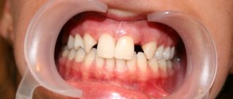
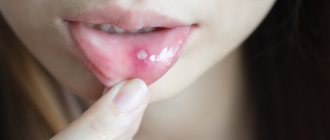
- when teeth are damaged, toothache, bleeding gums, and discharge of pus occur;
- in case of brain damage, symptoms depend on the location of the granulomas: paralysis, deterioration of vision, hearing, and smell may be observed.
Laboratory and instrumental diagnostics
A general blood test may show an increase in ESR, eosinophilia, leukocytosis, and a decrease in hemoglobin levels.
The use of various imaging methods (radiography, CT, MRI) allows us to identify foci of destruction up to 5 cm in size with clear boundaries without sclerotic changes, sometimes pathological fractures, flattening of the affected vertebrae (vertebra plana).
Pathomorphological examination is of decisive importance in the diagnosis of histiocytosis in adults and children. Microscopy reveals infiltrates of Langerhans cells (large oval cells with irregularly shaped nuclei), eosinophils, lymphocytes, and macrophages. Immunohistochemical analysis reveals the expression of CD1a, langerin, and S-100 proteins, characteristic of Langerhans cells. Electron microscopy allows one to see the Birbeck granules characteristic of Langerhans cells.
Differential diagnosis for EG is carried out with osteomyelitis, primary tumors or metastatic bone lesions, lymphoma, multiple myeloma, Papillon-Lefevre syndrome and bone cysts.
Figure 6. Birbeck granules, characteristic of Langerhans cells (electron microscopy data)
Diagnosis of granuloma
A large number of diseases can provoke the appearance of inflamed nodules. To determine the cause of their occurrence, the doctor examines the patient, studies complaints and medical history, then prescribes additional examinations. Their range may include :
- general blood analysis;
- blood chemistry;
- microscopic examination of smears from the genital organs (if granuloma venereum is suspected);
- biopsy of granuloma cells (examination of tissue under a microscope; allows you to determine the nature of inflammation);
- radiovisiography (computer x-ray; used to determine dental granuloma).
Treatment and prognosis
In some cases, eosinophilic granuloma does not require treatment, does not manifest itself in any way and goes away on its own within a few years. In case of severe symptoms and/or pronounced bone defects, different approaches and their combinations are used: surgical removal of the lesion (curettage, excision) or its radiofrequency ablation; prescription of cytostatics or a combination of cytostatics and glucocorticosteroids (including those administered into the pathological focus); radiation therapy in the presence of large lesions or lesions that compress neighboring tissues and organs. Correction of concomitant pathologies is also carried out.
Anti-cytokine drugs, anti-CD1a monoclonal antibodies, and trans-retinoic acid drugs are considered promising treatment regimens for histiocytosis X.
EG generally has a favorable prognosis, up to the possibility of spontaneous recovery, however, when transitioning to other forms, the course of histiocytosis from Langerhans cells can be more malignant - this happens, for example, when the bone marrow is involved (a rather rare phenomenon). In some cases, Taratynov's disease requires surgical intervention. After the course of treatment, patients must be under clinical supervision of an oncologist for 3 years with monthly examinations and x-ray examinations every six months.
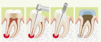
Conservative (medicinal) treatment of dental granuloma
This technique is used only for those cases in which the development of granuloma has not yet progressed far. The essence of conservative therapy for tooth root granuloma will be the intake and use of various drugs - antibiotics, antiseptics, drugs that have an anti-inflammatory effect. All medications are prescribed by a dentist and the treatment process takes place under the strict supervision of a specialist. This treatment option for granuloma is effective with early diagnosis of the disease and, if you strictly follow the doctor’s instructions, the granuloma will resolve within 1-2 weeks.
In some cases, conservative treatment of granuloma is complemented by root canal therapy. Working with tooth canals for root granuloma is indicated:
- For constant toothaches, the intensity of which does not subside from taking painkillers;
- Swelling of the gums, spreading to the cheek and jaw;
- Treatment of tooth canals is clearly indicated when large granulomas are detected - from five millimeters.
Clinical case
Based on Lam S. et al, Eosinophilic granuloma/Langerhans cell histiocytosis: Pediatric neurosurgery update. 2015
A 17-year-old young man was hospitalized due to an increasing scalp lesion over the past 6 weeks. The formation is painful on palpation and periodically bleeds due to ulceration, but no neurological deficit has been identified. CT and MRI revealed a large lesion in the frontal bone on the right, compressing the superior sagittal sinus. A total resection of the formation was performed, and the diagnosis of Langerhans cell histiocytosis of the skull bone was confirmed. At the outpatient stage, cytostatic therapy was carried out.
Figure 6. (a) CT examination without contrast - frontal scan (upper and middle part) and 3D reconstruction of the skull (lower part). (b) MRI scan. T1-weighted image in the coronal plane (top) and T2-weighted image in the sagittal plane
Sources
- Coppes-Zantinga A., Egeler RM The Langerhans cell histiocytosis X files revealed. Br J Haematol, 2002. - V. 116 - N. 1 - P. 3–9.
- Lam et al., Management of adult patients with Langerhans cell histiocytosis: recommendations from an expert panel on behalf of Euro-Histio-Net. Orphanet J Rare Dis, 2013 - V. 8. - N 72.
- Lam S., Reddy GD, Mayer R., Lin Y., Jea A. Eosinophilic granuloma/Langerhans cell histiocytosis: Pediatric neurosurgery update. Surg Neurol Int, 2015. - N. 6 (Suppl 17): S435 - S439.
- Langerhans' Cell Histiocytosis (Histiocytosis X). What is it? Harvard Medical School. Harvard Health Publishing. October, 2014. www.health.harvard.edu
- Sharma R., Singh R. et al. Langerhans cell histiocytosis (skeletal manifestations). Radiopaedia. https://radiopaedia.org/articles/langerhans-cell-histiocytosis-skeletal-manifestations-1
- Shea C. R, James W. D. et al. Langerhans Cell Histiocytosis. Medscape, 2021. https://emedicine.medscape.com/article/1100579‑overview
- Churilov L.P. Death on takeoff, or Who are you, Doctor Taratynov? Health is the basis of human potential: problems and ways to solve them, 2014. - T. 9 - No. 2 - P. 919–929.
- Yusupova L. A., Yunusova E. I., Garayeva Z. Sh., Mavlyutova G. I. Histiocytosis X. Practical Medicine, 2014 - T. 08 - No. 14.