In 1913, Nikolai Ivanovich Taratynov (1887–1919), a young doctor and employee of the Department of Pathological Anatomy at Kazan University, received material for research and clarification of the diagnosis. It was a fragment of tissue taken from a patient with a contusion of the cranial vault - some time after the injury, a granulomatous formation formed in this place.
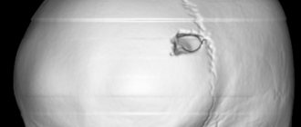
Figure 1. 3D reconstruction of skull bones with solitary eosinophilic granuloma https://radiopaedia.org/cases/eosinophilic-granuloma-skull
It was assumed that this was bone tuberculosis, but instead of the classic tuberculous tubercle, Taratynov saw accumulations of mononuclear cells (tissue macrophages or histiocytes) and eosinophils, as well as Charcot-Leiden crystals, which had previously been found in the sputum of patients with bronchial asthma. The morphological picture indicated “the existence of granulomas, clinically and macroscopically completely similar to tuberculous ones, recognizable only microscopically and consisting almost exclusively of eosinophils.”
Figure 2. Eosinophilic granuloma of bone: lesion of the hip in an 11-year-old girl (x-ray). Swelling and pain were noted for 3 months. https://radiopaedia.org/cases/langerhans-cell-histiocytosis-skeletal-manifestations
The previously unknown disease was named Taratynov's disease. Currently, this name is mainly used in Russian-language sources and has rather a historical meaning.
In the 1940s. similar cases were presented in articles by US doctors Sadao Othani, Joseph Ehrlich, Louis Lichtenstein and Henry Jaffe under the names “solitary granuloma of bone” and “eosinophilic granuloma of bone”.
Similar histiocytic infiltrates were described by other authors - Alfred Hand Jr, USA in 1893, Arthur Schüller (Austria) in 1915, Henry Cristian (USA) in 1920, Erich Letterer , Germany) in 1924, Sture Siwe (Sweden) in 1933, in connection with which the terms “Hand-Schüller-Christian disease” and “Abt-Letterer-Siwe disease” were introduced. However, later it was concluded that all of these are forms of the same disease, different in severity and localization of foci, called “histiocytosis X”
in 1953.
Figure 3 . Eosinophilic granuloma of the skull (X-ray) https://radiopaedia.org/cases/eosinophilic-granuloma-1
The answer to the question of what the histiocytes that form granulomas are was given in 1973 by pediatrician and morphologist Christian Nezelof (France), who identified them as Langerhans cells (a type of antigen-presenting cells localized in the epidermis). In this regard, since 1987, the name “histiocytosis X” was replaced by “Langerhans cell histiocytosis” ( LCH ).
The head of the Department of Pathology at St. Petersburg State University, Leonid Churilov, believes that Doctor Taratynov became the prototype of Doctor Zhivago. This bold assumption is supported by the coincidences in the life of Nikolai Taratynov and his family with the story of Yuri Zhivago: both victims of the revolution, military doctors, whose daughters were orphaned early and were raised by their father’s relatives. Nikolai Ivanovich Taratynov was shot and killed by a sailor, the brother of a patient who died in the hospital, in 1919 at the age of 32. You can learn more about Taratynov’s biography here.
Etiology
The question of the reasons for the development of granulomas remains open. One theory considers Langerhans' histiocytosis to be a neoplastic process. This is supported by such features as monoclonality of pathological cells (origin from a single pathological cell), increased expression of proliferation activators and factors inhibiting apoptosis. At the same time there are no genomic defects in the cells , and spontaneous remission of the disease is possible. At the site of granuloma development, activation of T-lymphocytes and T-regulatory cells with suppressor activity has been described. These signs indicate that other immunopathological processes may underlie the development of the disease.
Figure 4. Eosinophilic granuloma: pathological changes. In the last image, the S-100 protein expressed by Langerhans cells is colored. https://radiopaedia.org/cases/eosinophylic-granuloma-histology-1
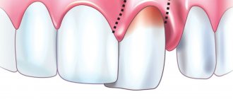
Removal of tooth granuloma
If therapeutic treatment of dental granuloma does not help, surgical intervention is used. From the outside, through the bone, access is made to the tooth root, the granuloma is scraped out, the place of its attachment is carefully processed, and the mucous membrane is sutured. In some cases, the infected root tip of the tooth is excised. Subsequently, the bone is restored, and the tooth continues to serve for a long time. Sometimes it is necessary to remove the entire root. This is also a common operation today. In this case, the desired root is excised along with the coronal part, and if the remaining roots are able to withstand the required load, a prosthesis is installed in this place.
If a tooth granuloma has been removed, but pain or inflammation remains, and it is painful to press on the tooth, you should contact a dentist in Moscow again. The situation when a tooth hurts after treatment or removal of a granuloma is common. The pain may persist for a long time, but you should make sure that there is no inflammation and that this pain is truly residual and not an indicator of another problem.
Clinical manifestations
Eosinophilic granuloma (EG), also known as Taratynov's disease, is a relatively benign variant of Langerhans' histiocytosis with the appearance of single foci in flat or tubular bones. The appearance of two or three or more lesions is much less common. Typically, Langerhans cell histiocytosis occurs in children and adolescents (up to 15–20 years), more often in boys (approximately 1.5:1). Frequency of occurrence: less than 1 person. per 100,000 population, which is 60–80% of all cases of LCH. In adults, eosinophilic granuloma, like hysteocytosis X in general, is much less common.
The most commonly affected bones are the skull bones, femurs, and less commonly, the pelvic bones, ribs, and vertebrae. There are also cases of the appearance of pathological foci in the thymus, skin, bladder, parathyroid glands, hypothalamus, lungs and gastrointestinal tract.
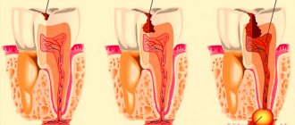
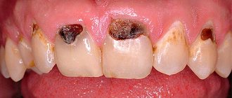
The clinical picture is determined by the location of the granuloma. An intraosseous formation may not cause any symptoms, but usually leads to the development of swelling, pain, and sometimes pathological fractures. When lesions appear in the jaw, tooth loss, damage to the mastoid process or temporal bone may occur with the development of otitis media. When the walls of the orbit are involved in the process, exophthalmos may develop. Sometimes polymorphic rashes are observed on the head, back, armpits, perianal and genital areas in the form of spots or plaques, small nodules or nodes with ulcerations. Common manifestations of Taratynov’s disease are common: increased fatigue, weakness.
Figure 5. Photo of the manifestations of eosinophilic granuloma on the skin - typical elements of purpura, must be differentiated from seborrheic dermatitis
Pathogenesis
Typically, pyogenic granuloma, also called telangiectatic granuloma, occurs at the site of a minor skin injury, such as a scratch or abrasion. The formation process is quite fast - it begins 1-2 weeks after a minor scratch and is benign; it can be tumor-like, granulomatous, rising above the skin and forming a structure the size of a lentil grain, cherry bone or nut.
The formation of pyogenic granuloma occurs in several stages:
- phagocytes accumulate in the area of minor injury ;
- then the transformation of cellular structures into macrophages and degeneration into granuloma occurs;
- Subsequently, an epithelioid pyogenic tumor is formed due to the transformation of macrophages and phagocytes into epithelioids, which additionally gives it the properties of a granuloma.
The structure is usually represented by granulation tissue in the form of an atypical proliferation of numerous small capillaries ( angioblastoma ), inflammatory infiltrate and endothelium; in later stages it can become lobulated. Then a regression stage is possible due to extensive fibrotic processes.
In most cases, botryomycoma occurs at the site of minor injuries, however, it can also occur without previous trauma; in this case, elevated tumor-like formations of a bright red or bluish color develop, which contain a large number of superficially located vessels, which gives it the features of a lobulated capillary hemangioma. Ulceration and damage to the tumor entail an increased risk of secondary infections and recurrent inflammation. It is important to remember that any benign neoplasm can degenerate into malignant.
Laboratory and instrumental diagnostics
A general blood test may show an increase in ESR, eosinophilia, leukocytosis, and a decrease in hemoglobin levels.
The use of various imaging methods (radiography, CT, MRI) allows us to identify foci of destruction up to 5 cm in size with clear boundaries without sclerotic changes, sometimes pathological fractures, flattening of the affected vertebrae (vertebra plana).
Pathomorphological examination is of decisive importance in the diagnosis of histiocytosis in adults and children. Microscopy reveals infiltrates of Langerhans cells (large oval cells with irregularly shaped nuclei), eosinophils, lymphocytes, and macrophages. Immunohistochemical analysis reveals the expression of CD1a, langerin, and S-100 proteins, characteristic of Langerhans cells. Electron microscopy allows one to see the Birbeck granules characteristic of Langerhans cells.
Differential diagnosis for EG is carried out with osteomyelitis, primary tumors or metastatic bone lesions, lymphoma, multiple myeloma, Papillon-Lefevre syndrome and bone cysts.
Figure 6. Birbeck granules, characteristic of Langerhans cells (electron microscopy data)
Treatment and prognosis
In some cases, eosinophilic granuloma does not require treatment, does not manifest itself in any way and goes away on its own within a few years. In case of severe symptoms and/or pronounced bone defects, different approaches and their combinations are used: surgical removal of the lesion (curettage, excision) or its radiofrequency ablation; prescription of cytostatics or a combination of cytostatics and glucocorticosteroids (including those administered into the pathological focus); radiation therapy in the presence of large lesions or lesions that compress neighboring tissues and organs. Correction of concomitant pathologies is also carried out.
Anti-cytokine drugs, anti-CD1a monoclonal antibodies, and trans-retinoic acid drugs are considered promising treatment regimens for histiocytosis X.
EG generally has a favorable prognosis, up to the possibility of spontaneous recovery, however, when transitioning to other forms, the course of histiocytosis from Langerhans cells can be more malignant - this happens, for example, when the bone marrow is involved (a rather rare phenomenon). In some cases, Taratynov's disease requires surgical intervention. After the course of treatment, patients must be under clinical supervision of an oncologist for 3 years with monthly examinations and x-ray examinations every six months.
How to make an appointment with a dermatologist
You can make an appointment with a dermatologist using the special form “Make an appointment with a doctor” - just enter your data so that the administrator can call you back and coordinate the time of the visit. During the consultation, the doctor will examine the clinical picture, prescribe tests if necessary, and select treatment. You can make an appointment with a dermatologist using the number. JSC "Medicine" (clinic of academician Roitberg) has a convenient location at the address: 2nd Tverskoy-Yamskaya lane, building 10. Nearby there are metro stations: "Mayakovskaya", "Tverskaya", "Novoslobodskaya", "Belorusskaya", "Chekhovskaya" .
Clinical case
Based on Lam S. et al, Eosinophilic granuloma/Langerhans cell histiocytosis: Pediatric neurosurgery update. 2015
A 17-year-old young man was hospitalized due to an increasing scalp lesion over the past 6 weeks. The formation is painful on palpation and periodically bleeds due to ulceration, but no neurological deficit has been identified. CT and MRI revealed a large lesion in the frontal bone on the right, compressing the superior sagittal sinus. A total resection of the formation was performed, and the diagnosis of Langerhans cell histiocytosis of the skull bone was confirmed. At the outpatient stage, cytostatic therapy was carried out.
Figure 6. (a) CT examination without contrast - frontal scan (upper and middle part) and 3D reconstruction of the skull (lower part). (b) MRI scan. T1-weighted image in the coronal plane (top) and T2-weighted image in the sagittal plane
Sources
- Coppes-Zantinga A., Egeler RM The Langerhans cell histiocytosis X files revealed. Br J Haematol, 2002. - V. 116 - N. 1 - P. 3–9.
- Lam et al., Management of adult patients with Langerhans cell histiocytosis: recommendations from an expert panel on behalf of Euro-Histio-Net. Orphanet J Rare Dis, 2013 - V. 8. - N 72.
- Lam S., Reddy GD, Mayer R., Lin Y., Jea A. Eosinophilic granuloma/Langerhans cell histiocytosis: Pediatric neurosurgery update. Surg Neurol Int, 2015. - N. 6 (Suppl 17): S435 - S439.
- Langerhans' Cell Histiocytosis (Histiocytosis X). What is it? Harvard Medical School. Harvard Health Publishing. October, 2014. www.health.harvard.edu
- Sharma R., Singh R. et al. Langerhans cell histiocytosis (skeletal manifestations). Radiopaedia. https://radiopaedia.org/articles/langerhans-cell-histiocytosis-skeletal-manifestations-1
- Shea C. R, James W. D. et al. Langerhans Cell Histiocytosis. Medscape, 2021. https://emedicine.medscape.com/article/1100579‑overview
- Churilov L.P. Death on takeoff, or Who are you, Doctor Taratynov? Health is the basis of human potential: problems and ways to solve them, 2014. - T. 9 - No. 2 - P. 919–929.
- Yusupova L. A., Yunusova E. I., Garayeva Z. Sh., Mavlyutova G. I. Histiocytosis X. Practical Medicine, 2014 - T. 08 - No. 14.
Symptoms
Usually botryomycoma does not cause significant discomfort, except for complexes regarding appearance. However, its injury and corresponding bleeding can be quite painful.
Pyogenic granuloma: distinctive appearance features
The appearance of pyogenic granuloma resembles millet grain, cherry bone or hazelnut, which is covered with crusts of a dirty brown color, in other cases with a papillomatous or smooth surface; a soft nodule is usually located under the neoplasm. It has a dense elastic consistency and an external smooth or, in most cases, uneven surface, which may have lobulated or papillary tissue patterns. The base can be thin or wide, bleed easily when injured, cause ulceration and necrotization processes with the formation of a hemorrhagic crust. Along the periphery at the base, a “collar” can be identified due to the exfoliating epidermis, as in the photo below.
Botriomycoma on the finger
Typically, neoplasms are single, having a soft-elastic structure with a characteristic painless or slightly painful organization. Externally, the formation is tumor-like, its size in rare cases is more than 1 centimeter in diameter, but can reach 3-4 cm.