Removing a foreign body from a tooth root canal is a manipulation aimed at removing fragments of dental instruments from it. Most often these are endodontic files designed to remove pulp. They are made from metal, which begins to rust over time, causing irritation to the canal walls. If the instrument is not removed from the canal in time, it can cause root cracking and loss of the entire tooth. In addition to dental instruments, orthopedic structures in the form of pins and inlays can also be considered foreign bodies. The need to remove them arises if the tissue around the root is inflamed or the patient suffers from pain symptoms after their installation.
Removal of the anchor pin and stump inlay in Moscow can be ordered at the dental department of CELT. Our clinic is multidisciplinary and has been operating in the paid medical services market for almost three decades. Our dentistry employs leading domestic orthopedists, orthodontists, surgeons and therapists, who have many years of experience in scientific and practical activities. They provide treatment with the conclusion of a contract and provision of guarantees. To find out the cost of removing a stump tab or pin from the root canal, go to the “Services and prices” tab in this section. To avoid misunderstandings, do not forget to clarify it with the operator of our information line or during a consultation with a doctor.
removal of a foreign body from a dental canal - 4,000 rubles.
At CELT you can get advice from a dental specialist.
- The cost of a consultation with a dentist-therapist is 1,000
Make an appointment
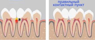
Types of foreign bodies that may end up in the root canal
Most often, foreign objects end up inside root canals during complex dental treatment. Depending on the situation, these could be:
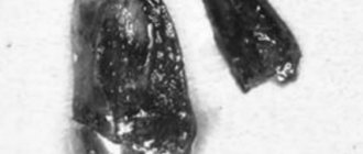
- Broken fragments of endodontic instruments (in particular, files for pulp extraction);
- Anchor fiberglass pins, which are specially installed to strengthen the tooth and its further prosthetics;
- Elements of stump inlays (in particular, their pin parts, which can break off during use).
Fragments of instruments end up in the canal during its treatment - as a rule, removal of necrotic pulp from it during:
- Carrying out manipulations with narrow or tortuous channels in which the instrument breaks, unable to withstand the stress of bending;
- Repeated use of hand tools whose tips may break off due to metal fatigue or defects.
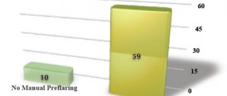
During endodontic treatment, instruments do not break very often, but such a possibility cannot be excluded. Today it is one of the pressing problems of dentistry. Treatment under a microscope helps reduce its risk to almost zero, in which the doctor is able to notice the broken fragment immediately and remove it in a timely manner.
How to make the right choice
There are certain requirements for material compatibility. So, for example, a crown on a tooth on a metal pin (anchor or regular thickness) should be metal-ceramic. If you place a ceramic crown here, then, firstly, the metal will become visible through the thin translucent ceramics, and secondly, the ceramics may crack due to different strength indicators with the metal.
Fiberglass, ceramic, and zirconium dioxide are suitable for aesthetic ceramic and ceramic-composite crowns. Under the zirconium dioxide prosthesis, you can place a rod made of the same material or metal.
Indications for removing the stump tab, pin, dental instruments from the canal
Removing a post, onlay or endodontic file from a root canal while keeping the tooth suitable for prosthetics is not an easy task. It requires the dentist to have skill and experience in carrying out such manipulations, as well as the availability of appropriate tools and equipment. The procedure is carried out in several stages and requires the removal of a denture or filling, if they have been installed. Only after this can you begin to directly remove the fiberglass pin from the tooth or any other structure. Indications for the procedure:
- Violations committed by the dentist during treatment;
- Development of inflammatory processes in the root area and periodontal tissues;
- Development of caries and pain in the patient;
- Destruction of the dental unit under the crown.
As for fragments of endodontic instruments, the patient does not experience any unpleasant sensations when they come into contact with them. They appear much later and are manifested by the following symptoms:
- Discomfort and pain when biting on a tooth and chewing;
- Swelling of the gums in the area of the recently treated tooth;
- The appearance of a fistula in the oral cavity;
- Suppuration of soft tissues.
The above is a reason to promptly seek professional dental care. The dentist will prescribe a visiography, which will help identify foreign objects in the filled tooth. Thanks to it, it will be possible to establish the presence of caries or inflammation.
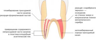
Particles from endodontic instruments must be removed, as they begin to rust and irritate the canal walls. As a result, the tooth root may crack and the tooth will have to be completely removed, since restoration in such a situation is not possible. Removing the instrument from the tooth canal is also necessary because:
- It will not allow it to be sealed properly, and as a result, the procedure will have to be carried out again;
- Its presence inside the canal is the cause of the development of serious inflammatory processes (if a fragment of a necrotic nerve remains under the fragment, it will rot).
It is for these reasons that refilling of root canals is carried out only after removing the instrument from the root canal.
Do you want to restore a tooth with a CEREC ceramic inlay?
8
or order a free call
Request a call
This is how the dental inlay for the lower canine turned out:
The inlay was fixed with double-curing cement Relix U200 (RelyX U200) - this is one of the most expensive cements, German cement. The patient was super satisfied. The restored tooth looks like its own:
All work FROM and TO was carried out in 1 appointment
, on a fresh tooth surface. Great job.
PS
As you can see in the video, the patient is facing the next stage of dental restoration. But we'll talk about this some other time.
Author:
Sergey Samsakov
orthopedic dentist
born 02/02/1989
Education:
2011 — Graduated from the Moscow State Medical and Dental University named after. A.I.Evdokimova
2012 — Internship in the specialty “Orthopedic Dentistry”, Moscow State Medical University named after. A.I.Evdokimova
2014 - Residency in Orthopedic Dentistry, Moscow State Medical University named after. A.I.Evdokimova
Indications for removing the stump tab, pin, dental instruments from the canal
The procedure is carried out in two ways:
- Simple - indicated when identifying broken fragments of pins. In the process, the endodontist destroys the filling by applying ultrasound to it. Thus, he provides himself with access to the required area. After this, he carefully loosens and removes the foreign body;
- Complex – indicated for identifying fragments of endodontic instruments. The process involves a dental microscope. The specialist prepares the root canal, loosens the discovered fragments and carefully removes them using a special device. If the foreign body is not visible in the lumen of the root canal, the procedure will require microsurgical intervention.
CELT dentists individually select treatment tactics and use modern equipment (in particular, a microscope and endodontic ultrasound attachments, which allow the procedure to be carried out as carefully as possible, without affecting healthy tissue). When choosing treatment, the nature of the patient’s complaints is taken into account, as well as the duration of the pathological processes, the identification of inflammatory processes and the location of the pathological focus.
Do you want to restore a tooth with a CEREC ceramic inlay?
Always at your service - Sergey Samsakov, orthopedic dentist with more than 10 years of successful experience, Moscow. Expert in 3D digital smile modeling, veneers and CEREC technology.
Patients come to me from all over Russia:
Removing a pin from a canal: risks and features
The method of removing the pin from the canal is also determined individually. First of all, it depends on the material from which the structure is made: gutta-percha and fiberglass pins are removed by drilling with special threaded needles. But titanium structures are grabbed with a special clamp and twisted as carefully as possible, trying not to damage the root. Other factors to consider when choosing a method for removing a post from a tooth:
- Type of endodontic structure;
- Material for fixing it in the root canal;
- Position of the problem unit in the dentition;
- Risk of complications;
- The thickness of the walls of the canal and the crown of the tooth.
As a rule, it is easiest to remove the core tab and pin from the frontal teeth. As for premolars and molars, everything is much more complicated here: even the choice of instruments is seriously limited... The fragility and thinness of the dental walls play a role, which significantly complicates the repeated procedure. There is a high risk of root perforation or fracture, which are always associated with complete loss of the unit.
If the pin was secured inside with cement or composite, it is recommended to use ultrasonic waves, thanks to which they can be removed without affecting the dental tissue. This is especially true, because after removing the tab, you can avoid tooth extraction only by maintaining its functionality. Moreover: a good prognosis for this must be long-term. Otherwise, it is better to immediately remove the entire unit along with the orthopedic structure.
The dentist’s standard algorithm for removing the pin is as follows:
- Removing the main volume of material with which it is secured in the root canal;
- Carefully removing its residues using ultrasonic waves;
- Removing the pin structure using a special endodontic clamp;
- X-ray of the tooth to monitor treatment and identify fragments that broke off during the removal of the pin.
Rehabilitation period and rules of care
As a rule, getting used to a crown on a pin occurs very quickly, because due to its small size and anatomical shape, it does not seem like a foreign body. At first, the tooth may ache a little, the gums around it may turn a little red due to installation errors - to relieve symptoms, rinse with a solution of soda or chamomile infusion.
While wearing a temporary crown and in the first week after installing a permanent one, you should chew food on this part of the dentition more carefully so that the denture does not fall off. When wearing any type of prosthesis, it is not recommended to eat hard foods, taffy, or chew nut shells or seeds. To keep the crowns from darkening and the gums to be healthy, you should brush your teeth with a brush and toothpaste 2 times a day, incl. special for prostheses. And also avoid brightly colored foods and drinks.
Types of pins and features of their extraction and root canal
According to the material of manufacture, the pins are:
- Fiberglass - made from woven glass fibers that are arranged in a horizontal plane and embedded in an epoxy matrix. They are quite durable, and their physical characteristics are as close as possible to the native tooth tissues. For these reasons, they are used for restorations of frontal units. They do not show through the crowns, and their removal is not very difficult;
- Anchor - made of titanium, precious metals or stainless steel. Thanks to their special properties, it becomes possible to reconstruct even an almost completely destroyed unit, provided that its root is well preserved. They are fixed in the canal using phosphate cement. Destruction of the latter is possible only with the use of ultrasonic waves;
- Parapulpal – made of dental stainless steel with a polymer coating. They are used to strengthen fillings and are installed directly into hard dental tissues, which eliminates the risk of infection or inflammation. At the same time, the proximity of their location to the surface leads to the fact that they break teeth and require re-installation. To remove them, the dentist drills out a small amount of hard tooth tissue around it with a drill, and then carefully unscrews the structure.
Anchor pins are most often removed due to the development of secondary caries, but situations are possible when it is displaced during the process of chewing food and causes damage to the prosthesis. In the latter case, you will have to replace the entire structure, sometimes correction is possible. Unfortunately, it happens that violations are detected at a stage when treatment of the unit will not give the desired result - it is only possible to remove it along with the pin.
How to install a crown
How to place a crown on a post? First, a translucent composite filling compound is applied to the rod and illuminated with a “blue” lamp. The doctor then takes impressions using impression material or an intraoral 3D scanner. Based on casts or virtual data, a model of the future prosthesis is formed, developed and manufactured by casting, pressing or milling on a robotic machine (CAD/CAM system). After the laboratory stage, the finished crown is handed over to the doctor, and it is placed on a pin (fixed with a cement composition).
While the permanent prosthesis is being manufactured, the patient is fitted with a temporary crown made of plastic or composite material. They have good aesthetics, are made in 1 day and are designed to protect against aggressive environmental factors.
Complications after removing pins from the root canal of a tooth
As a rule, experienced dentists calculate the risks associated with the procedure and in every possible way reduce them to zero. For example, removing a post made of a material such as zirconium carries a high risk of breakage due to its brittleness. For this reason, the specialist needs to act as carefully as possible, and upon completion of the procedure, monitor its quality. Other complications may include:
- perforation of the tooth root due to its mechanical damage;
- fracture of the tooth root due to the appearance of cracks in it and the retreatment procedure;
- damage to the pin itself and preservation of its fragments in the canal.
An important role is played not only by the occurrence of complications, but also by the availability of the resources of the endodontist in order to properly cope with them. Most often, perforation or root fracture requires removal of the tooth root and placement of an implant. At the same time, in some cases it is possible to use the mineral trioxide of the unit, the properties of which allow it to close the perforation and preserve the functionality of the unit, extending its service life.
Differences
- The pin puts pressure on the root of the tooth, which can cause it to become loose; there is pressure on adjacent teeth, and the inlay eliminates this load.
- Time of use - the pin can be used for up to 3 years, the inlay - much longer, almost 10 years.
- The pin is a single design, but the tab can separate.
- It is not difficult to make a pin, but to make an inlay, laboratory specialists are involved and special studies are carried out.
- The installation of the pin is carried out in one visit to the dentist; to install the inlay, you will have to visit the doctor at least twice.
- If the tooth has a crooked root, you can only use an inlay.
- The cost of an inlay is significantly higher than a pin.
- Preparing the tooth for the installation of a pin is relatively gentle; in order to install the inlay, a large amount of tissue is removed.
Is it possible to glue the bridge in place yourself?
What to do and how to secure a dental bridge if it falls out at home? Let us immediately point out that putting a fallen bridge back on your own is not the best idea. Since this can injure the supporting and neighboring teeth, harm the gums (if you suddenly decide to glue the bridge with pharmaceutical cement or household glue). The consequences can be unpredictable - up to the removal of “damaged” teeth or implants. Therefore, it is better to entrust your teeth to a professional dentist.
How much does a re-restoration cost?
How much does it cost to put a dental bridge in place if it falls out? If the supports do not need to be treated or removed, then fixation with new cement will cost approximately 1000-1500 rubles. If the screws on dentures on implants have come loose, then fixing them with new screws will cost about the same. If the patient needs to treat/remove supports, install new pins or inlays - and, as a result, make a new prosthesis, then the cost of restoration starts at 30 thousand rubles. And the final cost is calculated individually.
General overview
The pin is an artificial reinforcing element made in the form of a thin rod. The product is intended to extend the life of the restored unit. The rod is attached by insertion into the pulpless root passage.
Teeth with the nerve removed become brittle and susceptible to deformation due to increased loss of minerals. The pin acts as a support for fixed prosthetic structures. The product is implanted when reinforcement, replantation, bridge fixation, splinting for periodontal disease and other restoration procedures are required.