Teeth are bone formations whose main purpose is the primary mechanical processing of food. In most cases, an adult has 32 teeth. In addition to performing the chewing function, they influence the formation of human speech. The loss of baby teeth and the appearance of permanent teeth occurs after the baby’s fifth birthday - there are four types: incisors and canines, molars and premolars.
On one side of the upper jaw there are the following teeth: 1 canine and 2 incisors, as well as 2 premolars and 3 molars. The opposite side is similar to the upper one, and the arrangement of the lower jaw teeth is identical to the upper row of teeth. The condition of teeth indicates a person’s health, and if it is unsatisfactory, this may indicate some kind of disorder in the body.
READ ALSO: location of teeth in adults and their numbers
Human dental formula
Many people have heard that dentists call teeth by different numbers. Thus, each unit is assigned a specific number, which guarantees maximum accuracy in records of dental conditions or treatment procedures. The count is made starting from the middle of the dentition to the right and left sides. The anterior units - incisors - are No. 1 and 2, 3 - canines, 4-5 - premolars, 6, 7 and 8 - molars, the last of which is the wisdom tooth.
To determine exactly where the tooth is located, the jaw is figuratively divided into segments. The beginning of the numbering is the right side of the upper jaw of the teeth. Units are written as two-digit numbers, where the first determines the serial number, and the other determines the so-called segment. The upper right half of the teeth is designated by numbers 11-18, the left - 21-28, and the lower left segment - 31-38 and 41-48. Regarding primary teeth, the count is made starting from the front units on the left in a clockwise direction. They are assigned the numbers 50, 60, 70 and 80.
READ ALSO: structure of the human upper jaw and functions of its processes
There are several types of numbering that dentists use:
- With the square-numeric Zsigmondy-Palmer method, Arabic numerals are used for radical units, and Roman numerals are used for dairy units. Often used by orthodontists and surgeons.
- Haderup's method is based on the symbols "-" and "+", where the first denotes the lower and the other the opposite arc. Units are written in Arabic numerals, and in the case of dairy units, 0 is added.
- The Viola system uses Arabic numerals 1-8 for units and two-digit numbers for segments. This type is very convenient, which is why it is widely used by dentists around the world.
- The last system is universal. Each group of units is assigned a letter, number and segment number.
Anatomy of the maxillary canine
The incisors, dentes incisivi
, four on each jaw, have a crown shaped like cutting chisels that cut food into inappropriate sizes. The crown of the upper incisors is wide, the lower ones are twice as narrow. The root is single, compressed from the sides of the lower incisors. The root apex is deviated somewhat laterally.
Upper medial incisor
the largest of the group of incisors. The labial surface of its crown is convex in the transverse and longitudinal directions. It has 3 small longitudinal ridges, each of which ends at the chewing edge with a clove. On both sides of the median ridge there is one longitudinal depression. The lingual surface of the crown is concave in the longitudinal and transverse directions.
In the cervical region it has a tubercle, tuberculum dentale
, From which ridges extend, heading along the distal and mesial edge of the lingual surface to the chewing edge of the tooth. Of the 3 signs of a tooth, the sign of crown curvature is the most pronounced. The root is conical and longer than the crown; the lateral grooves on it are weakly expressed. It has 3 surfaces: labial and two approximal.
Upper lateral incisor
smaller than the medial incisor, from which it differs in the following features: on the labial surface of the crown there is often a median longitudinal groove, on both sides of which there is one small tuberculate elevation on the cutting edge of unworn teeth. On the lingual surface, the lateral ridges are usually better defined than those of the medial incisors. Often on this surface there is a depression located occlusally (below) from the dental tubercle. The mesial surface is longer than the distal one and passes into the cutting edge almost at a right angle, while the distal one forms a significant rounding. The sign of the crown angle is well expressed. The root is shorter than that of the medial incisor, compressed in the mesiodistal direction; in most cases it is straight and has lateral grooves. Its distal surface is more convex than the mesial one.
Medial and lateral lower incisors.
The lower incisors are the smallest in both jaws. In this case, the medial incisor is smaller than its distal neighbor. Both teeth have characteristics characteristic of all incisors. Their crown has the most typical chisel shape. On the front surface it is slightly convex in the longitudinal direction and flattened in the transverse direction, on the rear surface it is concave in the longitudinal direction and flattened in the transverse direction. The ridges are weakly expressed and sometimes absent. The root is significantly flattened.
At the medial incisor
there are no signs of curvature of the corner and root. To distinguish the right medial incisor from the left, a better defined lateral longitudinal groove on the root is important.
At the lateral incisor
the crown is wider than that of the medial one, and the root is more massive. At the same time, this tooth has quite clearly expressed signs of angle and root and weakly - curvature.
The structure of the upper and lower teeth
The elements of a human tooth are: crown, neck and root. The crown is visible to the naked eye - it is located above the gum. The connecting link between the tooth root and the alveolus is connective tissue. The neck of the tooth is located between the crown and the root.
READ IN DETAIL: dental formula for adults and children
From a histological point of view, the constituent basis of the human tooth is dentin, hidden under the enamel. Near the root, where there is no enamel, there are fibers that seem to hold the periodontium together. In the center of the unit there is a pulp, which is penetrated by blood vessels and nerve endings. With inflammation or caries, it makes itself felt with severe pain.
Incisor and canine crown
Both the lower incisor and its antagonist have similar components: the root and the crown. Slightly above the alveolar layer is the enamel crown of the tooth. The root of the tooth is located in the alveolar cavity under the gum (we recommend reading: how many roots does a person have in his teeth?). The large dimensions of the upper incisor are about 21-23 mm, while the length of the crown is 9-95 mm, and the width of the teeth is 4-6 mm, so the pressure on them is distributed evenly. The enamel is protected from physical damage and mechanical stress. The lateral (or lateral) incisor, in comparison with the medial one, has a huge root and a wide crown.
Function and role of fangs
The main function of the fangs is to tear and hold food. In addition, canines are the teeth that are least susceptible to caries. These are the most powerful and strong teeth, which bear quite a large load. If a person suddenly lost his fangs, then:
- perhaps he would have problems with diction;
- the entire load would fall on the weaker neighboring teeth, which in the near future would either become deformed or become loose;
- the dentition would lose its aesthetic appeal;
- the process of infection of teeth with caries has accelerated, since it is the fangs that are the barrier to the “creeping” of this problem from the molars that are most susceptible to it to the incisors;
- Tooth enamel is the hardest human tissue the body produces, but even it can wear down and wear down over time. Fangs are an excellent shock absorber for the contact of the upper and lower jaws; in their absence, tooth enamel would wear down several times faster.
What are baby teeth?
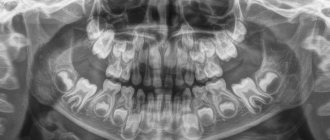
The formation of baby teeth begins when the baby is in the womb, namely at the 7th week of pregnancy. Teething begins when the baby reaches 6-24 months. The change from temporary units to permanent ones is carried out at the age of 5-13 years. The complete formation of a permanent dentition is completed by the age of 14. A deviation of no more than one year is acceptable. If the period of teeth replacement is exceeded, this indicates anomalies in the dental arch or jaw formation.
Structural features
Milk teeth are similar to permanent teeth, but are much smaller in size and have short, rounded roots. The thickness of dentin and enamel is also less. Due to low mineralization, teeth are susceptible to the rapid development of caries. It is worth noting that temporary units are characterized by a considerable volume of pulp and wide root canals, which facilitates the penetration of harmful microorganisms into the pulp.
The number of roots in baby and permanent human teeth is identical. When the time comes to replace temporary teeth with permanent ones, the roots of baby teeth dissolve, thereby making room for new ones. Their change is almost painless, since they are not strongly attached to the alveoli.
READ ALSO: The number of teeth of an adult healthy person is normal
Differences from permanent
Despite the similarities between milk and permanent units, there are a number of differences. First of all, this is the number of units - in contrast to permanent ones (32), temporary ones are only 20. In addition, children do not have premolars, and there is only one molar. There are also other differences:
- smaller crown
- thin enamel, white-blue color,
- dentin composition with a lower degree of mineralization,
- roots are branched and massive,
- deep fissures,
- the roots are bent closer to the lip, they are not so strong and shorter.
Description of the dentition
Incisor teeth.
The human jaw is symmetrical and contains the same number of teeth of each type. But there are certain anatomical features of the upper and lower jaw. Let's look at them in more detail. Incisors are the front teeth . A person has eight of them - 4 at the bottom and 4 at the top. Incisors are needed to bite food and separate it into parts. The peculiarity of the structure of the incisors is that they have a flat, chisel-shaped crown with rather sharp edges.
On anatomical sections there are three tubercles, which are erased throughout life. On the top of the jaw there are two central incisors - the largest of all the incisors in their group. The lateral incisors are similar in shape to the central ones, but smaller in size.
What is noteworthy is that the immediate cutting edge of the lateral incisor also has three tubercles, and often takes on a convex shape as a result of the development of the central tubercle. The root of the incisor takes the shape of a cone, and is flat and single. A distinctive feature of the incisor is that on the side of the tooth cavity there are three pulp tips corresponding to the tubercles of the cutting edge.
The anatomy of the upper teeth is slightly different from the structure of the lower dentition, so in the lower jaw everything is exactly the opposite. The middle incisors are smaller , unlike the lateral ones, and have a shorter and thinner root than the lateral incisors. The outer surface of the cutter is slightly convex, while the inner surface is concave.
The crown of the incisor is curved from the side towards the lips and is very narrow. The cutting edge has 2 angles - in the center, more acute, and inside - more obtuse. The roots have longitudinal grooves.
Chewing teeth and fangs
Fangs are used to break down food into smaller pieces.
The anatomy of the fangs is such that there is a groove on the inside of the crown; it disproportionately divides the crown into 2 parts. The cutting edge of the fangs has one pronounced and developed tubercle, this makes the cone-shaped crown often similar to the fangs of a predator. The canine on the lower jaw is narrower in shape, the ends of the crown are concentrated in the medial tubercle. The canine root is flat, inclined inward and the longest, unlike the roots of other teeth. A person has 2 fangs on both jaws . The lateral incisors with the canines form an arch, where the transition from the incisors to the chewing teeth begins in the corner.
Let us consider the structure of the small chewing tooth first, and then the large chewing tooth. Their main task is thorough processing of food . This function is performed by molars and premolars.
Premolars
The first premolar (No. 4 in the dental formula) differs from the incisors and canines in its prismatic shape, and there are convex surfaces on the crown.
The surface has 2 tubercles - lingual and buccal, with grooves between them. The buccal tubercle is much larger in size than the lingual one. The root of the first premolar has a flat shape , but with a slight bifurcation into the lingual and buccal parts.
The second premolar is similar in structure to the first, but its buccal surface is much larger, and the root has a compressed anteroposterior direction and a conical shape . In the first lower premolar, the chewing surface is inclined towards the tongue.
The second premolar is larger than the first due to the fact that both tubercles are symmetrical and equally developed, and the depressions in the enamel between them have the shape of a horseshoe. The root is the same as that of the first premolar. has 8 premolars in the dentition , four on each side (on the lower and upper jaws).
Molars
In the upper jaw, the first molar is the largest.
Its crown is similar to a rectangle, and the chewing surface is diamond-shaped with 4 tubercles. This molar has three roots: one straight - the most powerful, and two buccal - flat, deflected in the posterior direction. During the closure of the jaws, the first molars rest against each other and form a kind of “limiter ”; because of this, they undergo significant loads throughout a person’s life.
The second molar is smaller . The roots are the same as those of the first molar. The structure completely coincides with the location of the premolars described above.
On the lower jaw, the first molar for chewing food has five cusps. This molar has two roots - the anterior one with two canals, the posterior one with one. In this case, the anterior root is larger than the posterior one. In the lower jaw, the second molar is similar in structure to the first. The number of molars in humans is the same as the number of premolars.
The third molar is called a “ wisdom tooth ,” and a person has four of them in the dentition, two on each jaw. On the jaw below, the third molar has many variations in cusp development. As a rule, there are five of them. And, in general, in a person the structure of a “wisdom tooth” is the same as the structure of a second molar, but the root usually resembles a very powerful and short trunk.
Anatomical structure
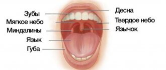
Going to the mirror, opening your mouth, you can examine your teeth in detail. A person sees only the vernal shell - these are hard enamel crowns. The protective layer reliably hides vulnerable internal tissues.
Incisors, canines, and molars have different shapes and numbers of roots, but they are united by a common structure.
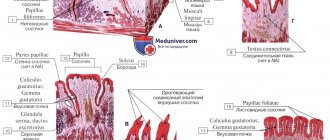
Let's take a closer look at the histological structure of the tooth in section.
- Enamel is a hard protective layer of a whitish-cream color, consisting of 96% inorganic substances. The fabric has increased strength, but is also fragile, prone to abrasion, and susceptible to adverse environmental influences. If the enamel is injured by the attachment of pathogenic microorganisms, caries develops. On the surface of the chewing teeth there are fissures, depressions and grooves. They most often accumulate food particles that are difficult to remove. This causes the development of the disease. When the pathology occurs, it gradually affects the healthy protective layer, leaving internal tissues defenseless.
- Dentin - located directly under the enamel, consists of 70% inorganic substances. Dentin is a hard tissue, but it is much more vulnerable than the surface protective layer. If the carious process reaches the dentin, the pathological process occurs rapidly, and in the absence of timely assistance from the dentist, inflammation of the neurovascular bundle occurs. When dentin is damaged, medium or deep caries develops. Signs of the disease are clearly visible - violation of the integrity of enamel and dentin, the appearance of pain sensitivity upon contact with negative environmental factors;
- The pulp chamber and root canals contain nerve endings and blood vessels and have a soft, loose structure. Thanks to the pulp, the tooth receives the necessary nutrients. It protects periodontal tissues from penetration of pathogenic microorganisms and promotes dentin regeneration. When the neurovascular bundle is damaged, an inflammatory process occurs. The person feels acute paroxysmal pain. Taking analgesics only briefly relieves the pain symptom. If pulpitis develops, you must immediately visit the dentist. If help is not provided in a timely manner, the dead nerve fiber will become a source of infection. Pathogenic microorganisms will penetrate into the surrounding dental tissues, and periodontitis will develop;
- Cementum is a hard tissue lining the outer surface of the root. Periodontal ligamentous fibers are attached to the cement, which securely fix the tooth in the alveolar socket.
The detailed structure of the tooth and methods of their treatment are described in the video: The crown part of the incisors, canines and molars is located above the surface of the gum, the root is hidden deep in the internal tissues of the jaw.
Second lower molar
Average age of teething: 11-13 years
Average age of root formation: 14-15 years
Average length: 19.8 mm
The crown is somewhat smaller in size than the first molar, and the more symmetrical second lower molar is characterized by a close arrangement of roots. The roots gradually converge distally and have closely spaced apices.
Exposure is made at the mesial portion of the crown with access extending only slightly distal to the central sulcus. After trephination of the cavity with a fissure bur with a cutting apex, an elongated ball-shaped bur is used to expand it until free access is achieved. The distal root angles often allow for a smaller opening than the lower first molar.
Particular attention should be paid to the shape of the mouth of the distal canal. A narrow oval orifice indicates the presence of a slit-like lumen in the distal canal, which requires more careful treatment.
All carious tissue, leaking fillings and denticles should be removed and replaced with a suitable temporary filling prior to endodontic treatment.
The lower second molar is most susceptible to vertical fractures. After preparing the access, but before starting endodontic treatment, the clinician should examine the floor of the pulp cavity using fiber optics. For mesial-distal crown or root fractures, the prognosis is extremely unfavorable.
To avoid vertical fractures after endodontic treatment, it is necessary to completely cover the cusps with a restorative structure.