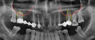
Perforation of the maxillary sinus during the treatment of upper teeth is damage to its shell when trying to treat root canals without prior examination. The penetration of infection provokes inflammation with symptoms of sinusitis, which cannot be treated with medication.
For more than 20 years, Doctor Levin has specialized in eliminating complications after unsuccessful dental manipulations in the area of the maxillary sinuses. Surgical treatment is performed by maxillofacial surgeons with ENT training.
Causes of sinus perforation during dental treatment
The maxillary sinuses or maxillary sinuses are located inside the upper jaw. They are separated from the oral cavity by a septum and the alveolar process, which contains the roots of the teeth. Treatment of the canals of such teeth is complicated by the risk of sinus perforation for a number of reasons:
- Anatomically thin septum Sometimes the height is only 1 mm. It happens that the teeth penetrate the cavity with their roots and only the sinus mucosa separates.
- Treatment of teeth with inflammation on the roots If there is inflammation on the roots of the teeth (periodontitis, cyst), the surrounding bone melts and becomes thinner.
- Excessive efforts by the doctor If the dentist does not calculate the efforts when passing the canals, the canal may rupture, damage the root, or perforate the bottom of the sinus.
Without a preliminary diagnosis, the endodontist, having no idea about the size of the sinus septum and the location of the roots, may not calculate the effort and damage the tooth and the thin bone layer that lies between the maxillary sinus and the root. As a result, infection penetrates into the sinus cavity, and fragments of instruments and filling material may fail. In our practice, there are cases when a foreign body in the sinus attracts secondary infections with the development of neoplasms - fungal colonies, cysts, polyps.
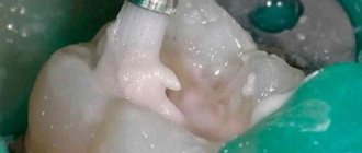
A fragment of an endodontic instrument in a tooth canal
X-ray of the sinuses: why and how they are done - MEDSI
Table of contents
- Indications for the study
- Contraindications to the procedure
- How the research is carried out
- Normal x-ray picture
- What can a radiologist find?
- Advantages and disadvantages of sinus x-rays
- Advantages of carrying out the procedure at MEDSI
X-ray
is a widely used type of examination using a special device that uses a special type of radiation. It allows you to visualize any pathologies, neoplasms, polyps or injuries. This type of examination also allows you to check how successful treatment or recovery from injury is.
Indications for the study
The doctor prescribes an X-ray of the nose in the following cases:
- Constant headaches concentrated in the upper part of the head and eye sockets
- Trauma to the nose, jaw, forehead
- Persistent nasal congestion
- The appearance of sudden discomfort in the eye area, bridge of the nose against a background of high fever and purulent discharge
- Periodic bleeding from the nose
This examination may also be prescribed before dental surgery in the upper jaw.
X-ray of the paranasal sinuses allows you to identify diseases and disorders such as:
- Malignant and benign neoplasms, cysts and polyps
- Ethmoiditis
- Frontit
- Sinusitis
- Injuries and fractures of the bone walls of the paranasal sinus
Contraindications to the procedure
The following contraindications exist for the use of this examination:
- During pregnancy and breastfeeding
- The patient's age is less than seven years (except for situations accompanied by purulent inflammation or a fracture of the sinus wall)
- Frequency of use - no more than two to three times a year
- Allergy to iodine (if a contrast study is necessary)
If indicated, MRI or ultrasound can be used instead of x-rays of the sinuses.
The radiation exposure of this method is low, so there are few restrictions on its use.
How the research is carried out
The procedure for performing an x-ray of the nasal sinuses is as follows:
- Before the procedure, it is necessary to remove various metal objects: jewelry, dentures, glasses, etc., to avoid image distortion
- The patient is placed lying or sitting on a special couch, which fixes the position of the head during the analysis
- The patient's upper body is then covered with an apron, which prevents it from being exposed to radiation.
- After this, within a few minutes a picture is taken in four projections:
- Side view - shows the boundaries of the frontal, maxillary and sphenoid sinuses; at the same time, the patient opens his mouth and touches the screen with his chin
- Chinocranial projection - allows you to examine the sphenoid and the walls of the frontal sinus
- Waters position - demonstrates the structure of the anterior part of the cells of the ethmoid sinus and the floor of the orbit, the maxillary sinuses; the patient presses his chin to the screen and throws his head back
- Posteroanterior projection - shows the ethmoid and frontal sinuses from above; the patient tilts his head forward and leans his forehead and nose against the screen
- If necessary, an examination with contrast can be performed - in this case, an iodine-containing solution is injected into the sinuses
The results of the study should be viewed by a qualified specialist who can correctly interpret the images.
Normal x-ray picture
By reviewing the X-ray results, your doctor can determine if there are any abnormalities. In normal condition, the image should show the following elements:
- The triangular nasal cavity with the nasal turbinates and passages should be symmetrically divided in half by a septum, with the turbinates appearing as shadow areas and the passages as lightened
- The triangular maxillary sinuses should have clear boundaries and be located on both sides of the cavity
- The space between the eye sockets displays the ethmoid sinus, separated by thin walls
- The frontal sinuses are visible above the eye sockets, which may have bony septa and different shapes
What can a radiologist find?
The examination result shows various abnormalities that indicate illness or injury. These are violations such as:
- A light, round spot outside the sinus border indicates the appearance of a cyst or other similar neoplasm
- Thickening of the walls, accompanied by a narrowing of the sinus lumen, is a sign of a chronic inflammatory process
- Fracture or distortion of the shape of the bone wall, dense fragments are a sign of injury
- Dense neoplasm - indicates the presence of a tumor
- Thickening of the mucous membrane, fluid in the sinus - these are signs of an acute inflammatory process
When conducting a contrast study, signs of sinusitis are also determined: both acute and chronic.
Advantages and disadvantages of sinus x-rays
Like any other diagnostic method, x-rays of the nose have positive and negative sides:
- Pros:
- Painless
- No surgical or endoscopic intervention
- Can be used by children from 7 years of age
- Minimum radiation dose
- Qualitatively demonstrates the formation of fluid in the sinuses
- The ethmoid sinus is not visible clearly and accurately
- Does not allow assessing the severity of inflammation
- Diagnostics may be hampered by overlapping bones on the image.
Advantages of carrying out the procedure at MEDSI
- MEDSI clinics conduct more than fifty types of radiographic examinations, including x-rays of the nose
- Only new generation expert-level systems with a low or individually calculated radiation dose are used, which ensure the accuracy and quality of the result
- To examine children, only the safest equipment with a minimum level of radiation is used.
- The results of the study are interpreted by an experienced radiographer.
- If necessary (trauma, acute inflammation, etc.), an urgent examination may be performed
- When making a complex diagnosis, a medical council is involved, which consists of specialists of different profiles
- If you are worried about headaches and difficulty breathing, do not wait for complications, make an appointment with an experienced MEDSI specialist by calling 8 (495) 7-800-500
How to prevent perforation
If patients with anatomical features of the location of the roots in the sinus are treated according to the standard protocol, the threat of rupture of the membrane is very high
The doctor must have the necessary information about the anatomical and topographical features of the patient and be prepared for an emergency situation.
- To prevent perforation of the sinus membrane, computed tomography is required. After studying the CT image, the doctor selects the optimal treatment tactics. But not every equipment allows you to identify all the nuances. In our Center, diagnostics are carried out using a modern 3D tomograph Sirona Galileos with ENT mode settings. Provides high quality images, allows you to evaluate the structural features of the upper jaw and sinus, calculate the height of the septum, find out the location of the roots, their anatomy.
- When working with the canals of teeth located in the sinus, all manipulations must be extremely careful. The dental canals are very thin, sometimes tortuous, the doctor must control every action. Endodontic dental treatment in our Center is carried out using a Seiler dental microscope. Multiple magnification expands the view, allows you to examine the entire length of the dental canal in great detail, and prevent the filling material from leaving the root.
- The experience of a specialist is of great importance so that he can give an adequate assessment from a CT image, choose tactics, and calculate efforts. Our Center employs highly qualified endodontists, with excellent clinical thinking and honed manual skills. Qualifications and skills allow us to prevent complications and act promptly in the event of an emergency situation.
We have minimized the likelihood of complications after dental treatment
. Advanced CT diagnostics in ENT mode and work using a dental microscope allow us to select competent tactics to prevent perforation of the bottom of the maxillary sinus.
Levin Dmitry Valerievich Chief physician and founder of the Doctor Levin center
After endodontic treatment, control CT diagnostics is mandatory! This preventive measure allows you to timely identify perforation (even if the doctor did not notice it during treatment) and take measures to eliminate the consequences.
If a piece of instrument or filling material gets into the sinus, removal of the foreign body must be prompt and urgent ! Otherwise, there is a high risk of infection, development of inflammatory processes and neoplasms.
Acute sinusitis. Symptoms Treatment. Prevention.
- home
- For patients
- Articles
- Acute sinusitis. Symptoms Treatment. Prevention.
»
»
»
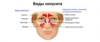
Some bones of the human skull are hollow formations, that is, they have sinuses inside (maxillary (maxillary), ethmoid, frontal (frontal), main (sphenoidal)). Inflammation of the mucous membrane of one or more of these sinuses is called the general term sinusitis (from the Latin sinus - sinus).
The largest sinus is the maxillary sinus.
The entry of a bacteria, virus or allergen into the sinus provokes the development of edema, the lumen of the sinus decreases, and an excess amount of mucus is formed, which disrupts the movement of air flow. Bacteria begin to multiply in the cavity, producing pus.
Sinusitis is an inflammation of the maxillary (maxillary) sinus.
Symptoms:
• Increased body temperature, chills.
• General malaise. • Runny nose (nasal discharge is usually mucous at first, and on the 5-7th day it becomes purulent). • Nasal congestion. • Sneezing. • Pain in the maxillary sinuses (infraorbital-buccal region), radiating to the teeth, forehead, and root of the nose. • Pain intensifies when bending the head forward, sneezing or coughing. • Nasality. • Decreased sense of smell.
Forms
According to the type of inflammation, they distinguish: • acute catarrhal sinusitis (viral) usually does not differ from a runny nose and is manifested by congestion, nasal discharge, and sometimes there is a slight heaviness in the buccal-infraorbital area. Acute catarrhal sinusitis can end in recovery or go into the second stage, that is, the stage of acute purulent sinusitis; • acute purulent sinusitis (bacterial) is characterized by the accumulation of pus in the maxillary (maxillary) sinuses. Facial pain becomes more intense, a headache occurs, and the patient’s general condition worsens significantly.
In any of the forms, sinusitis can be: • unilateral (inflammation of the sinus on one side); • bilateral (inflammation of the sinuses on both sides).
Causes
Acute sinusitis develops: • as a complication after acute respiratory infections; • in the presence of foci of infection in the nasopharynx (rhinitis (runny nose), tonsillitis (inflammation of the tonsils)); • against the background of disease of the teeth of the upper jaw (odontogenic sinusitis); • as a result of allergies; • as a result of injuries and wounds of the upper jaw (traumatic). A predisposing factor is also seasonal hypovitaminosis, that is, a state of decreased body tone due to a lack of vitamins (usually in the autumn-winter period).
Diagnostics
• Analysis of complaints and medical history: nasal congestion, thick nasal discharge (possibly green), headache or facial pain, aggravated by bending the head forward, nasal tone, increased body temperature, the presence of a runny nose or colds that preceded the disease, the fact of treatment by a dentist etc. • General examination: the presence of swelling in the cheek area; when pressing in the area of the maxillary sinuses, discomfort is noted - from increased sensitivity and mild pain to sharp pain radiating to the orbital area, etc. • Rhinoscopy - instrumental examination of the nasal cavity, during which signs of the development of the inflammatory process (swelling and redness of the mucous membrane, purulent discharge). • Endoscopic examination of the nose reveals pus in the middle meatus, which indicates the presence of purulent sinusitis. • X-ray diagnosis: in some cases, an X-ray of the maxillary sinuses shows the fluid level. • Diagnostic puncture of the maxillary sinus: the wall of the maxillary sinus in the nose is pierced with a special thin needle under local anesthesia. Then, using a syringe, the contents of the sinus are drawn out. When pus is obtained, the sinus is washed and a medicinal substance is injected into it. • Ultrasound of the paranasal sinuses is sometimes used as an alternative to x-ray examination. • Oropharyngoscopy (examination of the oral cavity) to identify carious teeth, assess the condition of fillings, etc. If necessary, a consultation with a dentist is indicated.
Treatment
Drug treatment: • vasoconstrictor drugs in the form of sprays or drops into the nasal cavity (drugs in this group relieve swelling of the mucous membrane and help remove stagnant fluid from the maxillary sinuses). The products are used in a short course, lasting 5-7 days; • nasal sprays containing antibiotics and steroid hormones (have an anti-inflammatory effect); • antihistamines, if the disease develops against the background of allergic reactions; • mucolytics – drugs that help liquefy the contents of the maxillary sinuses and, as a result, improve its excretion; • for purulent sinusitis, it is possible to prescribe antibiotics (in the form of tablets or injections).
Non-drug treatment. • Puncture (puncture) of the maxillary sinuses. The method allows you to quickly remove purulent contents (which leads to a rapid reduction in headache and facial pain, and improve your general condition) and inject medicinal substances directly into the sinus. Carrying out punctures of the maxillary sinuses in many cases allows you to do without prescribing antibiotics. • Rinsing the nose with saline, herbal and antiseptic solutions: The procedure can be carried out independently at home using special devices for a nasal shower, sprays or syringes; • Physiotherapy (treatment using natural and artificially created physical factors) is prescribed only when there is a good outflow of contents from the sinuses. • Taking general strengthening medications.
Surgical treatment is performed in the presence of orbital (eye) and intracranial complications. Treatment comes down to opening the purulent cavities and their sanitation, that is, removing the pus.
Complications and consequences
• Inflammation of the soft tissues of the face. • Spread of inflammation into the respiratory tract: inflammation of the bronchi (bronchitis), lungs (pneumonia), and ears (otitis). • Spread of the inflammatory process into the cranial cavity with the development of meningitis (inflammation of the meninges), encephalitis (inflammation of the brain substance) or brain abscess (formation of purulent cavities). • Purulent inflammation of the skull bones (a complication requiring surgical intervention). • Inflammation of the eyeball and its membranes, which can cause vision loss. • Inflammation of the trigeminal nerve (the large nerve in the face), accompanied by severe pain. • Sepsis is a severe complication that develops when the pathogen enters the bloodstream, with the further formation of secondary foci of inflammation in vital organs. • Risk of death.
The sooner you seek help from a specialist, the greater the chances of maintaining health and reducing the risk of complications!!!
Prevention
• Timely and adequate treatment of runny nose (rhinitis). • Treatment of nasal diseases accompanied by difficulty in nasal breathing (chronic rhinitis, deviated nasal septum). • Timely sanitation (treatment) of carious teeth. • Timely treatment of allergies. • Prevention of colds and strengthening the immune system:
hardening in the autumn-winter period; avoid hypothermia; taking multivitamin complexes in the autumn-winter period; wearing protective bandages during periods of mass morbidity, for example, influenza; balanced and rational diet (eating foods high in fiber (vegetables, fruits, herbs, etc.).
The information was prepared by an otorhinolaryngologist of the highest category, Doctor of Medical Sciences I. V. Raitselis
Sources: “Otorhinolaryngology”, manual for doctors V. T. Palchun, A. I. Kryukov, Moscow “Medicine”, 2001 “Otorhinolaryngology: national guide” edited by V. T. Palchun, GEOTAR-Media, 2008 G.
Signs if perforation was left without the attention of a doctor
Symptoms of inflammation after unsuccessful treatment occur in different ways - after a few days, weeks or even years. Depends on the sterility of the foreign body in the sinus, its ability to cause inflammatory processes, and the characteristics of the patient’s immune system. The following manifestations are possible:
- feeling of heaviness;
- pain when chewing;
- purulent and serous discharge from one nostril;
- elevated temperature;
- impaired sense of smell;
- pain when lightly tapped under the eye and in the nose area.
These are typical signs of odontogenic sinusitis caused by poor quality dental treatment. The key difference from rhinogenic sinusitis (when sinus infection occurs from the side of the nasal passages as a result of ARVI or influenza) is that signs of the inflammatory process are detected only on one side, where treatment was carried out.
In 1953, A. Proetz [1], based on his own, fairly thorough research, conducted a detailed analysis of existing ideas about the role of the paranasal sinuses.
For a long time it was believed that the gas composition in the paranasal sinuses is identical to that in the nasal cavity and when inhaling, air leaves the paranasal sinuses, and when exhaling, the cleanest and most oxygen-rich air enters the paranasal sinuses through the natural anastomosis [2]. Some authors considered the paranasal sinuses as one of the organs of gas exchange [3, 4]. Due to the virtual impossibility of installing and fixing measuring devices in the nasal cavity and paranasal sinuses, until a certain time it was impossible to study the patterns of gas movement in these anatomical structures. This led to the formation of incorrect principles of surgical treatment of many pathological conditions, in particular chronic rhinosinusitis. According to A. Proetz [1], “when inhaling, the pressure in the nasal cavity drops to 1/2000 of atmospheric pressure and 1/2000 of its volume leaves the maxillary sinus; at the end of inhalation, the pressure in the nasal cavity is equalized with atmospheric pressure and air leaves the nasal cavity enters the maxillary sinus in the same volume; when exhaling, the situation is repeated in the reverse order, so the air passes through the natural anastomosis four times.” The author noted that these air movements have scientific value, not clinical value. At the same time, he noted that complete renewal of air in the paranasal sinuses occurs in 1 hour. It is important to note that we were talking about the maxillary sinus as an air cavity, without taking into account the gaseous waste products of the epithelium of its mucous membrane. One of these products is nitric oxide [5].
Nitric oxide (NO) plays a huge role in the human body. The Nobel Prize for “the discovery of the role of nitric oxide as a signaling molecule in the regulation of the cardiovascular system” was awarded in 1998 to R.F. Furchgott, L.D. Ignarro and F. Murad. A special enzyme, NO synthase (NOS), which exists in the human body in the form of three isoforms, is responsible for the production of nitric oxide. Neuronal and endothelial isoforms of NOS produce low levels of NO and are directly dependent on the concentration of Ca2+ ions. The third isoform of NOS is activated by cytokines or bacterial lipopolysaccharides and is independent of Ca2+ titers [6]. The antibacterial, antiviral and antifungal properties of nitric oxide with the chemical formula NO are considered proven, as well as its participation in the immune system, in particular NO potentiates the phagocytic activity of macrophages.
The presence of a high content of the NOS enzyme in the epithelium of the mucous membranes of the paranasal sinuses (including the main sinuses) has been shown [5]. The inhibitory effect of nitric oxide on the growth of pure cultures of Staphylococcus aureus
, which causes the absence of any microflora in the paranasal sinuses normally [7]. In experiments on animals, an increase in the frequency of flicker of cilia of the epithelium of the nasal mucosa under the influence of NO was observed [8]. An increase in NO content in the nasal cavity was revealed as the age of the subjects increased, depending on the development of the paranasal sinuses [5].
In clinical studies using puncture and catheterization of the maxillary sinus with triple (repeated procedures after 17 and 24 hours) air sampling from the sinus and from the nasal cavity on the opposite side for 20 minutes, the average peak concentration of nitric oxide in the maxillary sinus was 7.4 mg/day. m3 [9], despite the fact that the maximum permissible concentration of nitrogen oxide in the room is 5 mg/m3 [10].
It has been shown that the concentration of nitric oxide in the main sinus is several times lower than in the maxillary sinus and practically corresponds to that in the nasal cavity [11].
All subtypes of NOS were detected in the main sinus mucosa and the possibility of continuous production of nitric oxide by epithelial cells of the sinus mucosa was proven [5].
In foreign literature, it has been repeatedly suggested that a decrease in NO content in the paranasal sinuses, along with occlusion of the natural anastomosis, plays an important role in the pathogenesis of sinusitis [12]. It has been shown that in children the level of nitric oxide in the nasal cavity decreases with the development of acute sinusitis and increases during antibacterial therapy [13].
A decrease in nitric oxide titers in the paranasal sinuses was noted when modeling sinusitis of various etiologies in experimental animals and an increase in nitric oxide titers [14].
M. Deja et al. [15] studied the level of nitric oxide in patients with maxillary sinusitis that developed during long-term artificial ventilation. The results obtained were compared with the level of nitric oxide in the maxillary sinuses in apparently healthy individuals. It has been established that against the background of purulent pansinusitis with fluid levels in the sinuses in patients on long-term mechanical ventilation, there is a significant (more than 70 times) decrease in NO titer in the maxillary sinuses compared to the norm (2554±385ppb*; p
<0.001) [15]. It is important to note that in this study, the authors paid attention to the level of nitrogen dioxide (NO2). A generally accepted property of NO is its increased reactivity when interacting with oxygen - upon contact with atmospheric air, NO is oxidized to NO2. The study [15] showed the absence of such interaction with the formation of nitrogen dioxide in the maxillary sinuses.
There is an opinion about the dual role of NO in inflammation [16, 17]. M. Marletta [18] showed that during sinusitis, NO is oxidized to more stable metabolites (nitrates and nitrites). It has been shown that in swabs of discharge obtained during operations on the paranasal sinuses in patients with chronic rhinosinusitis (with and without polyps), there is an increase in NO metabolites, which, according to the authors, indicates an increase in the production of nitric oxide in sinusitis [17] . An increased content of nitrates was detected in the discharge from the nasal cavity and polyp tissue in patients with polypous rhinosinusitis [19-21], an increase in the expression of the i-isotype of the NOS enzyme (iNOS) in biopsy samples of the mucous membrane of the inferior turbinate of experimental animals on the side of the introduction of a streptococcal culture into the sinus [ 22].
I. Alobid et al. [23] showed that during the use of oral and intranasal steroids in patients with nasal polyposis, there is a paradoxical increase in the NO content in exhaled air, which may be due to an improvement in the functioning of the ostiomeatal complex.
The use of NO donor drugs for inflammatory diseases of the paranasal sinuses has been experimentally substantiated. Thus, T. Runer and S. Lindberg [24] showed an increase in ciliated activity of the nasal epithelium in response to the use of a nasal spray based on sodium nitroprusside. B. Jian et al. [8] noted that when using an NO inhibitor, the ciliated activity of the epithelium decreases by 40%, followed by recovery after the use of sodium nitroprusside.
Experimental studies on animals have shown early recovery after modeling acute sinusitis with the combined use of antibacterial therapy and sodium nitroprusside [14].
In recent years, various hypotheses have been put forward about the regulation of gas composition in the paranasal sinuses and nasal cavity, as well as the participation of oxygen, carbon dioxide and nitric oxide in the pathogenesis of sinusitis [3, 4, 25]. W. Qian et al. [26] confirmed previous research results on the effect of exogenous administration of L-arginine on the production of NO by the mucous membrane of the nasal cavity and paranasal sinuses. Thus, after intravenous administration of L-arginine, the NO content in the air of the nasal cavity remains elevated for 1 hour. L-arginine increased the NO content in the nasal cavity both in the presence and absence of oxygen and did not affect the content of nitric oxide in the maxillary sinus, just like the oxygen content in the inspired air does not affect the level of NO in a given sinus. Inhalation of 100% oxygen completely stopped the formation of nitric oxide in the frontal sinus; this effect could not be neutralized by intravenous administration of L-arginine. In the nasal cavity and frontal sinus, nitric oxide production increased after intravenous administration of L-arginine and was blocked after inhalation of pure oxygen. NO concentration did not change with intravenous administration of L-arginine and inhalation of pure oxygen. The production of nitric oxide in the nasal cavity, frontal and maxillary sinuses sharply decreased when inhaling a gas mixture with a high content of carbon dioxide (6%) and without oxygen. However, in the presence of oxygen in the inhaled air, this effect obtained from carbon dioxide was neutralized. Thus, the study [26] confirmed the hypothesis about the influence of external influences on the gas composition in the paranasal sinuses. It was previously shown that the concentration of NO does not change in the paranasal sinuses in response to local anesthesia with 2% lidocaine, and, on the contrary, decreases with the use of xylometazoline [27].
As noted earlier, the gas composition in the maxillary sinus is characterized by an increased content of NO and is insensitive to changes in the gas composition in the nasal cavity or to the administration of L-arginine. To understand the patterns of functioning of the paranasal sinuses, one should return to the studies of A. Proetz [1], where quite simple models show that the paranasal sinuses are not actively ventilated (with each inhalation-exhalation, only 1/500 of the sinus volume is updated). For the first time at the modern level, these data were confirmed by G. Xiong et al. [28], who, using computer modeling of air flows in the nasal cavity and paranasal sinuses, showed that under normal conditions, due to the structure of the ostiomeatal complex, the air in the maxillary sinuses is not renewed. However, after virtual removal of the uncinate process, a pathological change in the direction of air flow in the nasal cavity during breathing occurs and air from the nasal cavity begins to flow into the maxillary sinuses.
C. Hood et al. [29] for this purpose, modeled air flows in the nasal cavity and paranasal sinuses, taking into account the presence of an additional anastomosis of the maxillary sinus. It was shown that 84 hours are needed to replace 90% of the air in the maxillary sinus in the presence of a single anastomosis. However, in this case, the results of previous studies that showed continuous production of NO were not taken into account. Understanding the results of this study and the continuous production of NO suggests that the gas composition of the air in the paranasal sinuses is fairly constant. Although the NO content in the maxillary sinuses can vary significantly between different people: from 160 to 21,875 ppb (average 6792 ppb) [11].
A distinctive feature of the work of C. Hood et al. [29] was that the authors analyzed changes in the directions and velocities of air flows arising in the natural anastomosis of the maxillary sinus during nasal breathing, depending on the structure (length, diameter, orientation of the long axis, shape, location) of the natural anastomosis, as well as the presence of an additional anastomosis. Thus, the authors showed that normally (in most cases) the natural anastomosis of the maxillary sinus has an ellipsoidal shape with a length of 6 mm and a width of 3 mm, the long axis of which is oriented parallel to the direction of the main air flow in the middle nasal meatus. In this case, in the natural anastomosis of the maxillary sinus, two vortex flows arise at a speed of 10–5–10–7 m/s. However, as the natural anastomosis increases, the second vortex flow disappears, and the speed of the remaining vortex flow can increase to 10–2 m/s. In the presence of an additional anastomosis, a pressure difference of approximately 0.1 Pa arises between them, which generates an upward air flow in the maxillary sinus at a speed of 10–7 m/s. With such an air flow, 90% of the air in the maxillary sinus will be renewed in 40 s, which, according to the authors of the study, can lead to pathology in the maxillary sinus. It has been shown that changes in the speed of mucus movement in the maxillary sinus, which occurs due to mucociliary clearance, do not affect the speed of air flow passing through the natural anastomosis.
According to C. Hood et al. [29], the surface area of the mucous membrane of the maxillary sinus is 24—50×10–6 m2, the surface area of the mucous membrane of the nasal cavity is approximately 10–2 m2. At these values, neither the nasal cavity nor the maxillary sinus can provide the known normal concentration of NO in the nasal cavity of 5-50 nL/min, as previously thought [30]. It is more likely that the presence of the NOS enzyme in the mucous membrane of the nasal cavity and paranasal sinuses provides approximately the same production of NO per unit area, although A. DuBois et al. [31] give values of 217–455 nl/min of gaseous NO in the maxillary sinuses. In any case, the proven critically low level of gas exchange between the nasal cavity and the paranasal sinuses is sufficient to maintain a high concentration of NO in the sinuses.
In another study by C. Hood et al. [32] conducted a virtual simulation of two postoperative conditions: with the presence of a single anastomosis with a diameter of 10 mm (sinusotomy through the middle meatus) and two anastomoses with a diameter of 3 and 6 mm (sinusotomy through the lower nasal meatus). It turned out that the two most common types of operations on the maxillary sinuses lead to a statistically significant decrease in the concentration of NO in the sinus from 9.1 to 100-1400 ppb. It has also been shown that only operated paranasal sinuses with enlarged anastomosis can maintain the concentration of NO in the nasal cavity due to possible diffusion in this case. Thus, the assumptions of R. Aust and B. Drettner [33] about the high rate of gas exchange between the nasal cavity and the paranasal sinuses normally turned out to be false, since the diameter of the natural anastomosis in the absence of an additional anastomosis makes it impossible both the diffusion mechanism and the active one (with each inhalation and exhalation) the passage of a mixture of gases from the sinuses into the nasal cavity and back. The absence of gas exchange between the nasal cavity and paranasal sinuses has been shown in a number of studies and is currently considered recognized by the European Society of Rhinology [11, 19]. A decrease in the concentration of NO in the operated paranasal sinuses entails two problems: a decrease in the ciliated activity of the ciliated epithelium and a decrease in the direct antibacterial effect of NO.
Over the past 5 years, three-dimensional modeling of air flows in the nasal cavity and paranasal sinuses, as well as dynamic changes in the content of nitric oxide in the paranasal sinuses during surgical interventions, have been performed using various computer programs: Mimics [34-38], vWorks [39, 40] , Amira [41]. In all of the above studies, approximately the same result was obtained - surgical expansion of the natural anastomosis of the maxillary sinus leads to an improvement in its ventilation and, as a result, to a decrease in the concentration of NO in the sinus. Thus, in the work of A.A. Voronin [42] analyzed the ventilation of the maxillary sinuses based on computed tomography data of a patient with a maxillary sinus cyst, as well as after surgical removal of the cyst through an artificial anastomosis in the lower nasal meatus. It has been shown that the presence of an additional anastomosis in the lower part of the maxillary sinus leads to pronounced turbulization of the air flow inside the sinus, expressed in the formation of a significant number of large-scale vortex structures.
J. Zhua et al. [43] conducted a study of computed tomography scans with computerized air flow simulation in patients undergoing balloon sinuplasty or uncinate process removal and found increased gas exchange in the paranasal sinuses with both surgical procedures. However, the study included patients with an additional maxillary sinus anastomosis.
Thus, the physiology of the nasal cavity and paranasal sinuses is extremely complex and dynamic. The mucosa of the upper respiratory tract contains a specific isotype of the NOS enzyme (iNOS) that responds to cytokine and bacterial stimuli. The concentration of iNOS is maximum in the ciliated epithelium of the paranasal sinuses, in particular in the maxillary sinuses. On the other hand, classical studies of sinus physiology, indicating the absence of any clinically significant gas exchange between the sinuses and the nasal cavity, have now been confirmed using highly accurate 3D digital models. The high content of iNOS in the paranasal sinuses, along with the lack of ventilation, maintains a high concentration of NO gas in the paranasal sinuses, which has also been confirmed in a number of studies using modern high-precision gas analyzers [44]. Currently, the normal role of NO in the upper respiratory tract is generally accepted: antibacterial, antiviral, antifungal action, as well as a stimulating effect on mucociliary clearance [45, 46]. It has been shown that with the development of pathology in the paranasal sinuses, a decrease in the concentration of NO in the sinuses occurs [47]. Based on the capabilities of modern computer technologies, it is necessary to conduct computer modeling at the planning stage of rhinosinus surgery. Such preoperative preparation will allow taking into account changes in air flow in the nasal cavity and paranasal sinuses, as well as avoiding pathological diffusion of gases with a decrease in NO concentration in the sinuses [48]. Further studies are needed on the physiology of the nasal cavity and paranasal sinuses, taking into account the humidity and thermal characteristics of the mucous membranes, as well as the nasal cycle, which may influence the maintenance of constant NO concentrations in the paranasal sinuses.
Parts per billion.
What are the dangers of sinus injury?
Since the complication does not always manifest itself immediately, symptoms of sinusitis that arise after a few years force the patient to consult a regular ENT doctor. Treatment, as a rule, does not bring results - in most cases the cause is not found. You start going from one doctor to another, and precious time is lost. The longer a foreign body is in the sinus, the higher the risk of tumor formation. If sinus perforation is left unattended, it can lead to:
- chronic sinusitis and sinusitis
- inflammation of the roots of adjacent teeth
- osteomyelitis of the jaw
- encephalitis and meningitis
Why, when symptoms of acute purulent sinusitis appear, you should urgently consult an ENT doctor:
Do not forget that “the nose grows on the head,” which means that any purulent diseases of the paranasal sinuses mean the presence of pus in the skull, in close proximity to the cavity of the orbits and to the brain, therefore complications can be extremely serious, with the most unpredictable consequences:
- Otitis
- Intraorbital complications (orbital cellulitis, orbital abscess)
- Intracranial complications (meningitis, encephalitis, brain abscess)
There is no need to try to cope with sinusitis on your own. The presence of a purulent process in the sinus indicates that it cannot empty itself and if time is lost, there is a high probability that the pus will make its way into one of the nearby organs.
Why you should entrust your treatment to the ENT Department of Dentistry
ENT dentistry is a comprehensive approach to the treatment of complications in the maxillary sinuses after dental treatment
The symbiosis of two areas - dentistry and otolaryngology - makes it possible to identify the cause, assess the situation, and select competent treatment tactics.
The ENT department of the Doctor Levin Center has been providing assistance for many years when problems arise after treatment and removal of teeth located on the border with the maxillary sinus. Surgical treatment is carried out by candidates of medical sciences, maxillofacial surgeons with otolaryngological training .
Patients come to the Center after a painful search for a solution to the problem. Repeated treatment by an ENT doctor in the hospital does not bring results. But a thinking patient should understand that if there is concern on the side of the sinuses where endodontic treatment once took place, you need to contact an oral and maxillofacial surgeon with ENT training. Only in this case can a comprehensive assessment of the situation be made and an adequate treatment plan drawn up.
Cyst of the maxillary sinus
Maxillary sinus cyst
is a benign neoplasm that appears on the mucous membrane inside the paranasal sinuses.
A maxillary sinus cyst is a rounded sac, with a membrane in the form of a thin film, filled with fluid. It happens that the cyst shell spontaneously ruptures, and this is manifested by copious clear discharge from the nose. Retention cysts
in the sinuses are formed due to prolonged disruption of the normal movement of air in the nasal cavity against the background of sluggish chronic inflammation (for example, allergies or chronic sinusitis). Odontogenic cysts occur in the sinus in response to inflammation in the area of the roots of the teeth of the upper jaw or as a reaction to the installation of dental implants.
WHAT IS THE DANGEROUS OF A PARONAL SINUS CYST?
Cysts of the paranasal sinuses are not tumors
, do not become malignant, and do not pose a threat to life.
A small cyst may be located in the sinus and not manifest itself in any way for many years. If the cyst does not grow and does not bother you, no treatment is required. On the other hand, if the cyst occupies more than half the volume of the sinus
, this leads to impaired ventilation and stagnation of mucus. A large cyst completely clogs the sinus anastomosis, preventing the normal movement of air and the outflow of secretions. There are often cases when, against the background of an acute respiratory infection, a maxillary sinus cyst suppurates - acute purulent sinusitis occurs, which is already fraught with complications. For this reason, large cysts are recommended to be removed.
TREATMENT OF MAXILLARY SINUS CYST
Cysts of the paranasal sinuses can be cured only in one way - surgically remove them.
. Once formed, the cyst will no longer be able to resolve on its own or with the help of medications. If the cyst bursts during a sinus puncture or is “cauterized” with a laser, it will grow again in the same place. To avoid recurrence of the cyst, it is important to carefully remove all its membranes. This is only possible under visual control, during endoscopic surgery.
ENDOSCOPIC MAXILLARITY IN THE ENT CENTER
At the ENT Clinic, treatment of paranasal sinus cysts is carried out only by the endoscopic method.
according to the principles of FESS - functional endoscopic surgery of the paranasal sinuses.
This method involves the most gentle approach through the natural anastomosis of the sinuses, small incisions, minimal damage to the mucous membrane and intranasal structures. Endoscopic surgery to remove a maxillary sinus cyst is called endoscopic maxillary sinusotomy
.
METHODS OF TREATMENT OF MAXILLARY SINUS CYST
Cysts of the paranasal sinuses can be cured only in one way - surgically remove them. Once formed, the cyst will no longer be able to resolve on its own or with the help of medications. Traditional methods of treatment (rinsing the nose and sinuses with solutions of salt or medicinal plants), nasal sprays and antibiotics are ineffective against cysts. Non-surgical treatments can temporarily shrink the cyst by draining the fluid. If the cyst is burst with a needle during sinus puncture or cauterized with a laser, it will grow in the same place again in 2-3 months. To avoid recurrence of the cyst, it is important to very carefully remove all its membranes. This is only possible under visual control, during endoscopic surgery.
MODERN OPERATIONS FOR THE TREATMENT OF MAXILLARY SINUS CYST
At the ENT Clinic Center, treatment of paranasal sinus cysts is carried out only by the endoscopic method according to the principles of FESS - functional endoscopic surgery of the paranasal sinuses. Endoscopic surgery to remove a maxillary sinus cyst is called endoscopic maxillary sinusotomy. This method involves the most gentle approach through the natural anastomosis of the sinuses, minimal damage to the mucous membrane and intranasal structures. The advantages of endoscopic surgery are that everything is done inside the nose - there are no swelling, incisions, stitches or scars on the face or under the lip after it. The endoscope gives the surgeon a good overview, a wide field of view and bright light, which allows the entire cyst to be removed completely without leaving any membranes.
HOW THE OPERATION WORKS:
- The anesthesiologist and operating nurse prepare the patient for surgery.
- The surgeon examines the nasal cavity with an endoscope and identifies the natural anastomosis of the maxillary sinus on the lateral wall of the nasal cavity.
- With the help of special instruments, the sinus anastomosis expands to approximately 9-10 mm.
- The internal contents of the sinus are examined with an angled endoscope.
- Special micro-forceps are inserted through the enlarged anastomosis, the cyst is captured and removed entirely along with the membranes.
- The sinuses are washed and examined.
- After the operation, special breathable sponge tampons are installed in the nose.
The result of endoscopic maxillary sinus surgery will be the restoration of the normal volume of the sinus, the natural outflow of mucus and the movement of air in it.
Dear patients! Please note that the stated cost of the operation does not include the cost of laboratory blood tests and other examinations. The cost of treatment depends on the chosen method of anesthesia (local or general) and the length of stay in the hospital room. Only a surgeon after a face-to-face consultation can tell you the exact amount in your specific case.
Endoscopic surgery is used in the ENT Center clinic to treat not only maxillary sinus cysts, but also chronic sinusitis of any nature, polyps and other sinus tumors.
ENDOSCOPIC SURGERY IS INDICATED FOR DISEASES:
- Cysts of the maxillary sinuses
- Polyps of the nasal cavity and polypous rhinosinusitis (from a single polyp to widespread polyposis).
- Chronic and recurrent purulent sinusitis (sinusitis, ethmoiditis, sphenoiditis, frontal sinusitis, or their combinations - polysinusitis, pansinusitis).
- Odontogenic sinusitis and foreign bodies of the maxillary sinuses (after treatment by a dentist and cementing material entering the sinuses).
INDICATIONS FOR ENDOSCOPIC SINUS SURGERY:
A maxillary sinus cyst accidentally discovered on a CT scan does not always require urgent surgery. If it occupies less than 1/3 of the sinus, does not complicate breathing and does not cause pain, observation by an ENT doctor is sufficient. You should think about endoscopic cyst removal if:
- One or both halves of the nose are always stuffy, so you have to breathe through your mouth.
- There is an excess accumulation of mucus in the nose and nasopharynx.
- The constant feeling of heaviness, pressure, distension in the sinus area or headache is disturbing.
- Acute purulent sinusitis occurs on the side of this sinus.
WHAT YOU NEED TO PREPARATE FOR ENDOSCOPIC OPERATION:
1. First, you need to undergo an examination by an ENT surgeon to make an accurate diagnosis and determine clear indications for surgery. 2. Be sure to do a CT scan of the nose and paranasal sinuses for individual planning of the scope of the intervention. CT scans also help the surgeon navigate the procedure. 3. After the ENT doctor has confirmed the need for surgery, you need to undergo a general preoperative examination on an outpatient basis and obtain a physician’s opinion on the absence of contraindications. You can undergo the examination both at the ENT Center clinic and at your local clinic. You can download the list of required tests in PDF format from this link.
REHABILITATION AFTER ENDOSCOPIC SURGERY IN THE NASAL CAVITY
- After endoscopic surgery, special sponge breathable tampons are placed in the nose, which the surgeon removes after 1-2 days. Tampons sometimes cause a feeling of pressure in the nose and a headache, these symptoms quickly disappear after removal.
- The patient spends a day in the hospital and goes home the next day after the operation.
- At the appointed time, the patient visits the surgeon on an outpatient basis: to remove tampons, rinse the nose and monitor healing.
- In the first week after surgery, the patient is not recommended to drink hot and alcoholic drinks, take a hot shower, or perform physical exercise (so as not to provoke dilation of blood vessels in the nose and nosebleeds). It is best to follow a home regime and avoid colds.
- As it heals over the course of 1-2 weeks, clots of mucus and crusts constantly form in the nose - this is normal. The doctor will prescribe saline solutions to moisten the nose and other medications to reduce swelling and speed up healing. Nasal breathing is completely restored gradually, usually by the 3rd week after surgery.
Diagnosis of perforation
Having an accurate understanding of the situation in the sinus, the maxillofacial surgeon chooses the correct tactics and method of eliminating the problem
As part of diagnostic activities, our Center carries out:
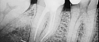
- X-ray allows you to assess the condition of the causative tooth and the quality of canal treatment
- Computed tomography in ENT mode allows you to get a complete picture of the condition of the maxillary sinuses
The modern Sirona Gallileos CT scanner guarantees high quality images , in which you can determine the location and size of the perforation, identify the presence and localization of foreign bodies, cysts and polyps (if they form), and assess the size of the inflammatory process.
How is the treatment carried out?
We respect the patient’s personal time and strive to carry out all activities comprehensively, in one day :
- Professional hygiene Preparation of the oral cavity to ensure sterility during surgery to avoid secondary sinus infection
- Surgery to eliminate inflammation Performed while you sleep; the method of access to the sinus is selected depending on the location of the foreign body and the presence of tumors
- Temporary prosthetics If the causative tooth had to be removed, a temporary crown or immediate prosthesis is installed to mask the defect
Surgical operations in our Center are performed under sedation. The patient does not feel anything, fears and worries are excluded. Sedatives put you into a controlled drug-induced sleep without falling into unconsciousness - this is not general anesthesia! They do not contain toxic components, act gently, preserving reflexes. The artificial lung ventilation device is not connected, hospitalization is not required.
The surgical intervention is completed by a mandatory CT examination , which is necessary to assess the quality of the work performed.
After 10-14 days, the patient is invited to the clinic for suture removal and a control CT image. A schedule of professional inspections is drawn up.
Results and discussion
Analyzing the diagnostic images, we drew attention to the following anatomical feature of the structure of the cribriform plate of the ethmoid bone: the low location of the central sections (olfactory fossa) relative to the lateral ones (ethmoidal fossa) (Fig. 1).
Rice. 1. CT scan of the skull in coronal projection. The low location of the olfactory fossa on both sides is determined (indicated by arrows).
P. Keros (1965) identified three types of location of the cribriform plate: high - the cribriform plate is located below the roof of the ethmoidal labyrinth by 1-3 mm, medium - by 4-7 mm and low - by 8-16 mm [6]. However, these measurements were made relative to the medial edge of the ethmoid bone. We, in turn, measured the depth of the olfactory fossa from the line that connects the lateral edges of the ethmoidal fossa of the cribriform plate to the lowest point of the olfactory fossa (Fig. 2). With this measurement, the general direction of the cribriform plate (horizontal or oblique), which also characterizes the features of the anatomical structure of the ethmoid bone, has a significant impact on the objectivity of assessing the depth of the olfactory fossa.
Rice. 2. CT scan of the skull in coronal projection. Landmarks for measuring the depth of the olfactory fossa are shown: a line connecting the medial edges of the ethmoidal fossa and arrows pointing to the lowest points of the olfactory fossa.
In 42 patients with SNL, a low location of the olfactory fossa was found - 7.2±1.8 mm below the level of the ethmoid. In 13 cases, attention was drawn to the asymmetrical location of the reticular plate, and the localization of the SNL corresponded to the side of the lower position of the olfactory fossa (Fig. 3). In 30 cases, fenestration of the cribriform plate was observed over 1-5 mm. The fistula was most often localized in the central sections of the perforated plate (in the area of the olfactory fossa).
Rice. 3. CT scan of the skull in coronal projection. Asymmetrical location of the olfactory fossa, lower on the side of development of the SNL (indicated by an arrow).
In one of the 34-year-old patients, SNL occurred after radical endoscopic surgery for right-sided polypous polysinusitis. A CT study demonstrated the absence of the anterior end of the middle turbinate and individual intercellular septa of the anterior cells of the ethmoidal labyrinth resected during the operation. In this case, the following variant of the anatomical structure of the ethmoid bone on the right was noted: the anterior cell at the base of the middle turbinate extended cranially and penetrated into the posterior sections of the frontal infundibulum; thickened mucosa and a horizontal fluid level were observed in it. In addition, there was a narrow bone defect (no more than 1 mm) of the lateral slope of the olfactory fossa on the right at the level of the posterior end of the cock's crest, opening into the cell of the frontal infundibulum described above. The perpendicular and cribriform plates of the ethmoid bone were covered with polyps with the presence of areas of periosteal ossification; local destruction of the septa of the ethmoidal labyrinth from the pressure created by the polyps was visualized.
Thus, in this case, the polyposis process led to the destruction of the ethmoid bone structures, the reticular plate in the area of the olfactory fossa was filled with polyps, which plugged the resulting bone defects, and after polypotomy, conditions were created for the leakage of cerebrospinal fluid (Fig. 4).
Rice. 4. CT scan of the paranasal sinuses of a 34-year-old patient: coronal (a) and axial (b) projections. The anterior end of the middle turbinate on the right is missing—a condition after its resection. Areas of periosteal bone formation and polyps are identified (indicated by a long arrow). The septa of the ethmoidal labyrinth are locally destroyed, and a defect in the olfactory fossa is revealed (indicated by a short arrow).
Ethmoidal meningocele was diagnosed in 2 patients. In one 37-year-old patient, a CT scan revealed a defect of up to 1.5 mm2 in the cribriform plate on the right in the area of the bottom of the olfactory fossa at the level of the middle cells of the ethmoid bone, through which an area of soft tissue density protruded between the nasal septum and the superior turbinate. At the same time, a small amount of cerebrospinal fluid density fluid was visualized in the right sphenoid sinus and sphenoethmoidal recess (Fig. 5).
Rice. 5. CT scan of the paranasal sinuses of a 37-year-old patient: axial projection (a) and reconstruction in the coronal plane (b): a defect in the olfactory fossa (indicated by arrow 1) and an ethmoidal meningocele with the contents of the cerebrospinal fluid density (indicated by arrow 2) are observed.
In one 45-year-old patient, a CT scan revealed a defect in the cribriform plate of the ethmoid bone on the right in the lateral sections with a length of up to 4 mm, the bone walls of the anterior cells of the ethmoid bone were thinned, and a soft tissue formation up to 10 mm in size was visualized in them (meningocele) (Fig. 6). In the right maxillary and frontal sinuses, and the nasopharyngeal vault, the contents of the cerebrospinal fluid density were determined.
Fig. 6. CT scan of the paranasal sinuses of a 45-year-old patient in the coronal projection. A bone defect of the cribriform plate (a, b; indicated by arrow 1), meningocele (b; indicated by arrow 2), and the contents of the cerebrospinal fluid density in the nasopharyngeal vault (c; indicated by arrows) were revealed.
In one 55-year-old patient, the occurrence of liquorrhea was caused by the presence of a mucocele of the anterior cells of the ethmoidal labyrinth, in which a thin-walled bone cavity was visualized on the right, which included a cell of the nasal tubercle and a ethmoidal bubble measuring 21x9 mm, filled with the contents of soft tissue density and extending to the perforated plate; its local destruction was noted in the area of the ethmoid fossa for about 4 mm (Fig. 7). The uncinate process and the infundibulum on the right were not clearly differentiated, and the frontonasal canal in the caudal sections was not traced. The right frontal sinus was hypoplastic and filled with pathological contents. A small amount of liquid content with a horizontal level was noted in the right maxillary sinus.
Rice. 7. CT scan of the paranasal sinuses of a 55-year-old patient: coronal (a) and axial (b) projections. a — local destruction of the reticular plate of the ethmoid bone at the location of the thin-walled bone cavity (indicated by an arrow); b — mucocele (indicated by an arrow).
In one 53-year-old patient, the development of SNL was caused by the presence of a space-occupying lesion in the right sphenoid sinus. On K.T. an enlarged (swollen) right sphenoid sinus was visualized, filled with a space-occupying formation with a density of up to 34 HU, spreading through the defect of the intersinus septum (mainly in the middle and upper sections, up to 11 mm in length) into the left sphenoid sinus (Fig. 8). The lateral wall of the right sphenoid sinus was destroyed for about 7 mm. Contents with air bubbles (mucus) were visualized in the posterior parts of the left sphenoid sinus. In the right maxillary sinus there was a small amount of liquid content of low density.
Rice. 8. CT scan of the paranasal sinuses of a 53-year-old patient in the axial projection. Destruction of the walls of the right sphenoid sinus (indicated by arrows 1 and 2) and a space-occupying formation (indicated by arrow 3) were revealed.
In some cases, it is possible to detect freely flowing cerebrospinal fluid through a defect in the cribriform plate, which looks like a linear area of lower density than the surrounding mucous membrane. In the presence of additional formations in the sphenoid sinuses and cells of the ethmoidal labyrinth, bone defects through which cerebrospinal fluid leaks occur are also clearly visualized. In such cases, information obtained from CT in the axial plane is sufficient.
In cases where defects in bone structures or dura mater were not clearly identified (44% of patients), nasal liquorrhea was diagnosed by indirect signs, such as thinning of bone structures without obvious destructive changes in combination with pathological contents of liquor density mainly in the posterior parts of the sphenoid sinuses and sphenoethmoidal recesses, as well as in the posterior cells of the ethmoidal labyrinth, less often in the maxillary sinuses. We performed dynamic CT examination in such patients. The primary examination was performed in the axial plane immediately upon admission of the patient to K.T.’s office. A repeat study was performed in the coronal plane 10-20 minutes after the initial one. Between the initial and repeat examinations, the patient was on the table in a supine or abdominal position. The time for repeat testing depended on the rate of cerebrospinal fluid flow: if more than 10 drops flowed in 1 minute, the test was repeated after 10 minutes, if less abundant, after 20 minutes. The amount of fluid in the sinuses listed above increased, and in some cases it was possible to trace a stream of cerebrospinal fluid (a linear area of lower density than the surrounding mucous membrane). In addition, we came to the conclusion that the very first anterior cell of the ethmoidal labyrinth, containing cerebrospinal fluid, corresponds to the level of localization of the fistula.
We did not perform CT cisternography on patients due to the rather high invasiveness of the procedure, the result of which was the entry of the contrast agent into the paranasal sinuses, which does not provide significant additional information, since the cerebrospinal fluid found in them during SNL has a natural contrast and is well differentiated against the background air without additional contrast. According to D.N. Kapitanova and A.S. Lopatin, CT cisternography can sometimes give false negative results due to an inadequate amount of contrast agent administered and poor timing of the study [7].
No hospitalization required
Operations in our Center are performed by operating teams of experienced maxillofacial surgeons with ENT training in a sterile operating room. The treatment is as gentle and minimally traumatic as possible; a 24-hour hospital stay is not required; you will go home the same day .
The intervention lasts about an hour, under sedation. After completion, the patient quickly returns to clear consciousness without unpleasant consequences or risk of complications. After 30-40 minutes you can safely go home. For patients with concomitant cardiovascular diseases, a day hospital is provided . You can lie down for the time necessary for recovery under the supervision of our anesthesiologist-resuscitator.
Material and methods
The article analyzes the CT results of 45 patients with SNL who were treated in the department of otorhinolaryngology of the Kursk Regional Clinical Hospital from 2000 to 2014. Among the patients there were 39 women aged 37 to 66 years and 6 men aged 34 to 66 years . The studies were carried out in axial and coronal projections with a slice thickness of 0.625-3.0 mm on a computed tomograph, followed by evaluation of diagnostic images on a workstation. All patients underwent biochemical analysis of nasal discharge.
Accelerated rehabilitation
After the operation, you will receive free medications and instructions with rules of behavior during the rehabilitation period. We have collected the necessary set of drugs to improve your well-being at home. Please take them as prescribed by your doctor and follow the recommendations to avoid unpleasant consequences.
For quick recovery after surgery, our Center offers its own method of rehabilitation in 1-2 days. The procedures allow us to minimize the formation of possible hematomas, swelling, and eliminate pain. The program uses:
Microcurrent therapy
A weak pulse current normalizes metabolic processes, activates ATP production, and improves tissue nutrition. Regeneration starts, healing accelerates, swelling decreases. Muscle spasm goes away.
PRP plasma therapy
Injections of purified and platelet-enriched plasma from one’s own blood trigger cellular regeneration processes using the body’s internal reserves. The manifestation of edema and hematomas is reduced.
Biostimulation of the face
Lymphatic drainage drugs D-NUCLEO and MesoSculpt C71 help reduce swelling. Active anti-inflammatory components improve blood flow, increase tissue nutrition, and improve healing.