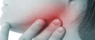
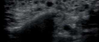
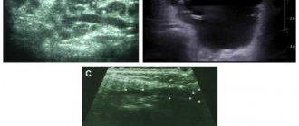
Ultrasound of the salivary glands is an ultrasound method used to assess the condition and diagnose diseases of this organ. It allows you to examine the excretory ducts, parenchyma, nearby lymph nodes and blood vessels. Using ultrasound, you can identify tumors, cysts, stones, inflammatory changes in the salivary glands, as well as determine the extent and severity of the pathological process. This diagnostic method is considered informative, safe and the most affordable.
Indications for the study
An ultrasound of the salivary glands is recommended for patients with the following complaints:
- feeling of fullness, acute pain in the parotid and submandibular area
- enlargement, hardening and tenderness of the salivary glands
- problems with swallowing
- feeling of dry mouth
- difficulty moving the lower jaw
Ultrasound examination of the salivary glands will help diagnose:
- benign and malignant neoplasms
- inflammation – acute and chronic sialadenitis
- salivary gland adenoma
- dystrophic processes in the parenchyma of the organ
- salivary stone disease
- anatomical anomalies in the structure of the salivary gland and its ducts
Since in parallel the doctor always examines nearby lymph nodes, he can additionally diagnose their inflammation - lymphadenitis.
Advantages of carrying out the procedure at the Central Clinical Hospital of the Russian Academy of Sciences
The Central Clinical Hospital of the Russian Academy of Sciences in Moscow receives doctors and candidates of medical sciences with many years of experience, specialists of the highest category. The clinic is equipped with modern expert-class equipment, which allows you to accurately and quickly make a diagnosis. The appointment takes place in comfortable conditions, without queues or long waits for examination results. Treatment is selected individually, taking into account the age and condition of the patient. If necessary, you can be consulted by doctors of related specialties.
You can sign up for an ultrasound of the salivary glands by phone or through a special form on the website. We will select a convenient time for you.
Fine needle aspiration biopsy in the diagnosis of cancer
Find out the cost of PTAB Thyroid gland
Sign up through your personal account
Biopsy (puncture) is a diagnostic method in which cells or tissues are taken intravitally from the human body and then examined microscopically.
In the structure of mortality in Russia, cancer is in second place after cardiovascular diseases. Cancer statistics in Russia show that approximately 1,500 patients with cancer are registered in our country every day. In total, at least 2.5 million patients with various forms of cancer are registered in oncology clinics in Russia.
The main reason for increased mortality from cancer in Russia is late diagnosis. An important reason for the late detection of malignant neoplasms is that people do not consult a doctor on time due to lack of knowledge in this area or insufficient funds. This leads to the fact that in Russia every third cancer patient dies within a year from the moment of diagnosis. For example, in the United States, more than 80% of patients pass the five-year survival mark.
In the majority of cancer patients, the pathology preceding thyroid and breast cancer is usually benign neoplasms. Nodules in these groups of patients can be detected by ultrasound. The widespread use of high-resolution ultrasound devices makes it easy to diagnose nodular formations, including small ones (up to 1 cm in diameter).
The main goal of a diagnostic search for nodal pathology is to identify patients with neoplastic changes in the mammary and thyroid glands, since it is this category of patients that is primarily subject to surgical treatment and, if necessary, radiation therapy, etc.
It is no secret that it is almost impossible to reliably determine the type of tumor (benign or malignant) using ultrasound; moreover, ultrasound is a rather subjective method in which the human factor (ultrasound diagnostic operator) is present. Significant variability in ultrasound diagnostic indicators depends on two main factors - the level of qualification of the diagnostician and the technical parameters of the ultrasound scanner used.
The insufficient information content of the used methods for studying diseases of the thyroid and mammary glands, combined with the need to clarify the diagnosis, forced clinicians to actively use invasive manipulations with morphological verification of the process.
Ultrasound-guided fine-needle aspiration biopsy (FNAB) is the most accurate, highly informative, safe method used in many diagnostic algorithms for focal diseases of the thyroid, breast and other superficially located structures.
The study of punctate allows not only to recognize the nature of the process (benign or malignant), to clarify the histogenetic affiliation of the neoplasm, but also to identify cancer in the early stages of development, when clinical manifestations of the disease are not yet present.
In patients operated on for thyroid cancer, if tumors are detected in the surgical site, a puncture biopsy provides a clear differential diagnosis between tumor recurrence, hyperplasia of the remaining gland tissue, or postoperative changes such as granuloma.
Main indications for TAPB:
- Single or multiple nodes in an organ, when their benign nature is in doubt.
- The presence of altered lymph nodes, when it is necessary to determine the source of metastasis.
- Focal formations of the mammary glands with characteristic signs of a tumor process, various cysts (atypical, simple). When a cyst is punctured, a therapeutic function is also performed, since its contents are removed as much as possible.
- Neoplasms of the salivary glands, soft tissues of the neck, in some cases, median or lateral cysts of the neck.
- Recurrence of the primary tumor
According to the American Association of Clinical Endocrinologists (AACE), fine-needle aspiration biopsy of nodules measuring 10 mm or less is not indicated unless ultrasound findings are suspicious for thyroid cancer and there is no high risk of cancer according to medical history. However, all these recommendations are not absolute and in certain situations leave the clinician the right to decide on the need for fine-needle aspiration biopsy, even in cases where nodular formations do not meet the above criteria.
Main contraindications to TAPB:
- blood diseases (blood clotting disorders can lead to bleeding, which may require urgent hospital treatment);
- the patient has mental illness, since TAPP can provoke an exacerbation of the underlying disease.
Methodology for performing FNAB of nodal changes in the thyroid and mammary glands
The fine-needle aspiration biopsy method is simple, as is drawing blood from the patient's arm. It has virtually no contraindications and can be performed on an outpatient basis without anesthesia. The introduction of anesthetics causes a deterioration in ultrasound visualization of the area under study, a change in the cellular pattern, and the anesthesia itself is as painful as the puncture. The patient is in a supine position. The skin surface is treated with a solution of 70% ethyl alcohol. An injection (puncture) is made through the skin into the tumor-like formation of the organ and the contents of the formation are aspirated. As a result of puncturing the formation under study with a needle, tissue fragments are collected. The movement of the needle and all manipulations are visually controlled on the screen of the ultrasound machine. After the tip of the needle is inside the formation, the doctor performs aspiration (sampling) of the contents of the node several times with a syringe. To obtain a sufficient amount of biological material, 2 - 3 injections are performed in different areas of the formation. The needle is then removed and the resulting material is applied to glass slides. The slides are sent for examination to the cytology laboratory. Next, these slides are specially stained, and a cytologist examines their contents under a microscope. By carefully examining the cellular material taken through a biopsy, the doctor will be able not only to determine the malignancy or benignity of the pathology, but also to identify the degree of damage to the organ. When planning surgery, the need for a biopsy increases. After the study, a written conclusion is issued. The duration of a puncture biopsy depends on the number of lesions being punctured. No special preparation is required to perform a fine-needle aspiration biopsy. The TAPB procedure is well tolerated by patients. A hypoallergenic sterile patch is glued to the injection site, which can be removed after 20 minutes. 5-10 minutes after the biopsy, the patient can lead a normal life.
Side effects and possible complications of TAPP
The occurrence of moderate pain at the site of TAPE is associated both directly with tissue damage in the manipulation area, and sometimes with the possible development of delimited hematomas, which resolve safely. Discomfort, pain in the neck, and headaches can be triggered by throwing back the head during TPA if the patient has cervical osteochondrosis.
Fine-needle aspiration biopsy is today the most valuable and informative method for preoperative differential diagnosis of various nodular formations. This is the only method that allows one to reliably confirm or refute the malignant nature of the process. Fine needle aspiration biopsy is critical in determining treatment strategy.
Using fine-needle aspiration puncture biopsy, you can obtain reliable and accurate information about changes in the tissues of the mammary and thyroid glands in order to identify the disease at an early stage and begin effective treatment.
FAQ
Is a fine needle aspiration biopsy really appropriate for me?
Fine needle aspiration biopsy is the only non-surgical method for determining whether a nodule in the organ being examined is benign or malignant.
The large tumor must be removed. In this case, am I also indicated for a fine-needle aspiration biopsy?
Yes, it is indicated to determine the extent of surgical intervention. Sometimes a node may have tumors of different histological structures. Knowing this in advance, the surgeon plans and carries out more competent treatment accordingly.
Should fine needle aspiration biopsy be ultrasound guided?
Yes, definitely! Tumors are often located close to blood vessels, neurovascular bundles and other organs. In order to avoid their traumatization during biopsy, the latter should be carried out under ultrasound control.
Can a node “degenerate” into a malignant one after a biopsy?
NO!!! Fine-needle aspiration biopsy does not cause malignancy (degeneration into a malignant neoplasm)!
Can a biopsy cause the tumor to spread?
There is no data in world practice to prove tumor spread after fine-needle aspiration biopsies.
The life of each of us depends only on ourselves. How to get maximum pleasure and minimum suffering from it? - try to maintain your health, strengthen your immune system, carry out the necessary prevention and treatment in a timely manner, and, accordingly, diagnosis.
Make an appointment for ultrasound diagnostics and an appointment with an oncologist-mammologist at the Clinic in Severny by calling +7.
Author of the article: ultrasound doctor Irina Yurievna Novikova.
