Author of the article:
Soldatova Lyudmila Nikolaevna
Candidate of Medical Sciences, Professor of the Department of Clinical Dentistry of the St. Petersburg Medical and Social Institute, Chief Physician of the Alfa-Dent Dental Clinic, St. Petersburg
It is very important to detect this form of pulpitis in time and cure it. Otherwise, the process of pulp necrosis will continue, which can ultimately lead to the formation of periodontitis, gumboil and forced tooth extraction.
Prevention of pulpitis is compliance with hygiene rules. We recommend using Asepta toothpastes, which reduce the number of bacteria, break down plaque, and gently whiten teeth.
It is worth noting that this type of pulpitis occurs quite often, and it affects both permanent teeth in adults and adolescents, and baby teeth in children. It can be either a stage of fibrous pulpitis in an acute form, or an independent primary chronic process, for which the previous acute stage is not characteristic.
Diagnosis of chronic fibrous pulpitis
The diagnosis is made not only on the basis of interviewing the patient and examining his oral cavity, but also based on the results of additional studies.
As additional diagnostic methods, the dentist uses:
- palpation: as a rule, it is painless near the gums;
- probing: as a result, the presence of an opening in the pulp chamber is determined;
- thermometry: by supplying cold water with a syringe, the presence or absence of a reaction to temperature stimuli is determined;
- X-ray diagnostics: helps to identify the communication of the pulp or carious chambers;
- percussion: the reaction, as a rule, is also absent;
- electroodontodiagnosis: exposure to current allows you to determine how much the pulp has retained its reality.
To obtain an accurate diagnosis, a qualified dentist uses several of these methods at once.
What diseases should be distinguished from chronic fibrous pulpitis?
During the diagnostic process, it is important to distinguish fibrous pulpitis from other diseases with similar symptoms.
- Deep caries. Unlike fibrous pulpitis, it is characterized by pain of a short-term nature, which disappears immediately after the cessation of temperature or mechanical influence.
- Hypertrophic pulpitis. It is characterized by long-term pain that occurs independently for no apparent reason. Upon examination, the patient feels pain and bleeding may occur.
- Gangrenous pulpitis in chronic form. This disease is characterized not only by bad breath, but also by extensive exposure of the pulp, which is detected upon examination.
- Chronic periodontitis. A patient with this disease complains of very severe pain, but no pain is felt on probing, but a fistula may be observed. An x-ray will also indicate periodontitis.
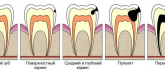
Classification of pulpitis
The clinical classification of the disease is simple and distinguishes the following forms of the disease:
- acute – begins suddenly and is accompanied by severe symptoms;
- chronic - can be either an independent disease or a consequence of acute pulpitis;
- exacerbation of the chronic form;
- condition after partial or complete removal of the pulp against the background of previous pulpitis.
In terms of prevalence, acute pulpitis is of two types: limited and diffuse. In the first case, only a small area of the pulp is involved in the inflammatory process. Diffuse pulpitis usually extends to the entire pulp and is a consequence of limited pulpitis.
Chronic pulpitis is divided into fibrous, hypertrophic and gangrenous forms. Each of them has its own clinical course.
Treatment of chronic fibrous pulpitis
Having assessed the overall picture of the disease, the dentist determines exactly how treatment will be carried out. There can be two of them:
- Biological (also called conservative). During this method, the dentist does everything possible to keep the pulp alive. Unfortunately, when it comes to a disease such as chronic fibrous pulpitis, this method is rarely used.
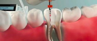
- Surgical. Used quite often. It consists in the fact that the pulp is completely removed and the tooth is filled. This can be done in one visit or two. In the latter case, to anesthetize the dental nerve, a special paste, for example, based on arsenic, is first placed in the tooth, and only during the next visit is the pulp removed. In this case, first remove its upper part, then the remains from the dental canals. After which they are processed and sealed.
With a timely visit to the dental clinic and proper treatment, it is possible not only to get rid of pain and associated discomfort, but also to save the tooth.
Professional oral hygiene, regular visits to the doctor and timely treatment of dental diseases can avoid the manifestation of this disease.
Reviews about our doctors
I would like to express my gratitude to the dentist Elena Nikolaevna Kiseleva and her assistant Svetlana - they are real specialists and at the same time sensitive, not burnt out by years of practice.
Thanks to them, I have been coming back here for many years. Thanks to the management for such doctors! Read full review Svetlana Nikolaevna
13.08.2021
I am very grateful to Evgeniy Borisovich Antiukhin for removing my three eights. Especially considering that the lower tooth was not the simplest (it was located in an embrace with a nerve). The removal took place in 2 stages, one tooth under local anesthesia, two under general anesthesia. I had no idea that wisdom teeth could be... Read full review
Sofia
28.12.2020
Clinical researches
Clinical studies have shown that for the effective treatment of chronic localized periodontal disease of traumatic etiology in young patients, a complex of treatment and preventive measures is required, which includes products for individual oral hygiene in the form of treatment and preventive toothpaste "ASEPTA PARODONTAL SENSITIVE" (JSC "VERTEX" , Russia) and mouth rinses "ASEPTA PARODONTAL ACTIVE" and "ASEPTA PARODONTAL FRESH", (JSC "VERTEX", Russia), which allows, within 6 months after completion of complex treatment, not only to improve oral hygiene and 55.37 % reduce inflammatory processes in the gums, but also reduce the number of relapses of localized periodontitis by 21.36%.
Sources:
- Prevention of recurrence of localized periodontitis in young A.K. YORDANISHVILI, Doctor of Medical Sciences, Professor, North-Western State Medical University named after. I.I. Mechnikov, Military Medical Academy named after. CM. Kirov, International Academy of Sciences of Ecology, Human Safety and Nature.
- Clinical studies of antisensitive toothpaste “Asepta Sensitive” (A.A. Leontyev, O.V. Kalinina, S.B. Ulitovsky) A.A. LEONTIEV, dentist O.V. KALININA, dentist S.B. ULITOVSKY, Doctor of Medical Sciences, Prof. Department of Therapeutic Dentistry, St. Petersburg State Medical University named after. acad. I.P. Pavlova
- Report on clinical trials to determine/confirm the preventive properties of commercially produced personal oral hygiene products: mouth rinse "ASEPTA PARODONTAL" - Solution for irrigator." Doctor of Medical Sciences Professor, Honored Doctor of the Russian Federation, Head. Department of Preventive Dentistry S.B. Ulitovsky, doctor-researcher A.A. Leontiev First St. Petersburg State Medical University named after academician I.P. Pavlova, Department of Preventive Dentistry.
Rehabilitation period and prevention
If the treatment was carried out in a timely manner and with high quality, then the discomfort and pain will disappear almost immediately, and the operation will not have complications. The preserved tooth will remain functional, and after restoration it will easily withstand quite severe loads.
Lack of timely treatment leads to the spread of inflammation and infection to periodontal tissue. To prevent fibrous pulpitis, it is imperative to follow the rules of hygienic oral care, as well as regularly visit the dentist.
Prevention of pulpitis and rules of tooth care after treatment of pulpitis
Prevention of gangrenous pulpitis is aimed at avoiding the recurrence of this inflammatory disease in the future. To do this, the patient needs to visit the dentist, treat caries and other dental diseases in a timely manner, and carry out caries prevention.
It should be noted that after treatment of pulpitis, the tooth may hurt for some time. From a dentist's point of view, it is absolutely normal for the pain to last two to three days. At this time, in order to somehow alleviate your condition, you can take some painkillers. After 48-72 hours after completion of treatment, the pain will go away on its own.
EXAMINATION OF THE PATIENT
Visual inspection:
The skin of the face and visible part of the neck is of natural color, there is no pronounced asymmetry of the face, there are no scars or sinus tracts on the skin.
Regional lymph nodes (occipital, postauricular, submandibular, submental, facial, cervical, subclavian) are not palpable.
The red border of the lips is bright pink, moisturized, there are no scales, crusts and other lesions, swelling, abrasions, or tears.
Examination of the vestibule of the oral cavity:
The mucous membrane of the vestibule of the oral cavity is pale pink, moist, edema, there are no morphological elements of the lesion.
The depth of the vestibule of the oral cavity is average (7 mm).
The condition and level of attachment of the labial frenulum is within the physiological norm, there are no vestibule cords, there is no blanching or separation of the gums from the necks of the teeth when the lower and upper lips, cheeks, and tongue are abducted.
The bite is orthognathic, there is no crowding or dystopic teeth.
Examination of the oral cavity itself:
The mucous membrane of the hard palate is dark red, moderately moist, and there are no pathological elements. The level of attachment and length of the frenulum of the tongue are within the physiological norm.
| P | |||||||||||||||
| 8 | 7 | 6 | 5 | 4 | 3 | 2 | 1 | 1 | 2 | 3 | 4 | 5 | 6 | 7 | 8 |
| 8 | 7 | 6 | 5 | 4 | 3 | 2 | 1 | 1 | 2 | 3 | 4 | 5 | 6 | 7 | 8 |
| P | P | P | P |
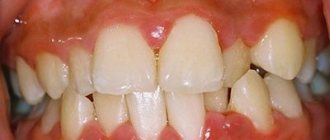
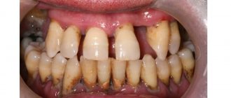
The shape of the dental arches, the shape and size of the teeth correspond to the physiological norm: the position of the teeth in the dentition is not changed, the lateral teeth are pale yellow, the front teeth are dark yellow, there are no changes in the thickness of the enamel, the shine of the enamel is preserved. Mobility of teeth I degree. On the oral surfaces of teeth 12, 11, 21, 22, 34, 33, 32, 31, 41, 42 and 43 there is supragingival and subgingival calculus.
To make an appointment with a doctor
- home
- Symptoms
- Tooth defects
- Pulpitis, causes, classification, clinic, prevention
Pulpitis, causes, classification, clinic, prevention
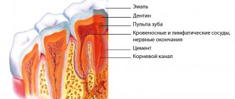
Pulpitis is considered to be the presence of an inflammatory process in the pulp in response to various irritants that come from the carious cavity. Microorganisms with their toxins often act as irritants.
The causes of pulpitis are recognized:
- traumatic factors, which include: exposure to toxins (in particular, phosphoric acid of inorganic cements and components of glass ionomer cements are recognized as strong toxic agents),
- fracture of a part of the tooth, accompanied by opening of the tooth cavity and exposure of the nerve, resulting in a violation of the trophism of the pulp;
- opening of the tooth cavity in case of unsuccessful preparation of a carious cavity, carried out at high speed without water cooling or with equipment that does not meet the standards (blunt bur);
- chemical exposure to medicinal substances, such as alcohol or ether, used in the treatment of carious cavities;
- dislocation or impaction of a tooth.
- poorly carried out treatment of a carious cavity in the stage of dental caries with partial removal of softened dentin, as well as a violation of the seal, which ensures the penetration of microorganisms under it,
- taking impressions of a ground tooth, which is why doctors insist on depulpitization when installing dentures.
The process of development of pulpitis goes through 3 stages successively: functional, morphological changes and the actual development of the inflammatory process, which can also be conditionally divided into stages of the disease, manifested in the transition of an acute focal process to a diffuse and chronic one. According to time intervals, the picture of the transition of the stages of pulpitis is as follows: after 3 days, focal pulpitis turns into diffuse, and after 12 weeks from its onset into the chronic stage. Chronicization of the process is characterized by deeper damage to the tooth and weaker clinical manifestations.
A complete picture of the scale of the pathological process and the need to immediately contact a dentist for qualified help is given by the classification of pulpitis:
- Acute forms of pulpitis: acute, focal, diffuse
- Chronic forms of pulpitis: chronic, fibrous, gangrenous, hypertrophic, exacerbation of chronic pulpitis.
Each form of pulpitis has its own clinic and, accordingly, a unique treatment. Clinical manifestations of the inflammatory process of the pulp are very diverse, which in turn is associated with local conditions in the oral cavity and the general condition of the body as a whole. Statistics show that 1/3 of pulp diseases occur at the acute stage, the remaining 2/3 are chronic pathological processes. The diagnosis of “pulpitis” is made on the basis of anamnesis, examination, probing, tapping the tooth, palpating the tooth, and the use of various tests. When interviewed, patients usually complain of pain symptoms arising from certain stimuli, often of a pulsating nature, accompanied by irradiation in the head area. In the chronic stage of the disease, the onset of pain is usually preceded by physical activity, exposure to an irritating agent (cold, temperature, food residues), nervous strain, overwork, and diseases of the body of a viral or bacterial nature.
Preventive measures for the development of pulpitis are periodic visits to the dentist, treatment of all carious teeth, exclusion of the tramming agent and good oral hygiene.
Clinical variants of pulpitis
Considering in general terms the clinical manifestations of the stages of development of pulpitis, a clear gradation of symptoms is noted.
Thus, acute forms of pulpitis are characterized by the presence of spontaneous pain without the influence of external stimuli. When exposed to the latter, pain intensifies and extends over time. In addition, night pain and paroxysmal periods of pain alternating with painless periods are characteristic.
Acute focal pulpitis
The clinic manifests itself in short-term localized, paroxysmal pain with long painless time intervals, prolonged pain when exposed to temperature stimuli. The intensity of pain occurs at night. Examination reveals a deep carious cavity and painful probing.
Acute diffuse pulpitis
The clinical picture is manifested by longer painful attacks, alternating with short pain-free intervals. The pain is more pronounced, radiating to the jaw, back of the head, temporal region, adjacent teeth, superciliary, zygomatic areas, depending on the location of the pathological process, and intensifies in a horizontal position. Probing a carious cavity is painful along the entire bottom, which cannot be said about percussion, which is often painless or slightly painful. Absolutely all types of irritants cause an attack of pain.
Chronic forms of pulpitis, compared with its acute manifestations, have less pronounced subjective symptoms. Common to chronic forms is a significant lengthening of the pain period, which does not disappear after the cessation of the stimulus. In addition to pain, there is discomfort in the oral cavity, which makes itself felt when eating, swallowing cold air, moving from a warm room to a cold one and vice versa, and bleeding of the gums of the affected side.
Chronic fibrous pulpitis
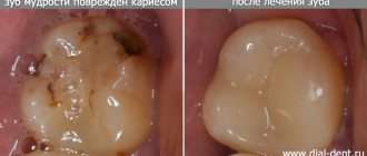
The clinic manifests itself in the appearance of paroxysmal pain from all kinds of irritants. The anamnesis indicates previously occurring pain with a gradual change in its nature from spontaneous to reflex. Examination by a dentist shows an open coronal cavity with a painful and bleeding pulp when probing.
Chronic gangrenous pulpitis
The clinic is manifested in the presence of a painful symptom of an acute paroxysmal nature with obligatory irradiation along the branches of the trigeminal nerve, often occurring when biting on a tooth, putrid odor from the mouth. An examination by a dentist shows communication between the carious cavity and the tooth cavity and pain on probing. The danger of this type of pulpitis lies in the formation of destruction of periapical tissues of varying degrees of severity.
Chronic hypertrophic pulpitis
The clinic may manifest itself in some subjective sensations (bleeding of the overgrown pulp, slight pain when eating) or the absence of them. Anamnesis indicates the presence of complaints characteristic of previous stages of the disease. Examination shows severe destruction of the crown and protrusion of hyperplastic pulp from the carious cavity.
Exacerbation of chronic pulpitis
The clinic is characterized by paroxysmal pain of a spontaneous nature, long duration, irradiation, which intensifies significantly when biting on a tooth. The anamnesis indicates the presence previously of one of the chronic forms of pulpitis. The examination reveals an open cavity in the tooth with painful probing.
Treatment of pulpitis
Treatment of pulpitis involves solving 4 problems: eliminating the source of inflammation, stimulating regeneration processes, preventing complications in the form of periodontal disease and restoring the shape and functionality of the diseased tooth.
Among the methods of treating pulpitis, two types should be distinguished: biological (conservative) and surgical. Treatment of pulpitis is best done under local anesthesia.
The biological method is used for reversible inflammatory processes of the pulp, when the viability of the pulp is preserved. The cause of such pulpitis can be injury or accidental opening of the tooth cavity. The method is most effective among young patients and is contraindicated in people over 40 years of age. The essence of the method is the opening, expansion and formation of a carious cavity as in caries, but the emphasis during treatment focuses on medicinal treatment of the cavity with non-irritating antiseptics, antibiotic drugs and proteolytic enzymes, followed by the application of medicinal paste to the bottom of the cavity in order to stimulate regenerative processes. Following the formation of a therapeutic pad, the cavity is closed with a temporary filling for up to 6 days. If there are no patient complaints, after the expiration of a week, the final filling of the tooth is performed.
A surgical method accompanied by the mandatory removal of the inflamed pulp and subsequent filling of the canal with filling material. The essence of the method is to cancel necrotic tissue, open the tooth cavity and remove the pulp. Pulp removal mainly occurs by applying arsenic paste for a day in single-rooted teeth and 2 days in multi-rooted teeth. This is followed by antiseptic treatment of the cavity and canals using cotton wool soaked in an antiseptic solution. Canal filling is carried out using a canal filler. The requirements for filling material boil down to its non-toxicity, radiopaqueness, slow hardening and resistance to oral fluid and saliva. Filling the canal is often accompanied by the use of metal and gutta-percha pins. Surgical treatment of pulpitis can occur in 1 or 2 visits, depending on the complexity of the pathological process and the advanced state of the disease. An earlier visit to a doctor for dental care will allow you to restore the functionality of the tooth with less loss, both anatomically and materially.
What leads to the occurrence of gangrenous pulpitis?
Most often, untreated purulent pulpitis in an advanced stage and the penetration of putrefactive microorganisms into the pulp can lead to this stage of the disease. The pulp can become infected both during the opening of the tooth during the treatment of caries, and as a result of various tooth injuries.
How quickly gangrenous pulpitis develops depends on the general condition of the human body, the condition of the periodontium, the duration of pulp irritation, the intensity of caries and other factors.
