Usually, the healing of the hole after tooth extraction occurs painlessly and does not cause any sensations in the patient. It would be good if everyone knew how this process works and how to speed it up.
Proper oral care after tooth extraction and following all the dentist’s recommendations will help speed up the healing of the hole and avoid complications.
Stages of socket healing by day
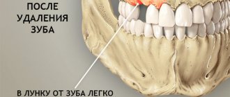
- The bleeding that occurs after tooth extraction stops after a few minutes , and a blood clot is formed that protects the wound from the penetration of microbes. Under the clot, damaged tissue is being restored, so it is so important to maintain its integrity until the hole heals completely.
- Day 1 – the processes of epithelization (healing) of the wound surface begin.
- Day 3 - signs of epithelization become noticeable, healing begins from the edges of the wound and moves towards the center.
- Day 4 - granulation tissue begins to form - a type of connective tissue that is formed in the body only during wound healing by secondary intention, when there is a strong defect in the skin.
- Day 7 - granulation tissue replaces part of the blood clot, it remains only in the center of the hole. Osteoid (bone) beams begin to appear, indicating bone formation. At the same time, epithelial tissue grows significantly from the edges of the gums.
- 14th -18th day - epithelization of the wound is completely completed, its surface is tightened and heals completely. By this time, the hole is completely filled with granulation tissue, and osteoid tissue is actively developing.
- After a month, a significant part of the hole is filled with new bone tissue. By the end of the second, sometimes third month, this process is completely completed.
- 4th month - bone beams in the newly formed osteoid tissue calcify, it matures: it becomes spongy and does not differ from the rest of the bone. At the same time, the edges of the socket dissolve, the alveolar edge in the area of the extracted tooth becomes thinner and lower than it was before surgery.
Long-term healing and tissue restoration
Before moving on to the question of how much the hole (more precisely, the tissues themselves) hurts after removal, let’s discuss how long it takes for it to heal. If the procedure is successful, bleeding stops after 20-30 minutes. This is followed by the stage of formation of a protective clot, after which all pronounced symptoms begin to rapidly decline. Thus, dental experts identify the following signs of normal healing:
- the appearance of a clot in the first couple of hours after surgery,
- tolerable pain in the area of removal,
- there may be a slight increase in temperature, swelling of the gums,
- discomfort when making swallowing movements,
- formation of a white film.
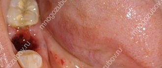
The photo shows the tooth socket immediately after removal.
These symptoms are considered normal, and their peak intensity occurs approximately 2 days after surgery. Already on the fourth day, all of the above manifestations should begin to subside. If the pain and swelling only intensify, and an unpleasant odor appears in your mouth, you should urgently consult a doctor.
Healing of the socket with gum inflammation
If tooth extraction was carried out against the background of inflammation, or the inflammatory process in the gums developed later, epithelization of the wound begins on the 10th -14th day, bone beams appear only by the 15th day. A significant part of the socket is filled with young osteoid tissue only by the end of the second month.
After a complex tooth extraction, when the gums rupture and the walls of the socket are traumatized, the edges of the gums cannot come together for a long time and the epithelization process slows down. Wound healing can only be completed after 1-1.5 months. In this case, the development of new bone tissue is delayed.
What happens to the gums during tooth extraction
To completely remove a tooth from its socket, surgeons have to traumatize the soft tissue of the gums. Before the procedure, the patient is given an anesthetic, since the extraction causes significant pain.
After the onset of anesthesia, the doctor bends the gum away from the tooth and wraps the neck of the element with special forceps. After this, the problematic unit is removed from the hole using rotational and rocking movements.
What happens to the gums after tooth extraction? The blood vessels that fed the pulp rupture, leading to excessive bleeding. Damaged nerve endings signal injury with severe sharp pains that appear immediately after the anesthetic wears off. The symptom worsens when talking and eating. During extraction, the ligaments that fix the root in the jaw alveolus are also damaged.
Surgical intervention is acutely perceived by the body, so the area of the hole begins to be intensively supplied with blood. An inflammatory process develops in the damaged area. This is necessary so that most of the leukocytes can penetrate through the blood vessels, thereby preventing infectious complications.
How to speed up the healing of a hole
To speed up the healing of the hole after tooth extraction, you need to follow these rules:
- Do not touch the blood clot on the socket with your tongue, much less with your hands or a toothpick, so as not to damage it. The presence of a clot is a guarantee of speedy healing of the wound.
- For three hours after visiting the dentist, you should not eat or rinse your mouth. Compliance with this point will also help maintain the integrity of the blood clot.
- · Drinks and food should not be too hot or cold for several days after removal. The food chosen is soft, without coarse inclusions, to prevent injury to the gums.
- You should not engage in heavy physical work for several days to avoid opening the wound and resuming bleeding.
- Since tobacco smoke and alcohol irritate the mucous membrane and inhibit the healing of the hole, you need to stop smoking and drinking alcohol for a while.
- To prevent infection from entering the wound and reduce inflammation after a traumatic tooth extraction, antiseptic baths are used. For them, solutions of chlorhexidine or furatsilin, infusions of sage, chamomile, and eucalyptus are used. The solutions are taken into the mouth, held for several minutes and carefully spat out.
- On the third day after removal, you can carry out antiseptic rinses with the same agents.
- Taking anti-inflammatory drugs (nimesulide, ibuprofen) will help not only eliminate pain, but also relieve the inflammatory process, which also helps accelerate socket regeneration.
- If the doctor has prescribed antibiotics, you should not refuse them, since eliminating the microbial infection shortens the healing time.
- When brushing your teeth, be careful not to touch the wound with the brush.
- To speed up healing, you can use Solcoseryl dental paste. It enhances intracellular energy exchange, due to which cell regeneration and restoration of damaged tissues are accelerated. Solcoseryl also creates conditions for the growth of granulation tissue. The paste is applied to a previously dried surface, then moistened with water. Before using the medicine, it would be more correct to ask your dentist about when to start using Solcoseryl after tooth extraction and how many times a day to apply it to the gums.
How is the removal procedure carried out?
The procedure is always performed under anesthesia, most often local anesthesia. However, if a complex extraction is ahead or the patient has an overwhelming fear of dental treatment, the operation can be performed under general anesthesia or sedation. After the anesthetic begins to take effect, the specialist moves the gum tissue slightly away from the walls of the tooth, and then grabs it using special forceps. If we are talking about the upper jaw, then the doctor presses on the instrument with his entire right hand. When it comes to dealing with the lower tooth, the specialist also works with his right hand, but mainly with his thumb.
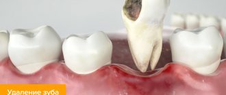
This is how a tooth is removed
Read more about how the procedure for removing upper teeth differs from the technique of extracting teeth from the lower jaw in our special article>>>
To extract a tooth from the gum and bone tissue, it is necessary to dislocate it. To do this, the doctor makes rotational and oscillatory movements. Regarding what is placed and what is used to fill the cavity after the hole is emptied, no specific manipulations are required here. The doctor only applies a sterile cotton swab to the wound, which must be held in the mouth until the bleeding stops completely. If a complex removal, for example, of a wisdom tooth, has been carried out, it will take more time for the tissue to heal, because the gum is first cut and then sutured, and this involves even greater trauma to the mucous membrane.
Next, the specialist gives important instructions on how to care for the hole and how to rinse it, tells how long it takes to heal and what to do if the wound does not heal for a long time. You need to listen to the dentist very carefully and then strictly follow his recommendations - this is the only way to protect yourself from the development of complications.
What to do when the hole has healed?
After the hole has healed and filled with full bone tissue, it is necessary to take care of restoring the dentition. The absence of even one tooth can lead to displacement of the remaining teeth, changes in bite, diction, aesthetic problems, and resorption (atrophy) of bone tissue at the site of the extracted tooth. A tooth can be restored using implantation, when an artificial root is implanted into the bone and a crown is installed on it. This method of prosthetics is by far the most comfortable and effective for the patient.
If bone tissue atrophy has already occurred, bone tissue augmentation is performed before installing the implant. The procedure allows you to perform high-quality prosthetics and restore the functionality and aesthetics of your teeth.
What determines the speed of regeneration?
All people want their mouth wound to heal as quickly as possible. But sometimes recovery takes longer. The speed of recovery processes is reduced:
- poor oral hygiene;
- associated jaw injuries;
- poor-quality tooth extraction (if there are root particles left in the hole);
- development and progression of gingivitis, periodontitis;
- decreased immunity;
- avitaminosis;
- diabetes;
- cavity infection;
- decreased blood clotting;
- hormonal disorders.
In order not to suffer from a slowly healing wound, prepare for the operation competently and follow the doctor’s instructions after it. Then very soon your gum will look like the healthy tissue surrounding it.
How to remove a tooth
There are two extraction methods used in dentistry: simple and complex. Their choice depends on which teeth are being removed - premolars and molars with tangled branched roots are removed using a complex method. It is very difficult to pull out such elements entirely due to the fact that the tooth socket is penetrated by retaining ligaments and alveolar processes. Errors during the procedure or insufficient experience of the specialist lead to serious complications. Therefore, even despite the acute condition, always find out in advance where you can have a tooth removed from a good doctor with positive recommendations.
Factors complicating the operation:
- complete destruction of the coronal part;
- high fragility;
- acute inflammatory diseases;
- Unerupted or misaligned wisdom teeth.
The technology of the procedure depends on which teeth are removed. In some cases, tissue incision and suturing are performed.
Root removal
How is the root of a tooth removed if the tooth is completely destroyed? Despite this situation, extraction is not always recommended. If, after diagnosis, the doctor determines that the root can be used for an inlay, then the tooth is restored with a crown. But if there is pain, an unpleasant smell and taste, or swelling has developed, then dental care is required urgently. In this case, the question of whether to remove the roots of the teeth is decided in favor of the operation, since the neglected condition can also lead to the loss of neighboring elements.
Regardless of the chosen technique, pain relief and X-ray control are required after removal. This allows you to make sure that there are no root fragments in the cavity, since it is often loose and can crumble under slight pressure from the forceps.
Possible complications
Signs of alarm should be considered an enlargement of the cheek, further spread of swelling, a persistent increase in temperature, increased pain, nausea, and weakness. If the healing process is disrupted, the following complications may occur:
- Cyst formation. It is a fibrous neoplasm filled with fluid.
- Flux. Formed after infection penetrates into the socket and then into the periosteum. The resulting inflammation is characterized by severe swelling of the cheek on the side of the diseased gum. There is severe pain and redness of the gums. The formation of flux requires immediate medical attention. Therefore, it is necessary to carefully protect the site of the extracted tooth from possible infection.
- Alveolitis. This is a complication that occurs during the inflammatory process of the hole in the jaw bone. The infection occurs due to a violation of the integrity of the protective blood clot. The onset of the disease is characterized by inflammation of the outer layers of the socket, spreading into the deep layers of the bone. Alveolitis is accompanied by aching pain during eating, swelling and redness of the gums. There is a putrid odor from the mouth. The patient feels chills, headache, and fever. The occurrence of the disease most often occurs during the extraction of molars located on the lower jaw. It is necessary to obtain medical attention in a timely manner to avoid the spread of infection to other organs. One of the dangerous complications of the disease is osteomyelitis.
Removing wisdom teeth is a more complex procedure, so gum inflammation often occurs after surgery. At the same time, discoloration or swelling of the gums should not cause concern to the patient. Often after surgery there are difficulties opening and closing the mouth. This is a consequence of surgery. To get to a hard-to-reach place, the doctor asks the patient to open his mouth as wide as possible. The pressure exerted on the tissues leads to their swelling. On the 3rd day, the discomfort usually goes away completely. The appearance of purulent contents in the hole, increased temperature, acute pain, heavy bleeding - all these signs require immediate contact with the dentist.
Treatment of dental granuloma with antibiotics and physiotherapy
Dentists often suggest treating dental granuloma with antibiotics. As a rule, this is only a temporary solution to the problem, since it does not eliminate the source of inflammation, and after some time the disease will return. Dentists successfully use physiotherapeutic treatment methods. For example, depophoresis works great for curved or complex tooth canals. Its action consists in the effect of a weak electric current on a suspension with copper hydroxide, which relieves inflammation, penetrating into all corners of the tooth.
Molar tooth extraction in children
Unfortunately, young children often develop diseases in both their primary and permanent molars. Worse, it is not always possible to heal them. Dentists remove permanent teeth for children only when there is no way to save them.
Primary molars are both treated and removed. If a permanent tooth begins to grow under a baby molar, then the baby tooth is always removed, since it interferes with the normal formation of the permanent tooth.
At the same time, dentists never agree to premature removal of baby molars. Such teeth are sure to heal. If you remove a baby tooth ahead of schedule, then the baby will develop a crooked bite.
There are situations when a child’s molar tooth must be removed. Such situations include:
- molar root cyst;
- presence of granuloma;
- inflammation of the tooth root;
- inflammation of the nerve of the lower jaw;
- severe destruction of the integrity of the tooth.
If there are root fragments left in the hole
If the wound edges were tightened with catgut, the patient’s treatment can be considered complete. If a non-absorbable material was used, the patient will have to return to the dentist to have the sutures removed after a week. During this time, it is necessary to monitor the condition of the gums, your own sensations and notify the doctor if:
- the operated areas constantly hurt;
- throbbing pain is felt.
This means that small fragments of tooth tissue, which may have gone unnoticed during extraction, rot in the hole. The doctor must prescribe a repeat X-ray for the patient, check the quality of the operation and find out the cause of the ailment. Inaction is fraught with the development of alveolitis (inflammation of the socket), osteomyelitis, phlegmon and other serious diseases.
Associated symptoms
Dry socket may be accompanied by symptoms such as:
- High temperature accompanies acute and purulent inflammation, a dangerous symptom that requires immediate attention from a dentist.
- Swelling of the cheek indicates the accumulation of pus, the development of a dangerous purulent form of the disease.
- Severe pain is an obligatory companion of alveolitis and increases with exposure of the bone and purulent inflammation.
Trigeminal neuritis
Today, traumatic neuritis of the trigeminal nerve is considered one of the most common causes of pain syndromes in the maxillofacial area.
As a complication, it occurs during the removal of permanent molars of the lower jaw, due to damage to the lower alveolar nerve in the mandibular canal. The apices of the roots of the lower molars are in close proximity to the mandibular canal and in some cases may be located in the canal itself. Sometimes, due to chronic apical periodontitis, the bone between the root apex and the wall of the mandibular canal is resorbed. When a tooth is loosened by an elevator, the lower alveolar nerve can be injured, which will lead to partial or complete disruption of the functions of the third branch of the trigeminal nerve. The result is pain in the jaw, numbness of the lower lip and chin, decreased or absent sensitivity of the gums, decreased electrical excitability of the dental pulp on the affected side. Usually all these phenomena gradually disappear after a few weeks.
Electroodontometry
Electroodontometry is the most effective method for assessing the functional state of the trigeminal nerve when it is damaged. EDI is based on a study of the reaction of the teeth of the lower jaw to electrical stimulation. The method is performed on all teeth of the lower jaw, with preserved pulp both in the affected area and on the opposite healthy side.
S.N. scale Fedotova (1997) to assess the severity of damage to the inferior alveolar nerve based on electrical odontometry data:
- mild degree - reaction of teeth with preserved pulp on the side of nerve damage within 20-40 μA;
- moderate severity – reaction of teeth to currents from 40 to 100 µA;
- severe degree - complete loss of pain sensitivity, reaction of teeth to currents above 100 μA
The use of EDI to diagnose traumatic injuries of the inferior alveolar nerve is impossible if:
- lower jaw teeth endodontically treated
- lower jaw teeth are covered with orthopedic structures
- metal elements of splinting structures are fixed on the teeth
- missing teeth
Locations for measuring electrical excitability of facial skin
Assessment of the area of paresthesia in traumatic neuritis of the inferior alveolar nerve
The zone of paresthesia is identified - impaired sensitivity of the skin based on a tactile test, photographed, followed by an assessment of the area of the paresthesia zone: points are drawn on the border of areas of normal sensitivity of the skin, the red border of the lips and the paresthesia zone, which are then connected by a continuous line. Zones of hyper-, hypo- and anesthesia were marked with different colors.
I - vertical lines:
- midline,
- a line passing through the outer edge of the philtrum,
- a line passing through the outer edge of the wing of the nose,
- pupil line;
II - horizontal lines:
- lip line,
- a line running along the lower edge of the red border of the lips,
- the border line between the chin and lower lip,
- a line drawn along the most protruding part of the chin,
- line of the border of the chin and submental areas.
Schematic representation of the areas for measuring facial skin paresthesia.
Each of the 12 resulting squares was assigned a score depending on the nature of the sensitivity disorder:
0-sensitivity is not impaired;
1-skin hyperesthesia
2-hypoesthesia of the skin;
3-anesthesia of the skin.
Next, the sum of points was calculated and divided by 12 (quadrants).
With the results:
3.0-2.1 – severe sensitivity impairment was diagnosed;
2.0–1.1 – moderate severity;
less than 1.0 – mild severity of the pathology being studied
Reasons for the development of pathology
There are two reasons for the development of granulomas on the root of a tooth.
1. Untreated pulpitis. The development of caries leads to the appearance of a deep cavity in the tooth. Pathogenic microorganisms enter the pulp, it becomes inflamed, and severe pain appears. Lack of medical care leads to the gradual death of the pulp. Bacteria penetrate beyond the tooth through root canals. A focus of inflammation appears at the apex of the root. We are talking about periodontitis.
A deep carious cavity in this case is not always observed. Inflammation can develop internally when secondary caries appears under the filling.
2. Poor quality endodontic treatment. Granuloma can develop at the root of a tooth in which root canal filling was previously performed. Usually there is underfilling: the doctor has not completely filled the canals with material. In the remaining voids, pathogenic bacteria develop, and the tissues surrounding the root react with inflammation.
These causes cause most cases of granuloma formation. But there are others, less common:
- poor quality orthodontic treatment;
- previous dental trauma;
- other inflammatory diseases - tonsillitis, abscess, etc.
In the latter case, the infection enters the tissues through the blood or lymph flow.
Ask a Question