Clinical picture of osteomyelitis of the tooth socket
The clinical picture of osteomyelitis of the tooth socket is characterized by complaints of acute throbbing pain both in the area of the socket and in the area of neighboring teeth. General symptoms such as weakness, hyperthermia, headache, chills, impaired performance and sleep are also observed. Swelling of the perimandibular soft tissues develops, the submandibular lymph nodes are enlarged, dense, painful on palpation.
With osteomyelitis of the socket of only one molar of the lower jaw, the inflammatory process can spread to the area of the masseter or medial pterygoid muscle, which, in turn, can cause difficulty opening the mouth. When examining the oral cavity, the doctor may detect a dirty gray coating on the bottom and walls of the socket. You can also smell a specific smell. Percussion of adjacent teeth is painful.
The mucous membrane in the area of the transitional fold is hyperemic and swollen. Palpation of the alveolar process from the buccal and oral sides is sharply painful both in the area of the socket and adjacent teeth.
The acute phase of inflammation lasts about 6–8 days, sometimes 10 days. Then the inflammatory phenomena decrease, the process becomes chronic. The general condition improves, body temperature decreases. Swelling and hyperemia of the mucous membranes also decrease, and then pain on palpation of the alveolar process, swelling of facial tissues and submandibular lymphadenitis disappear. After 12–15 days, the tooth socket is filled with loose, pathological granulation tissue, sometimes bulges out of the socket, and pus can be released when pressure is applied.
On the x-ray we see fuzzy blurred contours of the compact lamina of the alveoli; osteoporosis and bone destruction in the alveolar region are pronounced. In rare cases, after 20–25 days from the beginning of the acute period, it is possible to identify small sequesters.
Causes of limited osteomyelitis of the socket
1. as a complication of alveolitis. 2. as a result of decreased immunity
What to look for
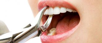
The most serious consequences that can occur after wisdom tooth removal:
- Alveolitis. This is an acute inflammatory process. It begins in the tooth socket and gradually spreads to other gum tissues. The most common reason for the development of alveolitis is improper care after removal.
- Paresthesia. Develops when the root of the jaw nerve is damaged or irritated. Manifested by a feeling of tingling, burning, numbness.
To avoid developing serious problems, monitor your oral health. The first symptoms are not difficult to replace:
- Bleeding from the socket. This is not always a sign of complications. Immediately after removal this is a normal reaction of the body. In the first minutes after tooth extraction, bleeding will be slightly heavier. The dentist treats the wound with antiseptics and packs it. After some time, the bleeding stops. Slight bleeding persists for 2-3 hours. This is also normal. You should pay attention to bleeding if it is profuse and begins several hours after removal. In this case, contact a specialist immediately.
- Swelling and pain. This is also a natural reaction. Pain occurs as the anesthetic wears off. Normally it lasts 2-3 days. At the same time, the gums swell slightly. You should sound the alarm if the swelling transfers to the cheek. In complex cases, the tumor can be very large. Severe swelling is one of the symptoms of alveolitis.
- Fever. On the first day after removal, the temperature rises to 37 degrees. Such an increase is within normal limits, especially if the operation was complicated. When the temperature lasts more than a day or rises strongly, this is a symptom of inflammation. To prevent the inflammatory process from spreading to neighboring tissues, visit a dental clinic.
- Suppuration. The inflammatory process, accompanied by suppuration, has pronounced symptoms. Suppuration is indicated by a putrid odor from the mouth and an unpleasant taste. When examining the tooth socket, you may notice gray plaque or white purulent contents. In this case, you need to go to the dentist immediately.
- Headache. Immediately after wisdom tooth removal, headache is normal. It is caused by stress and anesthetic drugs. If the headache is severe and is accompanied by fever, swollen lymph nodes, and weakness, we recommend visiting a doctor.
Dentistry for those who love to smile
+7
Make an appointment
Treatment of socket osteomyelitis
In the acute stage of the disease, therapy begins with revision of the hole. Under conduction or infiltration anesthesia, the tooth socket is cleaned of remnants of a blood clot, food and pathological tissue. Then the well is washed from a syringe with a weak antiseptic solution or a biologically active drug. The following drugs are used: staphylococcal and streptococcal bacteriophage, proteolytic enzymes, lysozyme. After this, a bandage with Alvogyl is applied to the wound.
To reduce inflammation and pain, dissection of the infiltrated area of the mucous membrane and periosteum is performed. An incision is made along the transitional fold 1.5–2 cm long, as well as an incision on the inside of the alveolar process. Drug therapy is prescribed: antibiotics, sulfa and antihistamines, analgesics, physical therapy (UHF, ultrasound, helium-neon laser). To increase specific immunological reactivity, phagocytosis stimulants are prescribed: pentoxyl, methyluracil, milife, lemongrass.
After relief of acute inflammation, treatment with multivitamins and stimulants of nonspecific resistance of the body is continued (methyluracil 0.5 g or pentoxyl 0.2 g 3-4 times a day, sodium nucleinate 0.2 g 3 times a day, milife 0.2 G). Concomitant ultrasound or laser therapy of the inflammation focus is necessary. Approximately 20 days from the onset of the acute inflammatory process, if the wound has not healed and sequesters are found on the radiograph, pathological granulations and small sequesters are removed with a surgical spoon, the walls and bottom of the hole are scraped out. The wound is washed with an antiseptic solution, dried and loosely packed with gauze soaked in iodoform liquid.
Dressings (treating the hole with an antiseptic solution and changing the iodoform gauze in it) are done every 2-3 days until young granulation tissue forms on the walls and bottom of the hole.
Prevention of socket osteomyelitis is the same as for alveolitis.
- Before the intervention, professional oral hygiene is necessary
- the doctor must inspect the socket and perform hemostasis by compression
- if more than two teeth are removed, apply sutures
- After tooth extraction, it is necessary to give clear recommendations to the patient
If, nevertheless, the appearance of alveolitis could not be avoided, and the patient complained of pain, it is necessary to stop the process as quickly as possible. Therefore, the patient must be informed about possible complications and motivated not to delay visiting the dentist if he experiences pain.
"Dental" sinusitis
Surely everyone has heard that you need to visit the dentist at least twice a year, otherwise fleeting preventive meetings with the dentist can turn into long and painful therapeutic appointments for both parties.
Moreover, often an otolaryngologist also joins this idyll. “What does he have to do with it?” - you ask. The answer is simple: an ENT doctor treats odontogenic or, more simply put, “dental” sinusitis, which quite often occurs as a complication of dental diseases.
How are bad teeth and sinusitis related?
And the connection here is purely anatomical. Sinusitis itself is an inflammation of the mucous membrane of the air sinus, which is located in the thickness of the upper jaw. The bottom or lower wall of this sinus is a layer of bone that separates the sinus from the roots of the upper teeth (molars and premolars, sometimes canines).
Trigeminal neuritis
Today, traumatic neuritis of the trigeminal nerve is considered one of the most common causes of pain syndromes in the maxillofacial area.
As a complication, it occurs during the removal of permanent molars of the lower jaw, due to damage to the lower alveolar nerve in the mandibular canal. The apices of the roots of the lower molars are in close proximity to the mandibular canal and in some cases may be located in the canal itself. Sometimes, due to chronic apical periodontitis, the bone between the root apex and the wall of the mandibular canal is resorbed. When a tooth is loosened by an elevator, the lower alveolar nerve can be injured, which will lead to partial or complete disruption of the functions of the third branch of the trigeminal nerve. The result is pain in the jaw, numbness of the lower lip and chin, decreased or absent sensitivity of the gums, decreased electrical excitability of the dental pulp on the affected side. Usually all these phenomena gradually disappear after a few weeks.
Electroodontometry
Electroodontometry is the most effective method for assessing the functional state of the trigeminal nerve when it is damaged. EDI is based on a study of the reaction of the teeth of the lower jaw to electrical stimulation. The method is performed on all teeth of the lower jaw, with preserved pulp both in the affected area and on the opposite healthy side.
S.N. scale Fedotova (1997) to assess the severity of damage to the inferior alveolar nerve based on electrical odontometry data:
- mild degree - reaction of teeth with preserved pulp on the side of nerve damage within 20-40 μA;
- moderate severity – reaction of teeth to currents from 40 to 100 µA;
- severe degree - complete loss of pain sensitivity, reaction of teeth to currents above 100 μA
The use of EDI to diagnose traumatic injuries of the inferior alveolar nerve is impossible if:
- lower jaw teeth endodontically treated
- lower jaw teeth are covered with orthopedic structures
- metal elements of splinting structures are fixed on the teeth
- missing teeth
Locations for measuring electrical excitability of facial skin
Assessment of the area of paresthesia in traumatic neuritis of the inferior alveolar nerve
The zone of paresthesia is identified - impaired sensitivity of the skin based on a tactile test, photographed, followed by an assessment of the area of the paresthesia zone: points are drawn on the border of areas of normal sensitivity of the skin, the red border of the lips and the paresthesia zone, which are then connected by a continuous line. Zones of hyper-, hypo- and anesthesia were marked with different colors.
I - vertical lines:
- midline,
- a line passing through the outer edge of the philtrum,
- a line passing through the outer edge of the wing of the nose,
- pupil line;
II - horizontal lines:
- lip line,
- a line running along the lower edge of the red border of the lips,
- the border line between the chin and lower lip,
- a line drawn along the most protruding part of the chin,
- line of the border of the chin and submental areas.
Schematic representation of the areas for measuring facial skin paresthesia.
Each of the 12 resulting squares was assigned a score depending on the nature of the sensitivity disorder:
0-sensitivity is not impaired;
1-skin hyperesthesia
2-hypoesthesia of the skin;
3-anesthesia of the skin.
Next, the sum of points was calculated and divided by 12 (quadrants).
With the results:
3.0-2.1 – severe sensitivity impairment was diagnosed;
2.0–1.1 – moderate severity;
less than 1.0 – mild severity of the pathology being studied
Treatment of neuritis
Treatment of neuritis in traumatic injuries must be timely and wait-and-see tactics are unacceptable. For mild damage to the inferior alveolar nerve, decongestant therapy (prednisolone, veroshpirone) is sufficient. In case of moderate severity of damage, drugs that improve the conductivity of the nerve trunk (neuromedin) are added to decongestant therapy. In case of severe damage to the nerve trunk, in the absence of positive dynamics in the restoration of sensitivity within 4 months, the patient should be referred for a consultation with a neurosurgeon in order to resolve the issue of the possibility of restoring the anatomical integrity of the nerve trunk.
Various reflexotherapy methods are widely used in the complex treatment of diseases of the peripheral nervous system. In the complex treatment of diseases of the peripheral nervous system, reflexology methods such as electroacupuncture and transcutaneous electrical neurostimulation are widely used.
Prevention of neuritis
Prevention of neuritis of the inferior alveolar nerve is the correct technique for removing teeth, correct diagnosis and correct reading of the radiograph, and a gentle technique for dislocating the roots of teeth in the lower jaw with an elevator.
Abscess after tooth extraction
An abscess after tooth extraction is a long-term complication of a tooth extraction operation that occurs as a result of contamination of the wound surface with microorganisms. When teeth are removed, not only the tissues surrounding the tooth can be injured, but also the mucous membranes of the mouth and cheeks. A fresh wound is a favorable environment and entry point for microorganisms. Therefore, under appropriate circumstances, an abscess may form in the soft tissues or in the hole, which over time and without treatment can “spill” with the formation of phlegmon in several areas.
Causes of abscess
The cause of an abscess may be failure to comply with the rules of asepsis and antisepsis directly during the tooth extraction operation. As a result of incorrect actions by the doctor, infection occurs in the tooth socket and subsequent suppuration. Also, infection of the socket and soft tissues can occur due to the fault of the patient himself if he does not follow all the dentist’s recommendations. The patient must strictly follow the prescribed procedures, otherwise repeated surgery cannot be avoided.
Prevention and treatment of tooth abscess
Prevention of the appearance of an abscess after tooth extraction is basic regular oral hygiene and following all the recommendations of the dentist. Also, preventive measures include visiting the dentist if you experience pain within a few days after tooth extraction. Treatment of an abscess consists of opening and cleaning the abscess cavity, removing purulent formations and prescribing anti-inflammatory therapy. Sometimes the abscess opens on its own, and the pain disappears. But this does not mean that the process has been stopped, so further treatment by a dentist is necessary. Otherwise, the infection may spread to contact areas and intensify the process.
Why can a lymph node become inflamed after tooth extraction and what to do about it?
An enlarged lymph node signals the occurrence of an inflammatory process in the body. Typically, during inflammation, the lymph nodes increase in size and some pain appears. Swelling of the lymph nodes of the neck, head and lower jaw is associated with the development of inflammation in the area of the extracted tooth. In this article you will learn how to avoid complications and how to help the body cope with infections faster.
Lymph nodes and where they are located
Let's start with the lymphatic system. It is represented by a collection of vessels of various diameters that permeate our body. The lymphatic system is closely related to the circulatory system (this plays an important role in the development of complications). Lymph, the fluid that flows through the lymphatic vessels, transports tissue fluid and protein molecules. There are large numbers of lymphocytes and granulocytes - cells that are the first to fight pathogens. They are produced in the lymph nodes; when an inflammatory process occurs in the body, the lymph nodes work more intensely, increasing in size.
Photo 1 shows inflammation of the lymph node
But how does this relate to tooth extraction?
Most of the lymph nodes are concentrated in the head and neck area. Surprisingly, it is true: inflammation in the lymphatic system in 60% of cases occurs due to dental pathologies. The process of tooth extraction is a very traumatic event. In addition, a large number of opportunistic microorganisms live in the oral cavity.
Attention: Opportunistic pathogens are those that are normally present on the surface and inside the human body, but do not cause harm to it. However, when conditions change, they can become pathogenic and lead to the development of diseases.
So, bacteria entering the wound formed after tooth extraction can cause an infectious process. The nearby lymph nodes of the jaw, neck and head will react most quickly to it, actively producing cells of the immune system. Therefore, the patient can feel enlarged lymph nodes, which in some cases can be painful.
Why does the problem occur after tooth extraction?
After tooth extraction, in 99% of cases the lymph node swells. Dentists identify two main reasons:
1. This is a defensive reaction to interference, and it is the norm. As a rule, after tooth extraction, the lymph nodes in the neck or under the jaw become enlarged.
Attention: When wisdom teeth, or numbers of eights, are removed, the lymph nodes in the neck become enlarged. The procedure is quite traumatic and affects a large number of tissues, so in 90% of cases the lymph node becomes enlarged. It may also be accompanied by fever, soreness, inability to open the mouth wide, and general malaise.
- Tooth extraction was carried out against the background of an existing purulent-inflammatory process. In some cases, tooth extraction is a necessary step in solving advanced dental inflammatory diseases of the oral cavity, which include:
- fistula;
- periodontitis;
- flux;
- granuloma;
- cyst.
- In this case, the inflammatory process can result in a number of complications, so it is recommended to fight the infection with the help of antimicrobial drugs, rinsing with furatsilin or chlorhexidine, potassium permanganate or salt solution, as well as applying lotions.
Attention: Prevention is easier than cure!
Reasons for the development of lymphadenitis.
So, we have already found out that when teeth are removed, especially wisdom teeth, the lymph nodes become inflamed. This process is called lymphadenitis. The development of lymphadenitis in the head and neck area is promoted by:
- tonsillitis (inflammation of the tonsils);
- laryngitis (inflammation of the larynx);
- stomatitis (damage to the oral mucosa);
- gingivitis (inflammation of the gums).
What to do if the lymph node under the jaw is inflamed?
When the lymph node under the jaw becomes inflamed, the following symptoms occur:
- a significant increase in the size of the inflamed lymph node;
- when pressed, a dense movable tubercle is felt;
- severe pain in the gums and inflamed lymph nodes, which can spread to the head and neck;
- limited jaw movements;
- difficulty chewing and swallowing;
- general weakness, malaise;
- sleep disturbance;
- chills, increased body temperature up to 40 ◦C.
In such cases, the doctor identifies the degree of inflammation and prescribes appropriate treatment.
The dentist identifies the source of pathology by palpation (feeling with fingertips) of an enlarged lymph node, which is easily detectable. In some cases, pressure even causes pain. It is interesting that the skin first acquires a reddish tint due to increased blood flow in the area of inflammation, and then changes to bluish.
The body’s immune capabilities play a major role in activating the lymph nodes under the chin and in the lower jaw area. Lymphatic tissue, as the main part of the immune system, protects the body from pathogenic environmental factors. However, there are some diseases in which the immune system itself suffers. They are called autoimmune.
Such diseases include, for example, systemic lupus erythematosus, rheumatoid arthritis and serum sickness. With these pathologies, the protective function of the body is sharply reduced and, for example, if a child suffers from this serious illness, the lymph nodes under the lower jaw can increase even when baby teeth erupt.
What complications may arise?
Complications of an inflamed lymph node can be:
- angina;
In the photo on the left - healthy tonsils, in the photo on the right - inflammation of the tonsils
- laryngitis or pharyngitis;
The photo shows dental flux
- laryngitis or pharyngitis;
In the photo on the left - healthy tonsils, in the photo on the right - inflammation of the tonsils
The photo shows laryngitis
- otitis.
Otitis. The photo shows an inflammatory process in the ear
They occur when pathogenic microflora spreads to other nearby areas. There is no way around this without a course of antibiotic therapy. In order not to struggle with complications later, it is necessary to diagnose and properly treat inflammation in a timely manner.
Complications of extraction
A separate group of complications are those that arise during tooth extraction. There may be, for example,
- alveolitis;
- flux;
- abscess;
- phlegmon;
- osteomyelitis.
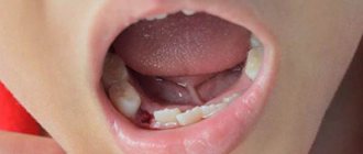
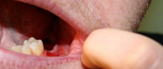
The most insidious of them is considered to be alveolitis, or inflammation of the socket of an extracted tooth. It develops due to a dentist’s mistake or when the patient’s postoperative regimen is violated. The cause of the development of alveolitis is the destruction of a blood clot that should form at the site of the extracted tooth. Without it, the hole becomes as if unprotected and pathogenic microorganisms enter it. This is how alveolitis develops.
The photo shows alveolitis (inflammation of the socket of an extracted tooth)
In addition, the tooth socket may begin to fester. Severe pain, swelling, uncharacteristic discharge and bad breath occur. It is necessary to treat the inflammation, since with its further progression, nearby tissues suffer: gums, periosteum, mucous membrane and others.
To treat alveolitis, you must immediately seek medical help. The dentist will clean the hole, removing all foreign elements, and treat it and other foci of infection that may also arise in the oral cavity with an antiseptic solution.
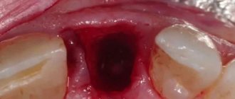
Dentists also consider dry socket to be a complication of extraction. As mentioned above, a blood clot must form, which will promote tissue restoration and wound healing. If the hole remains dry, then the risk of infection in the wound increases, the pain intensifies and an unpleasant taste appears.
With wisdom teeth, things are even more complicated - due to their distant location, the patient cannot always see that the blood clot has disappeared. Therefore, he seeks help only when pain develops. In the case of a dry socket, the doctor places a cotton swab on the wound, soaking it with antiseptics and an antibiotic solution. That is, a cotton swab literally replaces a blood clot. Thanks to this, the hole is protected and its regeneration is accelerated. The cotton swab must be changed every day until the hole is completely healed.
Treatment of lymphadenitis
Diagnostic measures aimed at eliminating the problem include:
- dental treatment
- drug therapy
- special regime.
1. Dental treatment involves getting rid of the causes that cause the lymph nodes to enlarge. The doctor may suggest:
- professional teeth cleaning;
- treatment or filling of root canals;
- sanitation of abscesses;
- cleaning the hole;
- resection of damaged parts of teeth;
- replacement of fillings and crowns.
Attention: After appropriate treatment, the lymph nodes do not return to normal immediately. Their increased size can last up to 2 weeks. If enlarged lymph nodes cause pain and discomfort to the patient, he may additionally be prescribed physical therapy, as well as choose medications.
2. Drug therapy consists of the dentist prescribing medications when inflammation is detected. Typically it is:
- antibiotics to eliminate pathogenic bacteria;
- immunostimulants and multivitamin preparations (to speed up the body’s recovery);
- home treatments in the form of lotions and rinses.
In case of fever, antipyretic drugs are prescribed, and in case of severe pain, which often accompanies dental surgery, analgesics are prescribed. Thus, etiotropic (antimicrobial drugs) and symptomatic (to relieve symptoms) therapy is carried out.
3. To speed up the relief of inflammation, the patient should follow a special regimen. These include:
- reduction of physical activity;
- in case of severe lymphadenitis, even bed rest is recommended; the need to drink up to 3 liters of warm liquid (herbal teas, fruit drinks and plain water help well - they help get rid of intoxication caused by inflammation as quickly as possible);
- rinsing (you can rinse your mouth with a decoction of chamomile or calendula);
- salt lotions (a concentrated salt solution is prepared, a large piece of cotton wool or a cotton pad is soaked in it, and it is placed behind the cheek where the dental procedure was performed. The hypertonic solution will help the cotton wool absorb metabolic products during inflammation).
Patients will have to make extensive adjustments to their teeth brushing. If there is inflammation in the oral cavity and, as a result, in nearby lymph nodes, it will not be enough to use only a brush and paste. It is recommended to use an irrigator and dental floss, and after all these procedures, use a mouth rinse.
Caution: Buy a paste that contains antiseptics such as chlorhexidine, chlorine, silver and zinc. It will provide you with additional sanitation of the operated area and will not allow inflammation to develop.
The basic principles that ensure effective treatment of inflamed lymph nodes are:
- good timely diagnosis;
- bed rest;
- the use of alternative medicine methods in the form of lotions and rinses;
- antibiotic therapy;
- adequate sanitation of the oral cavity.
Attention: Treatment of inflamed lymph nodes should not be delayed, as pathogenic organisms can enter the circulatory system, which will cause sepsis.
What medications are prescribed?
Often, antibiotic therapy for enlarged lymph nodes and inflammation includes penicillin antibiotics, for example, Amoxiclav, Ampicillin or Amoxicillin. In case of allergy to antibiotics of the penicillin group, antibiotics of other groups may be prescribed. The most effective of them are macrolides and fluoroquinolones.
Attention: The course of antibiotic therapy should not exceed 5, and in some cases – 7 days. Carefully read the instructions prescribed for this antibiotic and follow them! To avoid developing resistance, follow your doctor's recommendations.