From this article you will learn:
- why does the socket hurt after tooth extraction,
- what is alveolitis: photos and videos,
- How is alveolitis treated?
The article was written by a dental surgeon with more than 19 years of experience.
Alveolitis is a classic complication that occurs after tooth extraction and consists of the development of inflammation of the socket of the extracted tooth. Often alveolitis is also called “dry socket” (this is due to the fact that the alveolar bone in the depths of the socket is exposed due to the loss of a blood clot).
On average, alveolitis develops after tooth extraction in 3-5% of cases, but this applies to teeth of any location with the exception of wisdom teeth. When the latter are removed, alveolitis occurs in 25-30% of cases, which is associated with the greater complexity and traumatic nature of the removal process.
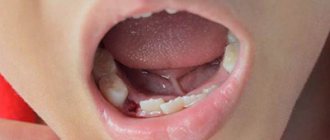
Dry socket after tooth extraction: photo
You can see what normal healing of a socket should look like (at different times from the moment of extraction) in the photo in the article: → “What a socket should look like after tooth extraction”
Alveolitis after tooth extraction: symptoms
As for the general symptoms, since alveolitis is not an acute inflammatory process, it usually does not cause fever or inflammation of the submandibular lymph nodes. However, when it lasts for a long time, patients often feel weakness, fatigue, and the temperature may rise (but not higher than 37.5 degrees).
- Patient complaints include aching or throbbing pain in the area of the extracted tooth socket (of varying severity - from moderate to severe).
Sometimes alveolar pain can also spread to other areas of the head and neck. When alveolitis develops, pain usually occurs 2-4 days after removal, and can last from 10 to 40 days - in the absence of qualified treatment. Sometimes the pain is so severe that even very strong analgesics do not help. In addition, almost all patients report bad breath and an unpleasant taste in the mouth.
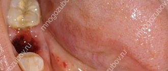
- When visually examining the socket, you can see an empty socket in which there is no blood clot (in this case, the alveolar bone in the depths of the socket will be exposed).
Or the socket may be completely or partially filled with food debris or necrotic disintegration of a blood clot. By the way, if the alveolar bone is exposed, it is usually extremely painful when touched, as well as when in contact with cold or hot water. In some cases, the edges of the mucous membrane converge so closely to each other above the hole that it is completely impossible to see what is happening in its depths. But when washing such a hole from a syringe with an antiseptic, the liquid will be cloudy, with a lot of food residue.
Dry socket after wisdom tooth removal –
Alveolitis after wisdom tooth removal may in addition have several more symptoms (in addition to those listed above). We are talking about difficulty opening the mouth or painful swallowing (24stoma.ru). Also, due to the fact that the socket of the 8th tooth is usually located deep in the soft tissues, suppuration from the socket often develops there (see video 2).
Alveolitis: video
In video 1 below you can see that there is no blood clot in the socket, there is exposed bone, and also in the depths of the socket it is filled with food debris. And in video 2 - alveolitis of the lower wisdom teeth, when the patient presses his finger on the gum in the area of 7-8 teeth, and copious purulent discharge comes from the holes.
Clinical diagnosis
Externally, the hole looks like a deep hole in the gum that has an unhealthy color. You can diagnose this disease yourself using the above symptoms, but visiting a doctor in this case is mandatory.
If severe pain, general weakness, redness of the gums and high fever appear in the socket area, you should suspect the development of alveolitis.
The dentist most often makes a diagnosis based on external signs. Visually, he is able to determine the degree of damage to the hole, on the basis of which the treatment method is selected. Additional blood tests may be required to confirm the diagnosis.
Based on the results obtained, the specialist prescribes appropriate medications to the patient.
The process of diagnosing a disease begins with an examination of the dentist and a survey of the patient. Already at the first pathological manifestations, you need to consult a doctor, because when the disorder starts, irreversible purulent-necrotic processes can arise and actively progress.
Immediately after removal, several preventive visits to the doctor are required. Usually, to identify alveolitis, it is enough to ask the patient about complaints and visualization; less often, radiography, computed tomography, and electromyography are additionally performed. When a disease occurs, the doctor detects a plaque with a green or yellow tint in the oral cavity.
Differential examination is not difficult, since the clinical picture of alveolitis is specific. It is important to distinguish it only from alveolar neuritis. But with neuritis, the temperature does not rise, the lymph nodes remain normal, there is no inflammation. Therapy proceeds faster, and the pathology itself is not dangerous if proper, timely assistance is provided.
Dry socket after tooth extraction: causes
There are many reasons why alveolitis develops. It can arise due to the fault of the doctor, the fault of the patient, and for reasons beyond anyone’s control. If we talk about the patient’s responsibility, then alveolitis can occur when -
- poor oral hygiene,
- the presence of untreated carious teeth,
- due to smoking after removal,
- when ignoring doctor's recommendations,
- if you rinse your mouth vigorously and simply rinse the blood clot out of the hole.
Alveolitis can also occur in women due to increased levels of estrogen in the blood during the menstrual cycle or as a result of taking oral contraceptives (birth control pills). A high concentration of estrogen leads to fibrinolysis of the blood clot in the socket, i.e. to degradation and destruction of the clot.
It is because of fibrinolysis that a blood clot is destroyed both with poor oral hygiene and with carious teeth. The fact is that pathogenic bacteria, which live in large numbers in dental plaque and in carious defects, secrete toxins, which, like estrogens, lead to fibrinolysis of the blood clot in the socket.
When alveolitis occurs due to the fault of the doctor –
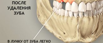
- If the doctor left a tooth fragment, bone fragments, or inactive fragments of bone tissue in the socket, which lead to injury to the blood clot and its destruction.
- A large dose of a vasoconstrictor in an anesthetic - alveolitis can occur if, during anesthesia, the doctor injected a large volume of an anesthetic with a high content of a vasoconstrictor (for example, adrenaline). Too much of the latter will result in the hole simply not filling with blood after the tooth is extracted. If this happens, the surgeon must scrape the bone walls with an instrument and cause socket bleeding.
If the doctor left a cyst/granulation in the socket, when removing a tooth with a diagnosis of periodontitis, the doctor must necessarily scrape out the cyst or granulations (Fig. 10), which might not come out with the tooth, but remain in the depths of the socket. If the doctor did not inspect the socket after extracting the tooth root and left a cyst in the socket, the blood clot will fester.
- Due to the large trauma to the bone during removal, this usually happens in two cases: firstly, when the doctor cuts out the bone with a drill without using water cooling of the bone at all (or when it is not cooled sufficiently).
Overheating of the bone leads to its necrosis and the start of the process of destruction of the clot. Secondly, many doctors try to remove a tooth for 1-2 hours (using only forceps and elevators), which causes such trauma to the bone with these instruments that alveolitis is simply bound to develop. An experienced doctor, seeing a complex tooth, will sometimes immediately cut the crown into several parts and remove the tooth fragment by fragment (spending only 15-25 minutes), and thereby reduce the trauma caused to the bone.
- If, after a complex removal or removal against the background of purulent inflammation, the doctor did not prescribe antibiotics, which in these cases are considered mandatory.
Conclusions: thus, the main causes of destruction (fibrinolysis) of a blood clot are pathogenic bacteria, excessive mechanical trauma to the bone, and estrogens. Reasons of a different nature: smoking, loss of a clot while rinsing the mouth, and the fact that the hole did not fill with blood after the tooth was extracted. There are also reasons that do not depend on either the patient or the doctor, for example, if a tooth is removed due to acute purulent inflammation - in this case it is stupid to blame the doctor for the development of alveolitis.
Why is pathology dangerous?
If you do not pay close attention to the problem, then within a few days after the operation severe pain will begin, indicating inflammation of the socket of the extracted tooth, that is, alveolitis. The disease is dangerous because, if treated improperly and not seeing a doctor in a timely manner, it can lead to the development of gumboil, when the cheek swells and an abscess appears on the gum. But flux is not the worst thing. Advanced alveolitis can lead to a purulent abscess, phlegmon of the soft tissues of the face and osteomyelitis of the jaw, damage to internal organs, sepsis (blood poisoning), and death.
Treatment of alveolitis -
If alveolitis develops in the socket after tooth extraction, treatment at the first stage should be carried out only by a dental surgeon.
This is due to the fact that the hole may be filled with necrotic decay of a blood clot; there may be inactive fragments and fragments of bone or tooth. Therefore, the doctor’s main task at this stage is to scrape it all out of the hole. It is clear that no patient will be able to do this on their own. Antiseptic rinses and antibiotics (without cleaning the socket) can only temporarily reduce the symptoms of inflammation, but do not lead to healing of the socket. But at a later stage, when the inflammation in the socket subsides, patients will be able to independently treat the socket with special epithelializing agents to speed up its healing.
Thus, the main method of treatment will be curettage of the hole, but there is also a second method - by creating a secondary blood clot in the hole of the extracted tooth. Read more about these methods...
Curettage of the tooth socket for alveolitis -
- Under anesthesia, a festering blood clot, food debris, and necrotic plaque from the walls of the socket are removed. Without removing the necrotic plaque and the disintegration of the blood clot (containing a huge amount of infection), any treatment will be useless.
- The hole is washed with antiseptics, dried, after which it is filled with an antiseptic (iodoform turunda). Usually, the turunda needs to be changed every 4-5 days, i.e. you will have to go to the doctor at least 3 times.
- The doctor will prescribe you antibiotics, antiseptic baths, and painkillers, if necessary.
Doctor's prescriptions after tooth socket curettage –
- NSAID-based analgesics (for pain),
- 0.12-0.2% Chlorhexidine solution for antiseptic rinses (2-3 times a day for 1 minute),
- Antibiotics: usually either Amoxiclav 625 mg tablets (2 times a day for 5-7 days) or Unidox-solutab 100 mg (2 times a day for 5-7 days) are prescribed. These antibiotics are better, but not cheap. Inexpensive ones are Lincomycin capsules 0.25 (2 capsules 3 times a day), but keep in mind that after this antibiotic, problems with the stomach and intestines develop more often.
Method for creating a secondary blood clot -
However, there are 2 situations in which a different treatment method can be used.
This method involves the creation of a secondary blood clot in the socket and, accordingly, if successful, the socket will heal much faster than after constantly placing iodoform turundas into it for 2-3 weeks. It is preferable to use this method only in the following two situations... Firstly, when you consulted a doctor immediately after, for example, you rinsed a clot out of the hole or it fell out on its own (i.e. when the hole is not yet filled with infection and food debris , and there is no necrotic clot disintegration or suppuration). Secondly, when the patient has a sluggish alveolitis for a long period of time, and the socket is filled with inflammatory granulations.
How this technique is carried out - if the hole is empty, then under anesthesia the bone walls of the hole are scraped out with a curettage spoon to create bleeding and the hole fills with blood (video 3). If the hole is filled with granulations, then they are carefully scraped out, i.e. do the same curettage (video 4). Then, in both cases, after the hole is filled with blood, an anti-inflammatory medicine (Alvogel) is placed deep into the hole, and several sutures are placed on the mucous membrane to bring the edges of the wound closer together. Antibiotics are immediately prescribed.
Curettage to create a secondary blood clot: video 3-4
Summary: i.e. In both the first and second methods, curettage of the hole is carried out in the same way, but in the first case, the hole heals slowly under iodoform turundas, and in the second case, a blood clot forms in the hole for the second time, and the hole heals, as it should do under normal conditions .
The main functions performed by such a “thrombus”
If a clot has formed at the site of the extracted tooth, then this is a good sign. It is this that protects a fresh wound from the aggressive environment of the oral cavity, where a large number of bacteria live. It also prevents saliva and food debris from entering injured tissues, protects them from the inflammatory process and related complications (alveolitis, abscess, gumboil, phlegmon, osteomyelitis).
The clot promotes the natural healing process of areas damaged during surgery. Moreover, some researchers claim that it even accelerates the process of tissue regeneration, and if it is not formed or falls out ahead of time, the rehabilitation period lasts longer.
What can you do at home -
After the acute symptoms of inflammation have subsided, there is no need for antiseptic turundas inside the socket, because they do not help the wound to heal (epithelialize) faster. At this stage, the best treatment method will be to fill the hole with a special Dental adhesive paste (Solcoseryl). This drug has an excellent analgesic effect (after 2-3 hours the pain will practically stop, and after 1-2 days it will go away completely), and also speeds up healing many times over.
Scheme of use - this paste is added to a hole that has been washed with an antiseptic and slightly dried with a dry gauze swab (completely filling the hole). The paste is perfectly fixed in the hole and does not fall out of it. There is no need to remove the paste from the hole, because... it slowly dissolves on its own, giving way to growing gum tissue. The only thing that may be required is to periodically add it to the hole.
How to rinse the hole from food debris -
In some situations (when the turunda has fallen out of the hole, and there is no way to see a doctor right away), it may be necessary to wash the hole. After all, after each meal, the hole will become clogged with food debris, which will cause new inflammation. Rinsing will not help here, but you can easily rinse the hole with a syringe.
Important : from the very beginning you must bite off the sharp edge of the needle from the syringe! Next, bend the needle a little and fill a 5.0 ml syringe with a solution of Chlorhexidine 0.12-0.2% (it is sold ready-made in every pharmacy for 20-30 rubles). Screw the needle tightly so that it does not fly off when pressing the syringe plunger! Place the blunt end of a bent needle into the upper part of the socket (do not insert too deeply to avoid injuring the tissue), and rinse the socket under pressure. If necessary, do this after every meal.
In principle, after this the hole can be dried with a gauze swab and treated with Solcoseryl. We hope that our article on the topic: Alveolitis after tooth extraction, symptoms, treatment - turned out to be useful to you!
Sources:
1. Higher prof. the author’s education in surgical dentistry, 2. Based on personal experience as a dental surgeon, 3. National Library of Medicine (USA), 4. “Outpatient surgical dentistry” (Bezrukov V.), 5. “Propaedeutics of surgical dentistry” (Soloviev M. .).
Pathology prevention measures
Every patient can protect himself from the occurrence of an unpleasant problem. To do this, you must initially choose professional dental surgeons who specialize in surgical interventions. At the stage of planning the operation, you need to inform the specialist about the diseases you have and the medications you are taking, so that the doctor, based on the data received, can draw up an individual treatment plan.
At home, you need to follow all the recommendations of specialists without exception. It is especially important to follow those that prevent damage and dissolution of the protective clot. Namely: you need to stop eating hot and solid foods, you shouldn’t engage in sports or physical activity, and you shouldn’t touch the wound with any objects or hands. For a while, you should stop smoking and drinking alcohol, as well as actions that lead to overheating, a rush of blood to the head and severe swelling of the tissues.