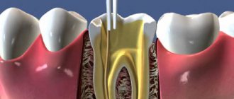
Filling the root canals of teeth is an endodontic dental procedure, the purpose of which is to close their lumen. Its “scientific” name is “obturation.” The process uses special materials, including ethonium and two-component pastes, as well as gutta-percha. The former is especially preferred as it fills the canal evenly and can be easily removed if the need for re-treatment arises. Filling with its use is called “three-dimensional obturation of root canals.” If the patient has indications, the procedure is combined with pin fixation. The latter is used to further strengthen the tooth if it is severely damaged and to provide prosthetics.
Tooth root filling in Moscow can be done at the dental department of CELT. Our multidisciplinary clinic has been represented on the domestic market of paid medical services for almost three decades. In our dentistry, we see orthopedists, surgeons, endodontists and therapists, who have many years of experience in scientific and practical work. Treatment is carried out according to international standards with the conclusion of an official contract and provision of guarantees. You can find out its price by going to the “Services and Prices” tab in this section. We regularly update our price list, but to avoid misunderstandings, please check the numbers with our information line operator or during a consultation with your doctor.
Root canal obturation - RUB 1,500.
Canal filling using lateral condensation method - RUB 2,200.
Application of an intracanal pin—RUB 2,500.
Three-dimensional obturation of one root canal with thermoplasticized gutta-percha - 4,000 rubles.
At CELT you can get advice from a dental specialist.
- The cost of a consultation with a dentist-therapist is 1,000
Make an appointment
Temporary and permanent obturation of root canals: what is it and why is it needed?
Filling the root canals of teeth is an endodontic dental procedure, the purpose of which is to close their lumen. Its “scientific” name is “obturation.” The process uses special materials, including ethonium and two-component pastes, as well as gutta-percha. The former is especially preferred as it fills the canal evenly and can be easily removed if the need for re-treatment arises. Filling with its use is called “three-dimensional obturation of root canals.” If the patient has indications, the procedure is combined with pin fixation. The latter is used to further strengthen the tooth if it is severely damaged and to provide prosthetics.
Tooth root filling in Moscow can be done at the dental department of CELT. Our multidisciplinary clinic has been represented on the domestic market of paid medical services for almost three decades. In our dentistry, we see orthopedists, surgeons, endodontists and therapists, who have many years of experience in scientific and practical work. Treatment is carried out according to international standards with the conclusion of an official contract and provision of guarantees. You can find out its price by going to the “Services and Prices” tab in this section. We regularly update our price list, but to avoid misunderstandings, please check the numbers with our information line operator or during a consultation with your doctor.
Clinical cases in which obturation is required
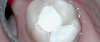
Tooth obturation is a procedure that is performed only on units that are subject to depulpation, that is, removal of the nerve. It is also impossible to do without it if the patient is preparing for prosthetics with crowns or bridges - the teeth that will act as a support for the dentures are exposed to it.
It is necessary to carry out sealing even when the crown of the tooth is destroyed by more than half. Endodontic treatment will help prevent the infection from spreading deeper, strengthen the roots and protect the internal cavities from the destructive process.
Temporary and permanent obturation of root canals: what is it and why is it needed?
Filling the tooth canals is a mandatory stage of their treatment, the purpose of which is to stop the inflammation of periodontal and pulp tissues. It is carried out for such dental pathological conditions as caries, periodontitis and pulpitis: the latter are often complications of carious processes that did not receive timely treatment. In addition, endodontic manipulation may be required when re-treating canals or in the process of preparatory measures for prosthetics. They themselves are quite complex, and therefore require the endodontist to have not only the appropriate knowledge and skills, but also equipment - a dental microscope (in some cases), as well as professional tools.
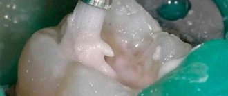
This approach is relevant, since the procedure for permanent filling of tooth canals involves working with its internal structures. They are the ones who ensure its functionality and nourish it, prolonging its life. Failure to comply with technology and making mistakes can lead to serious complications and – often – the loss of the entire unit... But thanks to proper treatment, a tooth can be saved, sometimes in the most difficult cases. Before starting the procedure, the dentist removes the necrotic pulp from the canal and expands it in order to extract all fragments of the affected tissue. The next step is disinfection and drying, and only then sealing.
As for temporary obturation of root canals, it is also carried out after endodontic manipulations are completed. In the process, the dentist also fills the canals with a special composition, but not in order to close the gap, but in order to cure them and eliminate the possibility of complications. As a rule, the medicine is placed for several days or even months, excluding the penetration of pathogens. This approach is intended to:
- stop the inflammation of periodontal tissues;
- ensure high-quality disinfection of root canals and dentinal tubules;
- guaranteed to clean them by removing microfragments of pathological tissues from them;
- stimulate the regeneration of periodontal tissues and bone tissue of the alveolar process;
- isolate the root canal and the processes occurring in it, if permanent filling in one visit is not possible.
Most often, temporary filling of root canals is practiced during the period of manipulation to eliminate periodontitis. Depending on the situation, it can last more than one month and requires that medications be placed directly into the canals.
Causes of urinary tract obstruction
Narrowing of the urinary tract cavity can be observed in people of different age categories. But if in children obstruction is most often congenital, then in adults in most cases it occurs due to disorders of the urinary system. The cause of obstruction of the urinary tract may be a mechanical defect in patency caused by a tumor, kidney stones, blood clots, foreign bodies (with a prolonged stay of the catheter in the ureter). Also, swelling that appears as a result of an inflammatory process in the bladder or ureter can cause a narrowing of the lumen of the canal. Obturation itself is not a disease, but only a condition indicating the occurrence of pathology in the body. Regardless of the etiology of obstruction, it can lead to the development of renal failure.
Indications and contraindications for dental root canal filling
Indications
- Pulpitis that cannot be cured with conservative methods;
- The need for re-treatment of the canal after previously performed procedures;
- Preparatory measures for prosthetics using stump inlays or a pin;
- Preparation for installation of a crown or bridge, provided that extensive grinding of hard tooth tissues is required;
- Treatment of a fractured tooth, provided that its pulp has been injured and died.
Contraindications
- Serious damage to periodontal tissues, which does not allow saving the tooth;
- Identification of a cystic formation that cannot be cured;
- Identification of any neoplasms in the area of the tooth root;
- Longitudinal root fracture;
- Too branched and curved dental canals;
- Their obstruction due to the lack of lumen or filling material that cannot be removed;
- Perforation of the root canal wall.
The last 4 points are relative contraindications. Treatment can be carried out by an experienced specialist using a dental microscope.
Preparation
The preparatory process is carried out taking into account the chosen method of sealing the root canal. The use of specialized equipment requires the selection of the appropriate tool.
For example, when installing an apical pin, you should select the appropriate size and trim its tip by 0.5-1 mm. Appropriate pluggers and equipment are also prepared.
Preparation for obturation:
- The use of specialized equipment requires the selection of the appropriate tool
cleaning all tissues affected by caries and installing fillings on the holes (except for the endodontic entrance);
- therapeutic procedures to eliminate periodontal problems;
- restoration work with the walls and surfaces of the tooth;
- if there is excessive salivation, the patient is offered the drug atropine (the solution is used to rinse the mouth half an hour before the onset of obturation).
Next, the dentist moves on to the next stage, which directly concerns the endodontic entrance:
- use of anesthesia (infiltration/conduction);
- opening access to the root canal (includes preparation of a cavity affected by caries, removal of the arch of the dental pocket where the nerves are located);
- extraction of tooth connective tissue and necrotic formations;
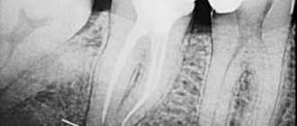
- determination of the parameters of the root canal opening (X-ray);
- treatment of the canal opening with medications and instruments;
- relieving inflammation (if any).
Experts do not recommend endodontic treatment to be carried out in one visit. In difficult cases, the period may drag on for several weeks.
Materials used for filling tooth root canals
There are special requirements for materials used for obturation of root canals; in particular:
- Compatibility with body tissues, minimizing the risk of rejection;
- Minimizing the risk of developing an allergic reaction;
- The presence of antiseptic and anti-inflammatory properties;
- Ease of insertion into and removal from the root canal;
- Elimination of shrinkage after hardening inside the channel;
- The ability to initiate regeneration processes in periodontal tissues.
- The ability to fully fill even small voids;
- X-ray detectability.
Obturators are usually divided into two groups:
- Fillers – pins made of metals, gutta-percha and fiberglass;
- Sealers are dental pastes and cements for fixing fillers.
Guttarpeche fillers are considered the most effective today, which give especially good results when combined with sealers. The latter are known for good adhesion to the walls of root canals and are able to provide reliable sealing if all conditions are met. In order for root canal filling to be carried out correctly, CELT dentists select materials for it on an individual basis, taking into account:
- The canal of which tooth (baby or permanent) requires obturation;
- Its branching, diameter, cross-country ability;
- The degree of destruction of the crown, the need to strengthen it;
- The patient's budget for the procedure.
List of materials used for the procedure
1. Gutta-percha and metal pins
Obturation of tooth canals today is most often performed using flexible gutta-percha pins. They are made of polymer, non-toxic, very elastic, and penetrate well into all branches. To fill one tooth, a doctor sometimes needs 20–30 pieces of gutta-percha pins. There are many techniques for working with this material, and doctors can always choose exactly the option that suits each specific patient.
Metal pins, in particular silver ones, are often used in endodontics, as they have a good antibacterial effect and plasticity. Due to their flexibility, they can penetrate the most inaccessible areas and branches.
Another type of pins is titanium. Due to their high strength and wear resistance, they are also in demand.
Important! Pins alone, as an independent measure, do not provide 100% tightness, so they are always used in conjunction with other fillers, most often sealers.
Liquid or paste sealers/cements
These are flowing, liquid or paste-like fillers, with the help of which doctors achieve hermetically sealed filling of all branches and cavities. They help ensure easier insertion of the pins and strengthen them. Among other things, they have good adhesive properties and harden quickly.
Currently, polymer sealers are most often used; they are very durable and are not subject to dissolution in tissue fluid. They also retain their tightness for many years, unlike natural cements based on zinc oxide, which were actively used until recently.
In some clinical cases, doctors also use cements with calcium hydroxide: they strengthen the tooth structure from the inside. If it is necessary to obturate mammary units, glass ionomer cements (GIC) are used.
Pastes based on zinc and eugenol
Pastes are inexpensive and easy to use; they have been used in dental practice for many years, but in recent years professional doctors have given them less and less preference, because they have a number of significant disadvantages. These include poor tightness, rapid shrinkage, allergic reactions and irritation of periodontal tissue. Another disadvantage is that the products do not penetrate into small branches of the roots, so they cannot guarantee one hundred percent tightness.
Complications that may develop after root canal obturation
Provided the procedure is carried out correctly, modern equipment and high-quality instruments are used, the risk of complications is minimized. Otherwise, the following complications are possible:
- Breaking off parts of the endodontic instrument and getting them stuck in the canal and, as a result, the presence of voids around them;
- Development of inflammatory processes and swelling in the cheek area on the side of the affected tooth;
- Perforation of the root canal wall;
- Allergic reaction to filling materials.
In all cases, the filling material will have to be removed, which is associated with a number of difficulties and the risk of complete tooth loss. After removing the fragments of endodontic instruments from the canal and cleaning it, the procedure will have to be repeated.
You just have to want it
For specialists in this case, the field of activity remains endodontic treatment in particularly complex root canal anatomy, as well as in particularly complex clinical situations, and for “particularly gifted” specialists - perhaps complex cases of root canal revision.
There is enough work for everyone. In any case, this would benefit “general-profile” therapeutic dentistry.
The article was provided by the magazine of the official publication of the Association of Dentists of Lower Saxony (Germany) NZB - Niedersächsisches Zahnärzteblatt (No. 6, 2011, pp. 31-34).
Translation by Inna Bichegkueva.
What is three-dimensional obturation? Its features and advantages
Gutta-percha obturation, also called three-dimensional, is considered the most effective method of filling root canals today. It ensures sealing of the entire root canal system, even if it is seriously branched. As a result, a reliable barrier appears between the dental cavity and periodontal tissues. The procedure solves several problems at once:
- Reliable sealing of the lumens of the dentinal tubules that extend into the lumen of the canal;
- Eliminating the risk of recurrent infection;
- Eliminating the risk of material resorption in the canal;
- Overlapping of the lumens of not only the main, but also the lateral and acesor canals;
- Creation of favorable biological conditions for tissue healing.
The advantages of this approach are obvious:
- Even more effective treatment of complicated caries;
- Painlessness, elimination of discomfort for the patient;
- Possibility of using a tab or pin;
- Guarantee that the canal will not become inflamed again.
It is worth noting that gutta-percha filling has been used in dentistry for several years now... But only today, thanks to a number of systems (“Gutta Core”, “ThermoFill”, others), it has become so effective, perfect and at the same time quite simple. As a rule, in the process of filling root canals, the endodontist faces a number of difficulties: from the individual characteristics of their anatomical structure to the development of inflammation of the tissues around the root.
Materials used for three-dimensional obturation
It is a special cross-linked thermosetting gutta-percha elastomer. It is made of gutta-percha in two different forms and combines its advantages. Its use makes the preparation stage for the pin as simple as possible, as well as removing the filling material from the canals, if the need arises. This material itself is quite strong, but under the influence of a certain temperature it becomes very plastic, which makes it easy to fill not only the central channel, but also its branches. Its composition is such that it will not dissolve under the influence of dentinal fluid. This also makes it quick and easy to remove.
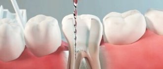
Three-dimensional obturation algorithm
3D root canal filling systems include everything you need and do not require the use of special tools. At the same time, the endodontist who works with them must know their features well and follow technological processes. Procedure steps:
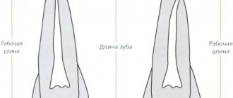
- Instrumental processing of the canal and its expansion to a value determined by the technology of working with a particular system (as a rule, the taper should be at least 6% for effective irrigation and obturation);
- Control of root canal measurements and selection of obturator size taking into account the taper involved in the process of instrument preparation. For control, the original verifier from the system with the appropriate size and taper is used;
- Introducing the sealer into the mouth or middle segment of the root canal, its uniform distribution during the process of introducing the obturator. It is important to avoid excess material as it may become trapped in the periapical tissues;
- Selection of an obturator, determination of its working length in accordance with the length of the root canal and heating in a special oven. Its slow introduction into the root canal and filling of all branches due to condensation at the mouth;
- Removing the media and handle and cutting the media directly above the mouth;
- Monitoring of the treatment performed by radiographic examination;
- Restoration of the crown part of the tooth by placing a filling or prosthesis.
Symptoms of urinary tract obstruction
Obstructions most often cause disturbances in the outflow of urine (even cessation) through the urinary tract, which is the cause of acute or chronic renal failure. Also, symptoms of a patency defect include pain, the intensity of which depends on the degree of development of obstruction. As the pathology progresses and homeostasis is disrupted, the pain becomes pronounced, with the manifestation of renal colic, and requires immediate medical intervention. If the obstruction develops slowly, renal function defects show little effect, pain may also be absent or quite mild or moderate and not cause much concern. At the same time, this condition also requires urgent assistance from doctors, since with chronic disruption of the outflow of urine, irreversible changes occur in the kidneys.
A favorable prognosis for obstruction of the urinary tract is possible only with its timely elimination and treatment of the diseases that caused it.
List of instruments used for obturation
An endodontist (endodontist) performing such a technically complex procedure must have a high level of professionalism. He also needs to have in his arsenal a set of specialized tools: a spreader for distributing and compacting gutta-percha, a canal filler, a gutta-condenser for filling canals and softening filling materials, pluggers for condensing gutta-percha, “GuttaEst” for heating and cooling gutta-percha.
Obturation methods
- obturation with cool gutta-percha - technology using both a pin and paste
- literal condensation of cool gutta-percha (use of several pins)
- thermomechanical condensation (has its drawbacks, so it is rarely used today) - high-speed screwing of gutta-percha into the root cavity
- the use of softened, cool gutta-percha is a method that does not require the use of a cementitious sealer
- filling with hot gutta-percha (for example, three-dimensional canal filling)
- “continuous wave” technique - gradual filling of the canal with parts of pins
Material requirements
Tooth obturation must be carried out with high-quality materials that meet certain requirements:
- moisture resistance,
- hypoallergenic and good biocompatibility,
- no shrinkage for many years,
- antibacterial effect,
- high adhesion to dentin,
- lack of interaction with tissue fluids,
- easy removal if necessary,
- good radiopacity,
- fast curing period.
Indications
Root hole obturation is recommended for the following dental problems:
- periodontitis (in any manifestation);
- chronic pulpitis;
- acute pulpitis.
When is there a need for filling?
Root filling is indicated for the treatment of a number of dental pathologies and for smile restoration by an orthopedic dentist:
- for pulpitis in adult patients: in children the rudiments of the permanent row are located very close, therefore endodontic treatment of the primary occlusion is not carried out (passing the canals with an instrument can be dangerous),
- in the treatment of periodontitis, cysts and granulomas: inflammatory processes or space-occupying formations under the apices of the roots can be treated surgically - osteotomy, for example. But this option may not be justified from the point of view of severe traumatism with a small pathological focus,
- when preparing a “support” for a prosthesis: artificial crowns, bridges or clasp appliances are attached to previously pulpless teeth in order to eliminate the risk of pulpitis or periodontitis while wearing the prosthesis,
- if the crown is destroyed by 50%: here a standard filling will not provide the necessary strength to the top of the tooth, so it is necessary to install a ceramic inlay, or strengthen the root part with a metal pin and install a crown on top. Endodontic treatment is also carried out to prevent the spread of infection.
Diagram of the difference between pulpitis and periodontitis
Design characteristics
Pins for the structure are made of various materials - silver, absorbent pastes, etc. Gutta-percha together with sealants is very popular.
The pins themselves can be of any shape - cone-like, thermoplastic mass, etc. The doctor selects them individually for each patient.
To accurately secure the pin, sealants made from calcium hydroxide, zinc oxide-eugenol, resin or glass ionomer materials are used.
This design is often used in the treatment of advanced forms of carious lesions (periodontitis, pulpitis, etc.). A pin can only be installed in a tooth that does not cause discomfort to the patient, otherwise additional treatment will be required.
For temporary obturation of primary teeth, pins made of chlorophenol-camphor-menthol composition are used. They are not particularly durable and are ideal for temporary fillings.