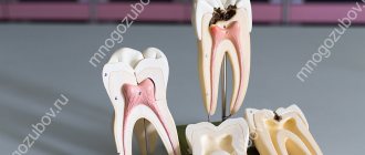
Root canal treatment is a procedure aimed at restoring and preserving a damaged tooth. The achievements of modern dentistry make it possible to perform it quickly, effectively and painlessly.
The discomfort of the canal treatment procedure is comparable to the usual filling process, but sometimes it is painful and therefore requires anesthesia. The procedure is also highly effective and less expensive compared to other alternative treatments. A cured and restored tooth, with proper care, can last for many years.
Root canal treatment methods
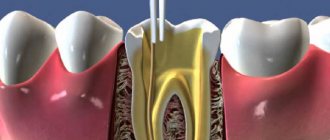
Two methods are used for root canal treatment: mechanical (machine) and manual. Both methods are aimed at expanding, and subsequently cleaning the canal from microbes and dentin crumbs. The treatment is completed by medicinal treatment of the dental cavity.
The purpose of these dental procedures is to make the dental canal suitable for subsequent filling. Initially, the dental canal has a large number of irregularities and micro-branches, which prevents the high-quality filling of the cavity with filling material, and this, in turn, leads to the development of complications. After the treatment, all irregularities are smoothed out, and the size of the canal becomes convenient for filling.
Indications for temporary filling of root canals
Temporary filling with a paste based on calcium hydroxide is indicated for chronic pulpitis with a closed tooth cavity that has 2 or more root canals; chronic apical periodontitis with a closed tooth cavity having 2 or more root canals; acute apical periodontitis of pulpal origin with a closed tooth cavity.
Temporary filling with a paste based on calcium hydroxide with iodoform is indicated for acute purulent pulpitis with an open and closed tooth cavity; chronic ulcerative pulpitis with an open tooth cavity; chronic hyperplastic pulpitis; pulp necrosis with open and closed tooth cavity; acute apical periodontitis of pulpal origin after the removal of acute phenomena; chronic apical periodontitis with an open tooth cavity.
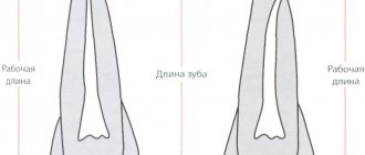
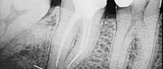
All patients in need of endodontic treatment are prescribed an X-ray examination at the stages of measuring the working length, filling the root canals, as well as in the long term.
Instrumental treatment, which involves eliminating the source of infection and forming the required shape of the root canal, is carried out using the “Step Back”, “Crown Down” or a mixed method.
After mechanical processing, a funnel-shaped canal is formed with a minimum diameter in the apex area and a maximum at its mouth; a balance is maintained between the diameter of the channel and the thickness of its walls; a constant position of the apical foramen is ensured; an apical stop is created to prevent the filling material from being pushed into the periodontium. Medicinal treatment is carried out, the walls of the root canal are dried using paper points.
In the treatment of pulpitis without symptoms of apical periodontitis (low contamination of the canals), the method of temporary filling of root canals with calcium hydroxide-based paste is used. In case of pulpitis with symptoms of periodontitis, chronic periodontitis, acute and at the acute stage of chronic periodontitis (after acute symptoms have resolved), the root canals are additionally treated with a 2% solution of chlorhexidine (exposure 2 minutes), after which they are temporarily filled with a paste based on calcium hydroxide with iodoform.
Method of temporary filling of root canals
Filling a canal with paste can be done either manually or using a canal filler.
Method of “manual” filling. Using a limiter, the working length of the canal is fixed on a K-file, K-reamer or on a special cannula - a syringe attachment. At the tip of the instrument, a small amount of paste is introduced into the equine canal up to the apex. Condense the paste using a damp cotton pad. The next portion of paste is injected to 2/3 of the working length of the canal.
Condense the paste in the same way. The next portion of paste is injected to 1/3 of the working length of the canal. Excess paste that has accumulated above the mouth is pressed into the canal using a cotton ball. The tooth cavity is hermetically sealed with a temporary filling for 48 hours.
Filling technique using canal filler. The canal filler, a size smaller than the last instrument used to expand the root canal, is fixed in the tip and the working part is immersed in the paste, retaining a small amount of material on the spiral. The instrument is inserted into the canal up to the apex, turned on at low speed (100-120 rpm) for 2-3 seconds, then the instrument is slowly removed from the canal while the drill is running.
The canal filler is again enveloped with filling material, inserted into the canal to 2/3 of the working length, the drill is turned on and the material is pumped into the canal, then the procedure is repeated. Excess paste that has accumulated above the mouth is pressed into the canal using a cotton ball. The tooth cavity is hermetically sealed with a temporary filling for 48 hours. If there is painful percussion, the canal is filled again at the next visit.
If the apical opening is wide or it was expanded during instrumentation, then the first portion of the paste is injected and condensed “manually” and only then the canal filler is used.
On the second visit, the remains of the temporary sealer are removed from the root canal using an H-file of the required size using scraping movements. Scraping is periodically alternated with irrigation of the root canal with an antiseptic solution using an endodontic syringe with a needle until the temporary sealer on the H-file is completely removed, after which the root canal is dried with paper points and filled using the sealer and gutta-percha points using the lateral condensation method. For permanent filling, according to indications, sealers are used, which contain calcium hydroxide with iodoform, zinc oxide eugenol, hydroxyapatite, and glass ionomer cement.
Assessment of the quality of endodontic treatment is carried out immediately after treatment and in the long term (after 6, 12 months). The criteria for clinical well-being are: absence of complaints; painless percussion; normal state of the transitional fold in the area of projection of the apex of the root of the tooth under study; positive dynamics or absence of pathological changes in the tissues of the apical periodontium on the radiograph.
Efficiency of temporary filling of root canals
Postgraduate student of the Department of Therapeutic Dentistry of BelMAPO O. V. Fedorinchik assessed the immediate results of endodontic treatment in study groups I (pulpitis without periodontitis) and II (with apical periodontitis).
After treatment, there were no complaints in 75.0% of cases in the control (without the use of calcium hydroxide); after temporary filling, this figure reached 95.2%. Within 1-2 days, complaints of pain when biting were reported in 11 cases (17.2%) in the control group; in the main group they were recorded in only three teeth (4.8%).
On days 3-5, in the control group, pain persisted in 5 teeth (7.8%), while in the main group it was absent.
In group II, complaints of pain after endodontic treatment were absent in the control group in 50.8% of cases, in the main group - in 85.7%. Pain when biting 1-2 days after treatment was noted by 22 patients (36.1%) in the control group and 6 (10.7%) in the study group.
On days 3-5, pain in the control group was 13.1%, i.e. 8 cases, and only 2 (3.6%) in the main group. Thus, when using temporary root canal filling, pain is significantly reduced.
The results of endodontic treatment after 6 months are presented in Table No. 1. In the main group, in 100% of cases there were no complaints of pain in the area of the treated tooth. In the control group, complaints of periodic pain were reported in one case. An X-ray examination in the control group revealed an expansion of the periodontal gap in the apical periodontal tissues in five teeth (7.8%), in the main group this figure was recorded in one case (1.6%).
When using a paste with iodoform for temporary filling, there were no complaints of pain in 98.2% of patients, and periodic pain was observed in only one tooth (1.8%). At the same time, in the control group there were no complaints in 72.1%, and periodic pain in the area of the treated tooth was bothersome in 17 cases (27.9%).
On the radiograph in the control group, there were no changes in the periodontium in 55.7% of cases, in the main group - in 80.4% of treated teeth.
At a follow-up examination after 12 months in group I (pulpitis), there were no complaints of pain in the control group in 95.3% of cases; in the main group there were no complaints in 100% of cases. Periodic pain in the area of the treated tooth was recorded in the control group in 3 cases, 4.7%. X-rays after 12 months showed no changes in the apical periodontal tissues in 85.9% of the teeth in the control group; in the main group, this figure reached 98.4%. In the apical region of 9 teeth (14.1%) in the control group, an uneven expansion of the periodontal fissure was noted, while in the main group these changes were recorded only in one case (1.6%).
In the main group II (apical periodontitis) there were no complaints in 100% of cases, in the control there were no complaints in 93.4%.
During X-ray examination, the absence of changes in the apical periodontal tissues was noted in 73.8% of cases in the control group. In the main group, this figure reached 92.9%. The expansion of the periodontal fissure in the area of the apical periodontium was preserved in 12 teeth of the control group (19.7%). In the main case - in 4 cases (7.1%).
Conclusion
Studies have shown that temporary filling of root canals can statistically significantly reduce the incidence of complications in both pulpitis and periodontitis. 6 months after treatment of pulpitis, the number of complications was minimal.
Complaints about pain and radiological changes in the area of the treated tooth were significantly less common in the main group. After 12 months, positive radiographic dynamics were more often observed in the main group, where a paste based on calcium hydroxide with iodoform was used.
LITERATURE
- Antanyan A. A. // Endodontics today. - 2007. - No. 1. - P. 59 - 69.
- Kazeko L. A., Fedorova I. N. Calcium hydroxide in endodontics: yesterday, today, tomorrow // Modern dentistry, 2009. - No. 2. - P. 4 - 9.
- Lopatin O. A., Fedorinchik O. V. The use of calcium hydroxide preparations in the treatment of complicated caries // Modern dentistry, 2007. - No. 3. - P. 33 - 37.
- Lutskaya I.K. Restorative dentistry: equipment, tools, auxiliary materials. - Rostov-on-Don: Phoenix, 2008. - 202 p.
- Lutskaya I.K., Martov V.Yu. Medicines in dentistry. - M.: Medical literature, 2007. - 384 p.
- Lutskaya I.K., Chukhrai I.G., Novak N.V. Endodontics: A Practical Guide. - Moscow: Medical literature, 2009. - 191 p.
- Bartlett D. A difference in perspective - the North American and European interpretation of tooth wear / D.Bartlett, K.Phillips, B.Smith // Int. J. Prosthodont. - 1999. - Vol. 12, No. 5. - P. 401 - 408.
- Basrani B., Santos JM et al. // Oral Surgery Oral Medicine Oral Pothalogy Oral Radiology & Endodontics. — 2002. — Aug; 94(2). — P. 240—245/
- Cwikla S., Belanger M., Giguere S., Vertucci F. // J. Endod. - 2005. - V.31, No. 1. - R. 50-52.
- Gomes B., Souza S., Ferraz C. // Intern. Endod. J. - 2003. - V. 36. - P. 267-275.
- Ingle JI, Bakland LK Endodontics. - Baltimore$ Philadelphia et al., 1994. - P. 230-241.
- Katebzadth N., Hupp J., Trope M. Histological signs of periapical inflammation after obturation of infected root canals of teeth in dogs // Journal of Endodontics 25, 1999: 364 -8.
A complete list of references is in the editorial office.
Carrying out the procedure
Before starting the procedure, the dentist prescribes an examination of the problem area using an X-ray machine to determine the extent of damage to the canals. Next, if necessary, an anesthetic is administered to relieve the person of pain during the procedure.
The procedure for performing root canal treatment:
- A special material is placed in the mouth to isolate the tooth from saliva (rubber dam);
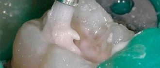
- The canals are processed and prepared for filling;
- Next, disinfection and treatment of the carious plane is performed;
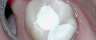
- At the end of the procedure, the tooth canals are filled with filling material.
After this, at the discretion of the doctor, a permanent or temporary filling is placed.
Mechanical method of channel processing
The mechanical method is divided into two processing methods: manual and machine. Both methods, due to rotation, remove chips from the walls of the dental canal, grinding the walls and expanding it to the optimal size suitable for filling.
Manual treatment is carried out using special instruments of different sizes, which the dentist carefully rotates in the dental canal to clean it. The size of the instrument is selected individually for each patient, taking into account the structural characteristics of the teeth and the degree of their damage.
Machine processing occurs due to the rotation of special nozzles made of flexible alloys, which ensures excellent cleaning of the canal and significantly reduces the risk of root penetration. This option is more effective and safer than manual processing, but is not always possible.
Medicinal treatment of canals
Medication treatment follows immediately after mechanical treatment; it is aimed at complete disinfection of the tooth canal. The procedure is carried out with antiseptic solutions using special needles, as well as using paper pins soaked in an antimicrobial agent. Next, the tooth cavities are washed with a stream of antiseptic. For treatment, medications containing chlorine-containing substances, iodine-containing preparations, hydrogen peroxide and others are used.
Modern antiseptics not only have antimicrobial properties, but also stimulate the processes of restoration of root tissues.
The main requirements for an antiseptic:
- the product should not cause irritation of the mucous membrane;
- the composition should quickly have a bactericidal and antimicrobial effect;
- the drug must penetrate deeply into the canals;
- the solution must remain effective for a long time.
Rehabilitation period
As with any dental surgery, it is recommended that you do not consume food or drink for at least two hours after a root canal procedure. In the near future, you should give up all solid foods and reduce your consumption of sweet and sour foods. It is also necessary to follow the doctor's recommendations, keep the mouth clean, and use special rinses. After endodontic treatment, it is recommended to undergo regular dental examinations, since a pulpless tooth is more susceptible to destruction than a tooth with living pulp.
Materials for temporary canal filling
There are conflicting opinions about the need to replace calcium hydroxide paste in the canal with new portions. HSChawla suggests that a single application is sufficient. A. Chosack et al. It is believed that repeated administration of calcium hydroxide is necessary after 1-3 weeks. Proponents of one-time fillings point to the fact that the hydroxide is only needed to initiate the reparative reaction, so no replacement of the drug is required. Many authors suggest re-introducing calcium hydroxide paste into the canal only when symptoms worsen.
The results of the study showed that Ca(OH)2 in its pure form is not always effective in destroying microbes, for example E.faecalis, C.albicans. A paste of calcium hydroxide with iodoform turned out to be more effective, penetrating into the tubules to a depth of more than 300 microns.
Calcium hydroxide paste with paramonochlorophenol and glycerol killed bacteria, including E. faecalis, within 24 hours of use. Calcium hydroxide in combination with 2% chlorhexidine gel has increased antimicrobial activity against resistant microorganisms.
The question of how long the hydroxide remains in the channel remains controversial.
In accordance with the recommendations of most companies, the duration of stay of the products in the tooth is up to 14 days, which creates some inconvenience in the work, namely, some patients do not complete the treatment, forgetting about the need to visit the dentist again. On the other hand, research results have shown that the paste creates an antibacterial effect after just 1 day of use. Calcium hydroxide causes complete inactivation of various types of microorganisms within 12-72 hours (Stuart et al., Estrela et al.). The combination of chlorhexidine and hydroxide inhibits the growth of E. faecalis 100% after 1-2 days of contact.
These results are confirmed by our own microbiological studies of the contents of the root canal after each of the three stages: standard mechanical and medicinal treatment; additional medicinal effects with a 2% solution of chlorhexidine for 2-3 minutes; temporary filling with calcium hydroxide or hydroxide in combination with iodoform for 48 hours. For this purpose, scraping was carried out from the walls of the root canals with an H-file, which was placed in a sterile tube with a transport system (2 ml of trypticase soy broth). The latter was delivered in thermal containers to the microbiological laboratory (40 samples).
0.1 ml of the prepared homogenizate was continuously inoculated onto a blood agar plate. Petri dishes were placed in a thermostat. Cultivation was carried out at 35-37 0C for 48 hours. When growth appeared, colonies were counted by determining CFU per 1 ml. Gram-stained smears were examined under a light microscope for the purpose of generic identification of microorganisms. In root canals with an initially low degree of contamination after standard mechanical and medicinal treatment, the number of microorganisms was 100 CFU/ml. Temporary filling of the canals with a paste based on calcium hydroxide made it possible to reduce the number of microorganisms to the level of 0[0/0] CFU/ml.
With an initially high degree of contamination, the contamination of the canals decreased significantly at each stage of the study. Thus, after standard treatment, the microbial number was 1,400 CFU, after drug treatment with a 2% chlorhexidine solution - 200 CFU, after temporary filling (for 48 hours) - 0 [0/100] CFU.
Thus, the use of additional medicinal treatment and temporary filling makes it possible to reduce the number of microorganisms in the root canal to a minimum.
Preliminary clinical trials have shown that reducing the residence time of calcium hydroxide in the canal under a temporary filling to 48 hours reduces the risk of incomplete treatment of pulpitis and periodontitis by 8-11%, and the combined use of calcium hydroxide preparations with iodoform reduces the number of complications by 5-25%.
Recommendations after root canal treatment
Root canal treatment with mechanical instruments demonstrates high success rates. After the manipulations, you must follow the recommendations:
- Due to the preservation of the reaction to irritants, it is worth excluding the consumption of hot, cold and spicy foods for a while.
- Maintain good oral hygiene - in addition to brushing your teeth in the morning and evening, use dental floss and irrigator.
- Do not eat solid foods (nuts, candies, etc.), because The tooth may be destroyed or the root canals may be damaged.
- Have a medical examination at the dentist (once every six months).
After the procedure, tooth sensitivity increases due to the tissue reaction to the filling material. The discomfort will disappear after a few days.
How painful is root canal treatment?
Unpleasant sensations in the tooth occur under the influence of microorganisms on the vessels and nerve at the root. Cleaning the canals and removing the inflamed pulp eliminates pain. With the use of modern anesthetics, endodontic treatment proceeds comfortably. Treatment of 1 tooth canal lasts about forty minutes (depending on the complexity of the root anatomy); a three-canal tooth may require about two hours. In our clinic, doctors carry out all procedures for root canal treatment at a reasonable price using a rubber dam to limit the working field and ensure comfort for the patient.
Cost of root canal treatment under a microscope
The patient should be prepared that root canal treatment using a microscope in Moscow will be more expensive than classical treatment. This service is not available in all clinics. The endodontist uses expensive equipment to increase access to the canal several times. The doctor first undergoes specialized training. All this affects the final price for root canal treatment in Moscow under magnification - from 7,000 rubles to 20,000 rubles. A repeated request for dental canal treatment using a microscope increases the cost of the service by an average of 3,000 - 5,000 rubles, depending on the number of canals.
Author:
Possible complications after root canal treatment
Sometimes treatment does not go smoothly and complications arise:
- Perforation is the appearance of cracks in the walls of the canals. The solution is treatment with medications and filling.
- Cheek swelling. The reason for the swelling of the cheek after root canal treatment is the penetration of the anesthetic into the periodontal tissues and mucous membranes, which easily absorb liquid.
- Tool failure. The files that process the canal walls are thin jagged needles. If it breaks during the necessary actions, the fragments are removed by specialized devices. Innovative tools are made from nickel and titanium alloys; they are more wear-resistant and durable.
- Adverse reactions to medications. The spectrum and their effects on topical anesthetics are minimized. Before administering drugs, the doctor must collect data on drug intolerance - so the possibility of allergies is close to zero. Moderate and minor adverse reactions are most often short-lived and can be overcome by replacing the drug.
- Other complications. Accidental swallowing of pieces of filling material, dust from teeth, and small instruments is now essentially non-existent due to the use of a rubber dam - a latex scarf that isolates one to several teeth from the oral cavity.
Treatment of permanent teeth at the stage of root canal formation
In this case, instrumental preparation is carried out similarly to the previous option. The specialist cleans the walls of the root canals with large H-files that have a safe tip. As in the treatment of temporary teeth, the internal cavity does not need to be given a conical shape, since at the stage of active formation the dentin layer remains very thin. In the process of removing infected tissue, the dentist needs to guide the instrument in such a way as to ensure the highest quality cleaning of the root canal, which expands towards the top.
Indications and contraindications
|
|
Treatment of tooth canals under a microscope
One of the most advanced methods in endodontics is root canal treatment under a dental microscope. Powerful optics increases the view many times over; given the channel diameter is no more than 1 mm, it is very difficult to process it efficiently without magnification.
Advantages of dental treatment under a microscope:
The doctor clearly sees the structural features of the canal mouths, every deviation from the direction, which is important when the root canal has a non-standard shape. The microscope makes it possible to preserve healthy tissue by removing only diseased tissue. Thanks to the targeted examination of tissue under the Labomed Magna dental microscope, which is available in the clinic, our highly qualified specialists will ensure the best outcome of the intervention, eliminating the likelihood of problems and complications.
Using a dental microscope allows the dentist to successfully solve any complex clinical problem. At the Implantmaster dental clinic, tooth canal treatment under a microscope is performed at affordable prices, which can be found on the website.
Read more about dental treatment under a microscope »