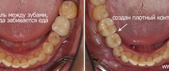
Caries on the side of the tooth, near the gum, belongs to a particularly dangerous type of caries. This type of caries destroys the tooth, starting from its base. Destruction of the deep layers of the tooth and massive damage to the dental canals are characterized by an increased destructive nature.
Despite the absence of pronounced distinctive features, the consequences resulting from caries on the side of the tooth near the gum are much more serious for the condition of the oral cavity than with other types of caries. That is why it is so important to make a timely visit to a specialist - a dentist.
Consultation with an orthodontist
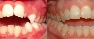
Parents came to the reception with a 12-year-old teenager. The boy's problem is that the fang does not grow in the same row with his teeth, but in front of the entire row. This does not look very nice, and also leads to poor hygiene on this side, since it is difficult to properly clean unevenly growing teeth. The teenager was treated by a district orthodontist in a public clinic for 1 year. The calculation was then made only from a panoramic image. The treatment was carried out using a plate, which was supposed to move the upper row of teeth apart to make room for the canine and, with the help of the anterior arch, direct it in the desired direction. Also, the goal of the district orthodontist was to correct the distal occlusion by changing the inclination of the upper anterior teeth. Over the course of a year, the upper row of teeth was moved apart by a plate to the point of extreme closure of the lower and upper teeth, but this did not provide enough space. Since the parents did not like the situation with the canine, and the previous orthodontist somehow doubted whether to make a new plate or put on braces, and whether it was necessary to remove the fours, the parents came with their son for a consultation with the orthodontist at Olga Baranova’s ORTHODONT PROJECT on the recommendation of friends.
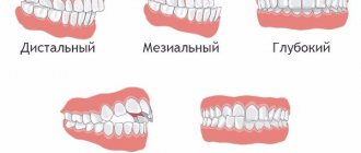
Upon examination, she indicated an incorrect bite, too straight position of the front teeth, crowding of teeth in both the upper and lower dentitions. In addition to simply meeting the upper and lower teeth, the cusps of the upper teeth should fit between the cusps of the lower teeth, forming a tight row. On one side, the child’s teeth did not close at all (there was a gap left), and on the other, the cusps rested on the cusps – which is wrong.
Parent questions:
- Do I need to remove teeth to make room?
- Should I make a new plate or put on braces?
- Should I put on braces now, or wait until the last chewing teeth grow in, the child becomes more conscious and brushes his teeth better, and is less shy?
- How long will the treatment take?
The orthodontist explained that it is quite possible to do without removing the fourth chewing teeth. Treatment on a plate will not give the expected result, since it is necessary to work on both the upper and lower jaws. Treatment should not be postponed, since the “sevens” are now emerging, there is potential for growth, and it will be easier to move the teeth. braces , both external and internal, if you want to make the treatment invisible. The approximate duration of orthodontic treatment is about 2 years, maybe less, but it’s difficult to say without additional calculations.
The parents and the teenager agreed to start treatment with braces at Olga Baranova’s ORTHODONT PROJECT. Having received a list of necessary examinations and consultations, they began preparing for treatment.
Video recording of the consultation (the recording is published with the consent of the parents):
Clinic doctors
To enable chewing teeth to perform their function, they have cusps and depressions in the tooth enamel (fissures). These formations form the relief necessary for grinding food. The depth of the grooves is approximately two and a half to three millimeters, but over time they become larger.
Types of fissures on the surface of teeth
Depending on the anatomical structure, there are four types of depressions on the enamel of teeth:
- Funnel-shaped.
Compared to other species, they are more open and, thanks to the access of saliva, are better mineralized. The shape itself with a rounded bottom helps wash away food debris.
- Cone-shaped.
The level of mineralization is lower and the likelihood of food particles accumulating is higher, so it is important to carefully monitor the quality of cleaning.
- Teardrop-shaped.
Such depressions on the chewing surface of the teeth are characterized by a narrow entrance and a wide bottom, which is why they are poorly mineralized and inaccessible for cleaning.
- Polypoid.
Their features are similar to teardrop-shaped cavities, but have an elongated shape.
Carious lesions in the grooves of tooth enamel
Since the grooves and grooves on the teeth present difficulties for hygiene, plaque accumulates there. And this is an excellent breeding ground for pathogenic bacteria. Microbes cause the appearance of carious cavities on the tooth enamel.
This type of caries is called fissure caries. It is not easy to recognize, especially at an early stage. Pronounced dark spots appear when the process is quite advanced. As the disease progresses, the lower layers of tissue are destroyed. Then another sign appears: the depressions on the tooth enamel and in the dentin layer become sensitive to the effects of cold, hot and sweet.
Treatment of depressions in tooth enamel
As long as the enamel is not seriously damaged, the process can be stopped with the help of fluoride and calcium-containing preparations, as well as sealants. In more complex cases, treatment of depressions in tooth enamel and dentin requires preparation and filling:
- the affected tissues are drilled out;
- the cavity is treated with antiseptics;
- the tooth is filled and restored;
- artificial material is brought to smoothness and shine.
Restoring the anatomical shape involves reproducing the tubercles and depressions on the tooth enamel for the full functioning of the organ.
Sealing fissures on the surface of teeth
This procedure is mainly performed on children and consists of sealing the grooves with special fluid materials. Before filling the fissures on the surface of the teeth with sealant, the doctor thoroughly cleans them. A brush, paste, sandblaster or ultrasound can be used for this. If necessary, the grooves are slightly enlarged to ensure that there is no plaque left inside.
The next steps are treatment with a solution of phosphoric acid and filling the recesses on the surface of the teeth with a sealing agent. The material hardens under the light of a curing lamp.
The final point is grinding and polishing. The resulting protective layer remains on the teeth for five to ten years.
Prices for fissure treatment depend on the severity of the problem and the technique used. In general, sealing is less expensive than preparation followed by filling.
Preparing for treatment
A panoramic photograph of the teeth was taken, an X-ray of the skull in a lateral projection (TRG-lateral), and impressions of the teeth were taken. The teenager underwent consultation with an osteopath, speech therapist, and pediatric dentist. Photometry was taken - the face and teeth from different angles.
Initial situation:
At a consultation with an osteopath , the osteopath did not identify any significant problems regarding the musculoskeletal system that could affect the bite. A small correction is proposed after each visit to the orthodontist in order to compensate for the influence of orthodontic equipment - to relieve tension in the bones of the skull, temporal joints, and neck.
At a consultation with a speech therapist , the condition of the muscles of the lips and tongue was tested. A decrease in tongue muscle tone and improper swallowing were revealed. A list of exercises is given that you can perform independently to train the muscles of the tongue and neck.
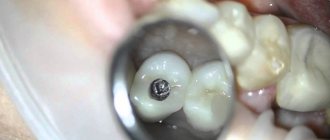
A consultation with a pediatric dentist revealed caries on one of the teeth, which must be cured before orthodontic treatment .
Consultations with specialists are provided free of charge. A feature of the treatment at Olga Baranova’s ORTHODONT PROJECT is an integrated approach to treatment - the interaction of different specialists aimed at eliminating all negative factors that disrupt the position of the teeth, which gives more stable results of orthodontic treatment .
The results of all consultations and examinations were sent to the orthodontist . The doctor carried out the necessary calculations, and on the second visit she explained the treatment plan, and also offered several braces to choose from.
The Damon self-ligating bracket system was chosen. In this bracket system, the braces are small in size with rounded edges. braces on the upper front teeth , which are less noticeable to others. It is better to use Damon Q metal braces to protect the upper teeth from excessive wear. When smiling and talking, the lower teeth are covered by the lip, and the aesthetics are not greatly affected.
After the brace system was approved, a treatment contract was signed and the cost of the brace system was paid. Payment for treatment can be made in full or in stages - half when fixing braces
and the balance in equal parts at each visit.
To maintain the health of the teeth while wearing braces, before fixing the braces as a gift, hygienic preparation of the teeth , including removing plaque using the Air Flow method, removing tartar with ultrasound, and polishing the surface of all teeth. The hygienist explained on a model the rules for brushing teeth with braces with a special orthodontic brush, showed how to use dental brushes, a single-tuft brush and special dental floss to clean braces and interdental spaces.
The next step will be fixing the braces , which is the beginning of orthodontic treatment.
Start of orthodontic treatment
Before installing braces, the patient must undergo professional teeth cleaning by a hygienist. The specialist cleans the teeth, removes all plaque (soft and hard), checks the condition of the gums, polishes the enamel and gives recommendations on how to care for your teeth while wearing braces. Patients who decide to start treatment with braces receive a starter hygiene kit, which includes an orthodontic brush, toothpaste, brushes, dental floss, monobrush, and wax.
Damon metal braces are fixed:
Treatment with braces at this stage is aimed at expanding the dentition in the areas of the 2nd and 4th teeth (special springs are installed on the brace system).
After 5 months, the next stage begins - the return of the fangs to the dentition. The second arch holds the anterior incisors in a stable position.
Stage of wearing orthoelastics (12 months after fixing braces):
The structure of the fangs
The canines of the upper jaw are shaped like an irregular cone. The cutting edge is shaped like a triangle, which is limited by three teeth - a pronounced middle one and two outer ones. The area of the tooth facing the lip (vestibular) has a convex shape and gradually approaches the lingual.
On the lingual part of the tooth there is a longitudinal ridge that divides the dental crown into two facets: the lateral part has the largest area. The upper canine has a pronounced crown curvature and root deviation.
The root of the tooth is quite thick and has a shape slightly compressed from the sides. Wider in anteroposterior dimension. The lateral and medial surfaces are very wide and have barely noticeable grooves.
The tooth has one, wide enough and accessible if treatment is necessary, root canal.
The canines located on the lower jaw are smaller in size than the upper ones, and the shape of the crown is close to the upper lateral (side) incisors. They are slightly narrower and longer than the upper canines. The medial surface is in a straight line with the root line. The cutting edge is less pronounced. The distal part of the cutting edge is longer than the medial one.
The labial surface of the lower canines is less convex and pronounced than that of the upper ones, and the lingual surface can be either straight or slightly concave.
The lower fangs have a slightly shorter tooth root than the upper ones, but they have absolutely no differences in shape. In rare cases, anatomical pathology occurs in which the root is divided into two parts.
The canines of both jaws have quite pronounced external differences in the dental crown, but at the same time the dental cavities are very similar.