15 August 2020
17221
0
2.3 out of 5
Herniated discs are one of the most common diseases that almost every person is at risk of encountering. However, they cannot be called harmless, since they can lead to the development of severe complications, including paralysis and disability. In addition, intervertebral hernias significantly reduce the quality of life, as they cause severe pain and disturbances in the sensitivity of various parts of the body, which can completely deprive a person of his ability to work and enjoy life. Therefore, when the diagnosis is made and the presence of a hernia is beyond doubt, it is necessary to begin treatment as early as possible in order to avoid the unpleasant consequences of the disease.
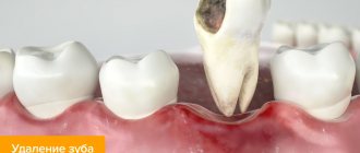
Resorption due to tooth extraction
After the removal of a tooth or several adjacent teeth, an area of edentulous tissue forms on the jaw.
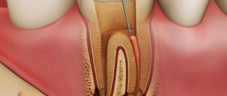
In the edentulous zone, the process of bone atrophy begins. Many patients in dental clinics, if they had been warned in advance that after the installation of removable and fixed dentures, bone tissue atrophy would continue, they would have chosen a different method of treatment and dental prosthetics. Bone resorption, also called atrophy, bone loss, dystrophy, is an irreversible process, but it can be prevented if you choose the right method of dental prosthetics. At the Nurdent clinic in the Kalininsky district, dentists recommend that their patients begin dental implant surgery as soon as possible after tooth extraction, so that the process of bone resorption does not go too far. Our dentistry near the Grazhdansky Prospekt metro station employs experienced orthopedic dentists who are ready to help the patient make the right decision about choosing the method of dental prosthetics.
Our doctors
18 years of experience
Baghdasaryan
Armen Evgenievich
Chief physician, dentist-orthopedist-therapist
Graduated from VSMA named after. N.N. Burdenko. Internship on the basis of MGMSU named after. A.E. Evdokimov in “General Dentistry”.
Clinical residency at the Moscow State Medical University named after. A.E. Evdokimov in “Orthopedics”.
More about the doctor...
5 years experience
Sadina
Ekaterina Vladislavovna
Dental therapist, surgeon
Penza State University Medical Institute, specialty “Dentistry”.
In 2021, she underwent professional retraining in the specialty “Therapeutic Dentistry” at the Moscow State Medical and Dental University named after A.I. Evdokimov.
More about the doctor...
8 years of experience
Arzumanov
Andranik Arkadievich
Dentist-orthodontist
Graduated from Moscow State Medical University. Internship - Moscow State Medical University at the Department of Orthodontics and Children's Prosthetics.
Residency at Moscow State Medical University at the Department of Orthodontics and Children's Prosthetics. Member of the Professional Society of Orthodontists of Russia since 2010.
More about the doctor...
Ways to Prevent Bone Dystrophy
To stop the resorption process, it is necessary to let the body know that the tooth root has returned to its place. This can be done in the only way: to perform a dental implantation operation by installing a titanium implant in place of the missing root. The implant, replacing the tooth root, takes over its function and stimulates the jaw bone, stopping bone resorption, which inevitably begins after tooth loss. Implants allow you to restore the functionality of the tooth, providing a natural process of chewing food. The visible part of the tooth is replaced with an abutment, which serves as a support for the crown.
Interdisciplinary reception
It often happens that malocclusions are much more complex and treatment with braces alone is not enough.
Often, treatment, for one reason or another, is delayed or does not go according to plan, or is initially planned incorrectly, which creates a lot of problems physically and psychologically for both the patient and the doctor. Therefore, I strongly recommend that you undergo a detailed diagnosis before orthodontic treatment. Calculation of casts, images, CBCT analysis, determination of central occlusion and central relationship of the jaws. According to the indications, axiography. This will allow you to make an accurate diagnosis. Accurate diagnosis = correct treatment plan. The orthodontist must have the skill, knowledge and ability to conduct interdisciplinary treatment. Your treatment will be monitored simultaneously by the orthodontist and related professionals: orthopedist, surgeon, gnathologist, periodontist, therapist, endodontist, and, if necessary, an osteopath. If necessary, during orthodontic treatment, a competent orthodontist with interdisciplinary experience will attract the necessary specialists to provide timely assistance. This work in conjunction allows you to speed up treatment and avoid complications. Treat previously undetected caries in a timely manner, rather than wait for further tooth destruction under braces until removal. Or, for example, if there are periodontal problems, then your doctor will treat in accordance with the treatment protocols of the periodontist, constantly communicating with each other.
Patients with TMJ disease must first undergo a full diagnostic with axiography, and, if necessary, therapeutic and diagnostic splint therapy from a gnathologist. This therapy allows you to put the joint in the correct position. This correct position may not seem entirely comfortable to the patient; the teeth may touch at completely different points than before. But it is only from this functional position (in case of TMJ pathology) that it is possible to begin orthodontic treatment, so that incorrect movements do not cause more harm to an already suffering person.
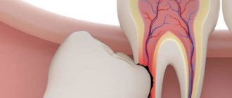
What is the danger of bone resorption in the frontal zone of the jaw?
The area where the incisors are located is a very thin bone, the degeneration of which occurs very quickly. Even if you install a bridge using, for example, fangs as a support for the “bridge,” after a while the gums will begin to collapse due to loss of bone, and the teeth will visually lose support. If the lost incisors are restored as a result of implantation, the implant replacing the root seems to give a command to the body that the tooth is in its place and resorption is not needed.
Treatment Options
Intervertebral discs are designed to protect the vertebrae when walking and other loads. They are a kind of flat capsule, inside of which there is a gel-like nucleus pulposus. The role of the outer shell is performed by the fibrous ring, and the endplates protect the intervertebral disc above and below.
Over the years, they wear out and become dehydrated, and regeneration processes slow down due to the diffuse method of nutrition (intervertebral discs do not have their own blood vessels). As a result, osteochondrosis develops. Against the background of progressive degenerative changes, the fibrous ring becomes unable to withstand the loads placed on it and becomes deformed. As a result, the disc protrudes, i.e., a protrusion is formed, and then a hernia. Such protrusions of the disc can compress the nerve roots passing in close proximity to it. The result of this will be irradiation of pain from the back to the legs, arms or disruption of the innervation of internal organs, which will cause the development of organic changes in them.
Depending on the size, the patient’s well-being, and the danger of the hernia from the point of view of the occurrence of severe neurological disorders, there are 2 main methods of treatment: conservative and surgical. Although surgery can radically solve the problem in the shortest possible time, many patients are in no hurry to go to the neurosurgeon’s table, fearing possible risks, and try to improve their condition using conservative techniques.
Non-surgical treatment of intervertebral hernia involves:
- the use of various drugs in the form of oral agents, injection solutions and all kinds of ointments or gels;
- physiotherapeutic procedures carried out in a special room at a medical institution with a certain frequency (often every other day): laser therapy, magnetic therapy, etc.;
- PPR is a method that involves injecting the patient into the affected area with his own purified blood plasma, in which the concentration of growth factors significantly increases (it has controversial effectiveness, and its effect on the body is not fully understood);
- massage and manual therapy sessions;
- daily physical therapy exercises, which at first also need to be performed under the supervision of a specialist;
- wearing orthopedic bandages, etc.
It is prescribed for at least 3 months, and often for a much longer period of time. At the same time, not only a doctor is not able to give a 100% guarantee that conservative treatment will bring a noticeable effect and will help the patient really get rid of pain and neurological disorders, and not temporarily suppress them with medications. Therefore, there are often cases when, when performing control MRIs to assess the effectiveness of conservative therapy, there are no significant changes in the condition of the intervertebral disc or even signs of a worsening of the situation are observed.
During conservative therapy, hernias may increase in size, which will ultimately require complex surgical intervention, whereas in the early stages of the development of the disease it could be easily dealt with using minimally invasive or percutaneous methods.
At the same time, the total cost of conservative treatment of intervertebral hernia can reach colossal amounts. We must not forget about the significant loss of time for carrying out all procedures and the patient’s labor costs, which, given the lack of guarantees of obtaining the desired treatment result, does not provide very good prospects.
This state of affairs does not suit either doctors or patients, which required the development of a new approach to the treatment of intervertebral hernias, such that it would be as safe as possible for the patient and at the same time allow the pathological protrusion of the disc to be quickly eliminated. The result was the creation of a method for mechanical resorption of hernia.
Bone atrophy of the lateral areas of the jaw
When molars are lost, a cosmetic defect such as sagging of the facial skin due to loss of bone tissue is observed. Another unpleasant complication can be considered a change in bite: in the front zone of the jaw, the teeth begin to “move apart”, the height of the face changes due to a decrease in the height of the jaw bones. A removable bridge prosthesis installed in the lateral area of the jaw not only does not stop the process of bone resorption, but also accelerates it. This is due to the fact that during chewing the denture puts pressure on the gum. To avoid strong pressure from the “bridge”, prosthetics should be carried out using a bridge-like prosthesis installed on implants. This way you can stop the change in bite and eliminate the risk of “sagging” of the face.
Treatment of extensive external and internal root resorption with MTA
Internal root resorption is usually asymptomatic and very rarely occurs in permanent teeth. Such lesions are usually discovered incidentally on targeted x-rays in the form of a single round or oval radiolucent enlargement of the canals and pulp space. The edges are smooth and clearly defined with a violation of the length of the canal. This resorption is characterized by a progressive loss of tooth tissue, starting from the root canal wall, sometimes reaching perforation.
There are many etiological factors, but the most common causes are infection, trauma, tooth fracture or treatment. Resorption processes can develop when the pH level shifts to the acidic side, during infection of the pulp, which leads to the dissolution of dentin and enamel structures by chelates. In cases of trauma, intrapulpal bleeding with the formation of clots is replaced by granulation tissue with giant multinucleated cells that resorb dentin. Very often, the cause of internal resorption is unified and classified as “idiopathic internal resorption.”
Untreated internal resorption can progress to external resorption or cause tooth fracture. Perforations are also a consequence of internal resorption. However, early diagnosis and treatment of such pathology is still possible. Complete removal of resorbed tissue from the root canal is essential to prevent long-term structural loss. However, endodontic treatment with cleaning, shaping and obturation of the canal with suitable material in such cases remains difficult, especially if the tooth damage is extensive. In many cases, removal is the only solution.
MTA was originally developed for such purposes, since conventional material did not have the necessary characteristics. MTA is also recommended for pulp capping, pulpotomy, open apex apexification, perforation treatment, and root canal filling. MTA has satisfactory characteristics including biocompatibility, radiopacity, difficulty in impregnation, high biological properties due to hydroxyapatite, high fracture resistance, resistance to compressive forces and microhardness.
Clinical case 1
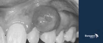
A targeted radiograph taken in 2009 of the asymptomatic upper incisors of a 14-year-old girl with a history of trauma revealed extensive internal and external resorption involving both upper central incisors (Figure 1).
Figure 1: Radiograph showing extensive external and internal resorption of the maxillary central incisors in a 14-year-old girl.
Initially, both teeth were planned to be removed due to extensive tissue loss. However, given the good periodontal condition and the patient's young age, the possibility of retaining 11 and 21 until the age of 18 was considered in order to replace the affected teeth with implants.
The teeth are isolated using a rubber dam without applying a clamp to avoid the possibility of horizontal fracture. Pulp tissue was removed through coronal access. The large communication between the resorbed cavity and the periodontium looked like an area of hemorrhage. After determining the working length, the canals are prepared with a size 70 H-file.
The apical stop was created using a 140 H-file using the step-back technique. All procedures were performed under copious irrigation with fresh 5.25% sodium hypochlorite solution (Farmacia Amazon, Brazil). Calcium hydroxide paste (Calen, SSW, Brazil) was applied to create an alkaline environment, dissolve remaining pulp fragments and control bleeding at the perforation.
After 7 days, the liner material was removed using irrigation with a solution of 5.25% sodium hypochlorite. The root canals were washed for 3 minutes with an EDTA buffer solution pH 7.4 (Odahcam, Dentsply, Brazil) with agitation with an instrument. Then MTA is mixed with sterile water until a grainy, sandy mixture is obtained. The resulting material is condensed into an open apex using a hand plugger, Hu Friedy #11. MTA, being hydrophilic, requires moisture, so absolute dryness of the field is not only not necessary, but also contraindicated. After this, a transparent plastic pin of size 3 (Luminex post, Dentatus, USA) coated with Vaseline was inserted into the canal lumen. After 24 hours, the pin is removed and the opened space is filled with gutta-percha (Dentsply Maillefer Switzerland). The filling was performed within 3 mm of the apex using a Pexit Plus sealer (Ivoclar Vivadent AG, Liechtenstein). All excess filling material was removed from the pulp chamber, which was then obturated with cement. The crown was finally restored with composite material.
Immediately after filling in February 2009, an x-ray was taken (Photo 2), confirming satisfactory filling of the root canals and resorptive defects. Clinical and radiological monitoring was carried out 24 months after treatment (Photo 3), demonstrating the functionality of the teeth without endodontic pathology. Despite the stability of the teeth and their functionality, discoloration has appeared to a gray tint. Composite veneers were made for the patient.
Photo 2: X-ray after treatment
Photo 3: After 24 months
Clinical case 2
A targeted x-ray of an asymptomatic maxillary right central incisor in a 26-year-old man with a history of trauma performed in June 2009 revealed extensive internal root resorption (Figure 4).
Photo 4: Pre-intervention X-ray: extensive resorptive process in the upper right central incisor in a 26-year-old man
The tooth is isolated with a rubber dam without a clamp. Pulp tissue was removed through coronal access. The small communication between the resorbed cavity and the periodontium looked like an area of hemorrhage. After determining the working length, the canal is prepared and obturated similarly to the case described above.
Ultimately, the crown was restored with composite. After treatment, an x-ray was taken to confirm satisfactory filling of the root canal and resorptive defect (Photo 5). After 2 weeks, the crown was made (Photo 6).
Photo 5: X-ray immediately after treatment
Photo 6: Vestibular view 1 month after crown fixation
Clinical and radiological examination after 24 months in February 2001 (Photo 7) and demonstrated the functionality of the tooth without endodontic pathology.
Photo 7: X-ray after 24 months
Photo 8: View 24 months after treatment
Clinical case 3
A targeted x-ray of an asymptomatic maxillary right central incisor in a 23-year-old woman with a history of trauma revealed extensive internal root resorption (Figure 9). The tooth is isolated with a rubber dam without a clamp. Pulp tissue was removed through coronal access. The large communication between the resorbed cavity and the periodontium looked like an area of hemorrhage.
Figure 9: Pre-treatment X-ray showing extensive internal root resorption in the upper right central incisor
After determining the working length, the canal is passed through a 25 size H-file. The apical stop was created using a 40 H-file using the step-back technique. All procedures were performed under copious irrigation with fresh 5.25% sodium hypochlorite and piezo ultrasound. Calcium hydroxide paste (Calen, SSW, Brazil) was applied to create an alkaline environment, dissolve remaining pulp fragments and control bleeding at the perforation.
After 7 days, obturation was carried out exactly according to the scheme with the previous two cases. The crown was restored with composite. An x-ray was taken (Photo 10), confirming satisfactory filling of the root canal and resorptive defect. After 2 weeks, the crown was made. A clinical and x-ray examination was carried out 38 months later in February 2011 (Photo 11), demonstrating the functionality of the tooth and the absence of pathology in the endodontic tissues.
Photo 10: X-ray immediately after filling
Photo 11: 38 months after treatment
Discussion
The prognosis for internal resorption is very favorable in cases where resorption has not affected the majority of the tooth volume. However, with the use of high-quality reinforcing materials, even a tooth with a large defect can be saved.
Treatment consists of complete removal of all affected tissue. Several materials have been used to fill resorptive defects: hydrophilic polymers, zinc oxide eugenol paste, zinc acetate cement, amalgam alloy, and thermoplastic gutta-percha injected or condensed. Some of these materials are not able to impart the required strength to the tooth. For the cases described, MTA was used due to its obstructive and reparative qualities, as well as mechanical strength. It is made of hydrophilic particles that harden in the presence of liquid and blood. The only disadvantage of MTA is the difficulty of its removal during repeated endodontic treatment, as well as the placement of a pin. To prevent these undesirable effects, the central segment is obturated with gutta-percha.
The exact mechanism of root enhancement by MTA remains unclear. There is very little material available to study the qualities of MTA.
There is no reliable data on the issue of MTA fillings and crowns. Further research is needed to clarify this.
Teeth change their shade after using MTA, so after working with this material, as a rule, a crown is required. Further research is required to overcome this shortcoming.
Conclusion
MTA can be successfully used to mechanically strengthen weakened teeth and treat internal root resorption.
Author: Salma BA Abdo
Completely edentulous
When all teeth are lost, the resorption process accelerates significantly. And in addition, there is a change in the structure of the jaws: the muscles slightly change the places of attachment to the bone. An unnatural sagging of the lips begins, and wrinkles appear. Changes in the structure of the jaws lead to disruption of chewing function, which negatively affects the health of the entire body, since a person cannot eat properly. Due to impaired chewing of food, the process of its digestion changes. This is the direct relationship between missing teeth and human health in general.
With complete edentia, some patients choose removable dentures for both jaws. But since prostheses do not stop the process of bone resorption and even accelerate it, after some time it is necessary to adjust the size of the prosthesis, changing its thickness, in order to compensate for the loss of bone tissue. However, if you install a removable bridge supported by implants, the bone crest of the gums is preserved, since the pressure of the “bridge” on the gums is eliminated. In the absence of tooth roots, resorption continues, but its rate slows down.
Etiology of the disease
The most common reasons for the development of resorption are:
- Injuries to dental tissues;
- Death of pulp tissue;
- Periodontal diseases and treatment;
- Whitening teeth that have undergone endodontic treatment.
There are also a number of reasons that are less common. These include compression created by:
- ectopically erupted teeth;
- neoplasms of benign and malignant nature;
- cysts.
The pressure created by them leads to damage to the periodontal ligament and cement, which protect the tooth root from resorption.
In our clinic you can get a free dental consultation!
Content:
- A little about baby teeth
- How does resorption of the roots of baby teeth occur?
- Factors influencing resorption
- Types of resorption
- Resorption disorders 5.1. Causes of premature resorption 5.2. Why is resorption delayed?
When speaking about the resorption of baby teeth, dentists mean the natural process of resorption of their roots. This is necessary to ensure that temporary units drop out in a timely manner and make room for permanent ones. It is thanks to the resorption of the bone bases that when examining in detail the fallen children's fangs, molars and incisors, it seems that they have no roots.