6763
Increasingly, people turn to dental clinics with problems regarding the appearance of their teeth. Patients are concerned not only about changes in the shade of the enamel, but also about its deformation.
The main cause of such manifestations is considered to be dysplasia of enamel and dental tissues, which not only reduces the aesthetics of a smile, but can also lead to complete tooth destruction.
What it is?
Enamel dysplasia is a group of diseases of non-carious origin, characterized by abnormal development of dental tissue.
This pathology is congenital, developing during the intrauterine development of the fetus, or is provoked by genetic factors.
Abnormal tissue development begins during the formation of primordia. Symptoms of the disease are not age specific . They can appear both during the period of first teething and in adulthood.
Dysplasia can affect the entire dentition, but is most often localized in its anterior section. Violation of integrity with the same intensity is observed in all segments of the tooth: cement, dentin, enamel.
Photo: tooth enamel dysplasia in a child
How does it manifest?
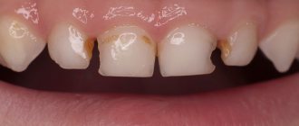
This pathology has certain signs that allow it to be differentiated from other diseases affecting the enamel:
- change in the color of the enamel, which takes on a gray tint;
- thinning of the enamel, which results in increased tooth sensitivity;
- partial or complete absence of the enamel layer ;
- uncharacteristic shape of the cutting part, which includes many notches and chips;
- heterogeneity and surface deformation. Most often, longitudinal grooves and cracks are found on the enamel during visual inspection, and ribbing is detected during instrumental examination;
- the appearance of irregularly shaped white spots , which darken as the disease progresses. They first turn yellow and then turn brown;
- symmetrical arrangement of spots on the teeth of the same name in one jaw;
- surface uniformity in the spot area , here the enamel is dense and smooth, but over time it becomes dotted or grooved;
- the affected area is not stained with caries markers ;
- in the area of the pathological spot, tissue loss , leading to their gradual destruction;
- Pain syndrome may due to exposure to local irritating factors. Once the source is eliminated, the pain disappears.
Due to the disruption of dentin formation, the teeth take on an irregular shape: cone-shaped, barrel-shaped, pear-shaped.
Who is at risk?
Among the provoking factors are:
- late toxicosis or pathologies that caused a pronounced disturbance of metabolic processes during pregnancy;
- endocrine diseases (affecting metabolism) during the active growth of a child in the first years of his life;
- delayed reaction to taking certain medications;
- inappropriate living conditions;
- wrong approach to feeding a child.
Symptoms
In the initial stages of the disease, it is impossible for a non-specialist to recognize it. All irregularities can only be identified by a dentist during a special examination. After thoroughly drying the surface. Only in this way will he be able to see the hidden spots, irregularities and grooves.
As the pathology develops, it manifests itself with the following symptoms:
- atypical lightening or darkening;
- the formation of yellowish, creamy or milky spots with clear contours - this is similar to the initial stages of caries;
- pain occurs on contact with sour, cold and hot;
- there is pain when exposed to air.
With this pathology, the enamel layer will always be smooth in the affected areas. Its peculiarity is that it does not give any reaction to special dyes.
Why is it dangerous?
Hypoplasia has only cosmetic disadvantages and causes inconvenience to the child by increasing the sensitivity of the enamel layer. Over time, serious complications appear.
These include:
- rapid development of deep caries - its development is due to the fact that it is easier for microorganisms to infect teeth with defective enamel;
- deep tissue damage by the degenerative process of dental tissue, which over time spreads to nearby areas of dentin and pulp;
- Often children subsequently develop a malocclusion.
For these reasons, treatment of hypoplasia must begin as early as possible, without waiting for the situation to worsen.
Classification
When diagnosing pathology, the dentist relies on a certain classification, which distinguishes several types, depending on the area and nature of the localization.
There are:
- Root. The enamel surface is not damaged, but has a slight deviation in color.
Dentinal canals are present in small quantities, obliterated into the tooth cavity and have a crescent shape.The roots of the affected teeth are shortened and narrowed, which leads to their premature loss. The surface of the root part is dysplastic.
- Coronal .
It is characterized by a change in the color of the enamel, which gradually acquires a yellow tint. In the area of the coronal part, pathological abrasion of the enamel is observed, which begins to expose dentin. Most often, pathological areas are located in the central part of the tooth or closer to its cutting surface. - Odontodysplasia .
It is characterized by severe thinning of both enamel and dentin. Pathology can equally affect both temporary and permanent teeth. Most often, teeth are irregularly shaped and smaller in size. In some cases, denticles are found in the cavity.
Another 3 types are distinguished by the nature of enamel lesions:
- Spotted .
It is expressed in the appearance of white round spots on any surface of the front and lateral incisors. The spots have clear boundaries and a shiny surface. Over time, it becomes rough. The thickness of the tissues of the affected area does not differ from the thickness of healthy enamel. In most cases, the spots do not change throughout life. - Erosive .
It is distinguished by cup-shaped or oval-shaped depressions formed as a result of damage to the enamel. The recesses are characterized by their overall width and depth. The bottom of the affected area is covered with thinned enamel, through which dentin is visible. Further development of the pathology leads to complete loss of enamel in the area of thinning. - Sulcata .
With this form of the disease, grooves are formed on the vestibular surfaces of the teeth. The groove is located parallel to the cutting part of the teeth. In the area of the furrow, the enamel is thinned or completely absent.
For those who should choose Pilot metal braces, read in a separate publication.
In this article we will talk about the causes of tooth enamel hypoplasia in children.
Here https://orto-info.ru/zubocheliustnye-anomalii/okklyuzii/perekrestnyiy-prikus.html we will discuss whether braces are effective for crossbite.
Diagnostics
For a qualified dentist, recognizing the disease is not difficult. Damage to the enamel layer has characteristic clinical manifestations that are easily determined visually.
For this purpose:
- examination with drying of the tooth surface (particular attention is paid to the bottom of the formation, it should not be rough);
- counting the number of formations, they must be multiple;
- staining the affected areas with methylene blue, while the affected areas are not stained.
Differential diagnosis of pathology is carried out with carious lesions at the initial stage.
Causes
three main factors that cause enamel dysplasia :
- Diseases of a hereditary nature .
Most often, they lead to darkening of the enamel and its thinning. Basically, pathology begins to appear in children already on their baby teeth. As a result of the pathological process, it becomes thinner over the entire surface, leading to dentin exposure. In hereditary diseases, tooth enamel is yellow immediately after eruption. - General diseases, occurring in an acute form, leading to disruption of the metabolic processes of mineral components.
As a rule, due to such reasons, dysplasia can be focal in nature and affect single specimens.This pathology is most often complicated by carious processes, which are characterized by rapid progression.
- Pathologies of the skeletal system , leading to disruption of the process of tooth formation due to a lack of bone-forming elements. Such diseases include rickets, periosteal dystrophy, marble disease.
Crooked baby teeth
Baby teeth serve a child for only 6–8 years, so their position does not always require correction, however, the state of the baby occlusion can be used to judge how correct the eruption of permanent teeth will be.
Gaps between baby teeth are normal, but rotation and crowding are no longer the case.
Rotation of baby teeth
The same applies to the bite: it should not be asymmetrical or open, the lower teeth should not completely overlap the upper ones. In such a situation, you need to consult an orthodontist.
The doctor may determine:
- Why did the disorder occur at such an early age? Are bad habits to blame (for example, feeding from a bottle at an age when a child should already eat with a spoon), poor posture, some congenital characteristics, etc.
- How can this be corrected? Refer the child to an orthopedist for additional examination or simply advise parents on how to organize nutrition.
- When should you be examined again? Follow-up in orthodontics is very important; appointments are usually recommended every 6–12 months. Then you can see in dynamics how the condition of the teeth changes.
Consequences
Almost all types of dysplasia are characterized by complications and serious consequences.
If left untreated, dysplasia leads to at least an aesthetic defect . In more complex cases, dentin is exposed and infections occur.
Such teeth are most often affected by caries, which quickly reaches the pulp chamber, provoking purulent inflammation. In this case, not only hard tissues, but also adjacent soft tissues can be damaged.
Teeth affected by dysplasia are fragile and susceptible to easy chipping, which disrupts their integrity and shape.
In most cases, without appropriate therapy, the pathology leads to complete destruction of the tooth or its severe loosening, which is an indication for removal.
Treatment of amelogenesis imperfecta
Unfortunately, it is not possible to eliminate the hereditary factor that determines amelogenesis imperfecta. Therefore, treatment of amelogenesis imperfecta is aimed at preserving teeth for as long as possible. As a rule, tooth restoration with crowns is used. If necessary, the bite should be corrected to reduce tooth wear. The correct selection of toothpaste will help reduce hyperesthesia and improve oral hygiene.
Meet our tooth fairies and check prices.
Remedies
Several types of methods are used to treat dysplasia. Some of them are corrective in nature, others are aimed at solving the problem by general strengthening of dental tissue .
Treatment methods are selected depending on the extent of the lesion and the intensity of the symptoms.
You will learn about the features of treatment of dentin dysplasia from our next review.
In the next article we will talk about the pros and cons of Clarity Sl self-ligating braces.
At this address https://orto-info.ru/zubocheliustnye-anomalii/zubov/formyi/konicheskie-i-shilovidnye.html you will find information about the sequence of treatment for spiky teeth.
Therapeutic
This group includes treatments aimed at visually correcting the appearance of enamel .
The following methods are used for this:
- Composite restoration. It is used for small foci of dysplasia. The procedure is performed under local anesthesia with preparation of damaged areas.
After preparing the cavities and the surface of the tooth, it is treated with an adhesive agent and a light-curing composite is applied. Most often, Consize and Evicrol are used for this.These materials allow you to accurately convey the appearance and structure of healthy enamel. They are highly durable and do not stain over time.
- Veneers. For extensive lesions, it is recommended to install veneers. They are thin ceramic plates that accurately replicate the relief of the vestibular and incisal parts of the teeth.
Before fixing the veneers, a small grinding is carried out, which is necessary to visually reduce the volume. The overlays are installed using special glue or conventional composite.In appearance, they are no different from the surface of real teeth and protect thinned enamel from the aggressive effects of external factors.
- Prosthetics with crowns .
Used in cases of volumetric destruction. Either single crowns or bridges are used, which are made from individual casts.
For one of the methods for eliminating enamel damage, watch the video:
General strengthening
General restorative treatment is indicated for minor manifestations of dysplasia in the spot stage, characterized by gradual modification.
Therapy consists of remineralization and includes the use of the following drugs :
- klamin _ Used orally, 1 tablet per day for a month. The drug must be taken 20 minutes before meals.
- calcium glycerophosphate . Just like the previous remedy, it is indicated for use for 1 month. The product should be taken no more than 1.5 g per day;
- multivitamin complex, for example, Kvadevit or Complivit. Take no more than 2 tablets per day;
Also prescribed:
- electrophoresis with a solution of calcium glycerophosphate . Conduct at least 10 sessions 3 times a year;
- enamel treatment with sodium fluoride.
How to treat
Dental hypoplasia today is considered a process that cannot be cured or reversed. There are no medications that can relieve symptoms. As for the treatment of dental hypoplasia, it is usually symptomatic. It is also possible to reconstruct the tooth enamel.
If the lesions are small, then you can do nothing, since the child or teenager’s teeth will not hurt. If there is erosion on the enamel, or there are deep stains, then the tooth needs to be filled using composite materials.
If there is no enamel on the surface of the teeth, the doctor may prescribe orthopedic treatment or prosthetics. But it is very important to remember that if such a disease or symptoms are identified, it is important to initially consult a specialist. You cannot self-medicate, otherwise serious consequences may occur.
Prevention of fluorosis
The only possible way to prevent and prevent further development of fluorosis is to stop drinking water with a high fluoride content; in case of severe air pollution, move to another area. Fortunately, in Russia, only 2-3% of the population lives in areas where fluorosis is endemic (that is, in areas where the fluoride content in water can cause the development of fluorosis in children and adolescents). In the Leningrad region this is the Volkhov district.
make an appointment with a pediatric dentist online or by phone: +7(812)3310000