Structure and functions
The structure of molars includes two parts:
- pulp, that is, soft tissue;
- group of hard tissues - dentin layer, enamel, cement.
Anatomically, the following areas are also distinguished:
- crown part protruding above the gum tissue;
- neck in the form of a border between the crown and root system, clinical and anatomical parts are distinguished;
- root system that provides fixation, located deep in the bone structure.
Eights belong to this group of teeth, but they may not be present in everyone. They do not carry any special functional load; if problems arise during development or eruption, removal of wisdom teeth is recommended.
Location of premolars
If the fangs are involved in tearing off pieces from the whole product, then the premolars are the rudiments of the chewing group of teeth. They are called small radical elements. They are located behind the fangs in two units, and outwardly they vaguely resemble them. The premolar units have 2 cusps on the chewing surface. This promotes good food grip, and the wide crown successfully chews food. The peculiarity of these rudiments is that they are absent in the primary dentition, but in the permanent dentition there are eight of them. In addition to grinding food, small radical units are capable of crushing it and roughly grinding it.
A characteristic feature of these masticatory organs is the presence of 2 tubercles on top of the crown. They are called buccal and lingual. They have a single root. The first premolar rudiments on both jaws are key stable units, while the second are variable organs that often undergo reductional changes. Some medical scientists view premolars as canines connected to each other. The upper and lower premolar units are structurally different.
Features of the lower premolars
The premolars of the lower jaw have a specific anatomy, a more rounded shape, similar to a barrel. Its crown is significantly inclined towards the tongue. The organ is stable, solid, and has excellent resistance. It is able to withstand high loads and considerable pressure when chewing. Units have one root. Characteristic features of premolar rudiments:
- Weak expression of the lingual tubercle, which is part of the median ridge.
- The furrow between the elevations is weakly expressed.
- The lingual tubercle, which looks like a semicircular belt, is slightly separated.
- Its convexity faces the tongue.
- The sharp tip is located on that tubercle that stands out well.
- The groove divides the lingual tubercle into 2 parts.
The length of the first unit is on average 22 millimeters. Usually she has one channel. It is very rare that two canals that converge are diagnosed. The largest cavity is in the area below the neck. The canal has an oval shape and ends in a narrowing. The root is characterized by a distal deviation. The length of the second rudiments is from 20 to 24 millimeters. In most cases there is only one root. There are organs with two roots. One root can have 2 independent channels. There is one channel inside 1 root. Near the top it can be divided into 2 separate passages.
Features of the upper premolars
The premolars of the upper jaw always have crowns with pronounced angles. Their edges are clearly aligned, and the main elements of the crowns are well defined. The first premolar organ has 2 roots, as well as:
- The average length is 21 millimeters.
- Most often there are 2 roots and two canals.
- There can be one root with one channel.
- Sometimes there are two canals in one root.
- It is rare that a unit has 3 roots and three canals.
One third of the roots have a distal bend. The direction of the organ cavity is buccal-palatal. It is located quite deep, on the same level with the neck of the rudiment. Its covering is a thick layer of dentin. The shape of the canal mouths is funnel-shaped. This allows you to freely enter the canal when opening the cavity. Second units:
- The average length is from 2 to 2.4 centimeters.
- Often such organs have 1 root and one canal.
- There may be 2 roots and two canals.
- Rudiments with 3 roots and three canals are very rare.
Typically, the canals of such teeth have two orifices. The canals are connected and also opened by a single apical foramen. 20 percent of units have 2 holes. The cavity of the chewing organ is located at the level of the neck itself. The shape of the channel is slit-like.
Upper molars
For the upper row of the jaw, the molars have the following anatomical surfaces:
- The buccal part is convex, with a prominent groove that is most susceptible to caries. When performing hygienic cleaning, it is recommended to pay special attention to this area.
- The chewing side has some differences and a large number of tubercles, which increases the role of this group in performing the chewing function. One of these features is the triangular trigon, which unites the following types of tubercles - protocone, metacone, paracone.
- The palatal part is distinguished by such a characteristic feature as the Carabelli tubercle, located on the border of the surface from the side of the palate and the medial area. In rare cases it has a separate root.
- The mesial part has a convex or straight line between the cementum and enamel. The root system is complex and can be barrel-shaped, divergent, or cylindrical.
- The distal one resembles the medial one and requires special attention when cleaning (dental floss is recommended to completely remove any remaining food).
When do molars erupt?
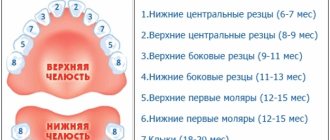
Molars are the last teeth that erupt in the child’s lower and upper dentition. As a rule, the first units appear on the lower jaw at the age of 12-18 months, a month later molars erupt on the upper row. The second molars appear in a child between 24 and 30 months of age.
Replacement of primary molars of the lower jaw occurs at the age of 6-8 years. In place of the upper units, molars grow only by the age of 9-12 years. However, these are average indicators and may vary depending on individual characteristics.
The timing of eruption depends on the following factors :
- hereditary predisposition;
- the general health of the child;
- congenital features;
- climatic living conditions;
- nature of nutrition;
- gender of the child.
Lower molars
Teeth of this type are located in the distal region of the lower row. They are distinguished by the presence of several anatomical surfaces:
- The buccal side is characterized by a narrowing towards the neck, divided into two or three parts with one and two grooves. The line of the enamel-cementum area is concave or convex, sometimes has an irregular shape. The roots are conical, cylindrical or trumpet-shaped.
- The chewing part has tubercles for chewing. There are also distal, medial-buccal, medial-lingual and other types of tubercles (there are five main ones).
- The lingual part has two convex uniform parts located on the lingual chewing line. There are no enamel streaks, the border is smooth and clear.
- The mesial side has a lingual slope, the contour line is straight or convex. The enamel-cementum border is straight. There is a slight tilt towards the cheek.
- The distal one is distinguished by the presence of a tubercle shifted towards the cheek or along the midline. The root is narrower than the medial one and has a wedge-shaped shape.
Diseases
The following diseases develop more often:
- Caries is a destructive process, as a result of which a cavity appears inside. Most often, the lesion affects the chewing surface. At the initial stage, there is a change in the shade of the enamel and pain. For treatment, it is necessary to remove the affected tissue and perform a filling.
- Pulpitis is a condition in which pathological processes affect the pulp and cause inflammation. Causes: infection, injury, or incorrect previous treatment.
- Cysts form in the root area and are small formations with purulent contents. There are usually no symptoms; the problem can be detected during examination or when the mass increases significantly in volume.
- Periodontitis is a condition in which inflammation develops at the root apex. The reasons are failure to carry out timely treatment, violation of the filling installation technology. Depending on the condition and form, three stages are distinguished - sluggish, chronic, acute.
- Pericornitis is a pathology in which inflammation of the periosteum occurs. Most often it is diagnosed in adults, when the teeth of eights appear.
Molars perform an important function, ensuring the formation of a bite and chewing food. To protect against the development of diseases of these units of the series, regular visits to the dentist for routine examinations are recommended.
Molar placement
The molar large units are located on the back of the jaw. Each jaw has six of them. They decrease in size from front to back. That is:
- the first rudiment is the largest;
- the second one is smaller;
- the third tooth is small;
The molar units are located most posteriorly in the arc. They are also called extreme. The number eight, that is, the wise tooth, grows in the period from 18 to 35 years. It happens that he does not appear at all. Row anomalies and bite defects can form in the absence of individual units or groups of rudiments. Often this concerns the end organs. Remember that normally there should be 28 units without eights, and with them 32 organs. The primary dentition has eight outermost units, the permanent dentition has twelve. In the molar group, the sixth units are key, and the 7s are variable, the most variable being the eights.
If caries treatment is carried out in a timely manner, the chewing organs will last a long time. The health of the whole body, the functioning of the gastrointestinal tract, liver and kidneys depends on this. Therapy should begin at the white spot stage, while the cavity has not yet formed. Prevention of caries is of great importance. To do this, you need to visit the dentist at least once every 6 months, carefully care for your oral cavity, and follow the dentist’s recommendations. Modern medicine has technologies the use of which can save even a dilapidated organ. Therefore, the key to health is timely treatment after a complete diagnosis and correct diagnosis.
It is important from childhood to teach a child to brush his teeth and treat the oral cavity. Children should understand that the main cause of caries is pathogenic bacteria living in dirty plaque. Therefore, trips to the hygienist will not allow the active proliferation of pathogenic microbes, subject to regular preventive cleanings.
Structural features
Those who looked at their teeth in the mirror noticed that they all had a different design. The structure of the crowns depends on:
- from the arrangement of organs in a row;
- depending on the shape of the unit;
- depending on the size of the rudiment;
The anatomical structure of the mammary organs is basically the same as the permanent units. True, temporary rudiments have distinctive features.
- They are smaller in size;
- The width of their crown prevails over the height;
- In the cervical area, the enamel layer is thickened;
- The shade of the enamel is bluish;
- The roots are widely spaced and short;
In addition, in the alveolar arch the temporal units are arranged steeply vertically. This is influenced by the fact that behind the roots there are already rudiments of permanent teeth. The upper units have 3 roots, one of them is lingual and 2 are buccal. The crowns have a cuboid shape, on the chewing surface of which there are 3 or more tubercles. The groove that separates the tubercles has two parallel lines and one perpendicular. The crowns of the rudiments on the lower jaw are somewhat elongated, and the fissures are located almost cross-shaped. These chewing organs have 2 roots. In eights, the roots often merge, the shape of such a root is conical. It happens that wise teeth grow with 4-5 roots. Since the upper and lower eights are close to each other, occasionally they can grow together.
To keep the chewing organs healthy, dentists may prescribe dental remineralization. This procedure restores the enamel layer well due to its saturation with deficient elements. The enamel ceases to be porous, acids do not penetrate its pores and do not contribute to the leaching of useful minerals. This event prevents and eliminates caries at an early stage, improves the aesthetics of crowns with fluorosis, reduces the sensitivity of units, brightens crowns and normalizes the microflora of the oral cavity.
Features of molars
Large molars differ from other groups not only in external characteristics, but also have other features :
- these are the largest units in each dental arch;
- have the largest chewing surface;
- have a powerful root system;
- the upper molars are slightly larger than those on the lower jaw;
- have a stronger surface coating;
- able to withstand weight up to 75 kg;
- upper molars have 4-6 canals, while lower units generally have only 3 canals;
- maxillary molars have 3 roots (palatal, buccal-medial, buccal-distal), less often 4;
- the chewing teeth of the lower jaw have only 2 roots;
- wisdom teeth are the very last molars, which may appear by the age of 50 or not appear at all;
- the last molars are very often located not in the chewing arch, that is, they can grow horizontally or with a slight slope, located buccally or towards the tongue or palate;
- wisdom teeth have from 1 to 5 chewing cusps;
- the last molars often have a complex root configuration (curvature, fusion).
All this entails the peculiarities of treatment of such teeth. The presence of a large number of complex root canals, as well as difficult access compared to other groups of teeth, requires highly qualified doctors and modern equipment for high-quality therapy.
In dental practice, molars are considered the heaviest units, especially wisdom teeth. Proper care and regular preventive examinations will help keep them healthy. When the first signs of damage to the molars appear, we recommend that you immediately sign up for a consultation at our KAS+ dentistry. Timely detection of dental problems guarantees their high-quality treatment with minimal risk and long service life.