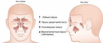
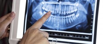
Although doctors do not have a consensus on the functions of the paranasal sinuses, if problems arise with them, any person experiences discomfort. In most cases, a standard medical examination using a special instrument allows you to find out the cause and prescribe treatment. In some cases, your doctor may prescribe X-ray of the paranasal sinuses
– a simple and affordable procedure.
Briefly about sinusitis
Sinusitis is inflammation of the mucous membrane in the maxillary sinuses. Nathaniel Gaymore was the first to describe these sinuses: the name came from his name. And Leonardo da Vinci was the first to draw the maxillary sinuses.
Essentially, sinusitis is sinusitis caused by various reasons, which are associated with infection in the sinuses and the accumulation of pus in them. It can be rhinogenic, odontogenic, traumatic, allergic.
Rhinogenic occurs due to untreated rhinitis, when the infection penetrates from the nasal cavity into the sinuses. Odontogenic sinusitis develops as a result of infection through a diseased tooth and inflamed tissue around it.
Traumatic rhinitis is a consequence of damage to the nasal septum, maxillary sinus or jaw.
Finally, allergic rhinitis occurs due to allergies to pollen, animal dander, dust mites, medications and other allergens.
Acute sinusitis lasts less than 3 months, recurrent acute sinusitis occurs up to 4 times a year, chronic sinusitis lasts from 3 months or more. An exacerbation of chronic sinusitis is also possible, when new symptoms are added to existing ones.
Do you need an x-ray?
If a survey of complaints, anamnesis, and identification of clinical symptoms is not enough to make a correct diagnosis, then doctors prescribe an x-ray.
X-rays have their own contraindications: pregnancy, breastfeeding, young age of the patient, repeated procedure.
An X-ray of the children's nasopharynx is performed only when other diagnostic methods have failed. The procedure itself is not dangerous for the child. Maxillary sinusoids up to 16 years of age are similar to light spots, have a dense structure, and do not appear hollow. Using the image, you can get an idea of the accumulation of masses of pus, excessive enlargement of the pharyngeal tonsils, and the development of sinusitis in the acute stage.
Decoding the testimony
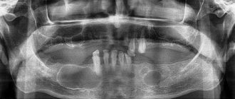
The radiologist deciphers what an x-ray of the paranasal sinuses shows. First of all, the condition and location of bone structures and cartilage is assessed. The liquid on the x-ray image appears as an intensely darkened horizontal boundary. Thickening of the mucous membrane indicates swelling in the paranasal sinus. You will receive a transcript of the research results in the form of a conclusion, but the final diagnosis is made by the doctor. Usually, when a pathology is identified that requires a more detailed study, another diagnostic method is prescribed.
Preparation for the procedure
Before starting the procedure, you must remove your outer clothing, metal and other jewelry, earrings, piercings, and remove dentures. Next, you need to tell the doctor whether similar procedures have been performed previously, whether there are dental implants, fractures of the nose or facial bone.
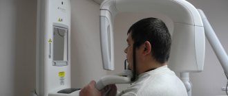
During the diagnosis, the patient must stand still and not move. He should rest his nose and chin on the stand of the X-ray machine, which has been previously adjusted to the patient’s height. After this, the doctor will take several pictures from another room and give further instructions to the patient. During the procedure, the patient will sometimes need to hold their breath for a few seconds (no more than 10 seconds). The radiologist will take pictures in two projections, and sometimes while lying down.
The whole procedure will take about 10 minutes.
What does sinusitis look like in the picture?
Healthy paranasal sinuses in the picture are similar in shade to the eye sockets. If inflammation develops, they acquire a darker shade with whitish, “milky” contents. The usually smooth edges of the sinus may be curved, and thickening may be observed locally or along the entire edge of the sinus. This picture indicates that pathology has developed in the sinuses.
An otolaryngologist and a radiologist interpret the X-ray. Photos of the sinuses highlight potential abnormalities and darkened areas. The analysis can determine: the presence of potentially affected areas - darkened or sub-darkened areas, whether there is sinusitis - lightened areas of the sinuses.
After treating a runny nose and congestion, the doctor can also determine the nature of the disease - sluggish, stopped, chronic, acute, the presence of tumors or cysts.
CT and x-ray of the maxillary sinuses
X-ray examination is an effective method for diagnosing the human body, including the condition of the ENT organs. If a person has a stuffy nose, there is discharge from it, or the timbre of his voice has changed, then this is an indication for an examination. X-rays are considered one of the most effective ways to detect sinusitis. With its help, it is possible to diagnose various pathological changes even at an early stage of their development, which is extremely important when prescribing appropriate treatment.
An examination is prescribed if the following symptoms are present:
- Nasal discharge (sometimes with pus).
- Problems with nasal breathing. As a rule, only one nostril is blocked.
- Frequent headaches.
- General fatigue, trouble sleeping and loss of previous activity.
- Nosebleeds.
- Soreness and swelling of the forehead.
If these symptoms are present, the doctor may suspect the presence of an inflammatory process, but it is necessary to determine the exact cause of this inflammatory process, because similar signs exist with polyps and a deviated septum. Therefore, an x-ray of the maxillary sinuses is a mandatory procedure, which allows us to determine the degree of development of the pathology, the presence of neoplasms and other aspects.
Content
- Why do you need an x-ray of the nose for sinusitis?
- How can you see sinusitis on an x-ray?
- How is an x-ray of the maxillary sinuses performed?
- X-ray in childhood
- How to determine sinusitis in an adult patient from an image?
- Computed tomography for sinusitis
- Is it possible to determine sinusitis without a picture?
Why do you need an x-ray of the nose for sinusitis?
Vivid symptoms will help the doctor make an appropriate diagnosis, but without an accurate diagnosis, further treatment will be impossible. It is important to first examine the condition of the sinuses, and then determine the medications and physiological procedures that can get rid of the disease.
When diagnosing, it is extremely important to know what sinusitis looks like on an x-ray. With this examination method, soft tissues are not visible, but bone structures are clearly visualized. If in the picture the shade of the paranasal sinuses and eye sockets is identical, then there is no inflammation. If there is purulent content, then visually it looks like large darkened areas.
An x-ray of the sinuses with sinusitis allows you to determine a number of pathological changes:
- Localization of lesions. They appear as dark spots.
- Presence of cystic formations. They have fairly clearly defined boundaries.
- The inflammatory process and the degree of its development. In the picture they are visible as white spots. Accordingly, the brighter they are, the stronger the inflammatory process.
- Various changes. You can see the level of filling of the nasal sinuses with pus, thickening of the mucous membrane and other pathological changes.
How can you see sinusitis on an x-ray?
Below you see a picture of sinusitis before and after treatment. The radiograph is interpreted by the attending physician or radiologist.
In the absence of pathological changes and abnormalities, the image will show the following information:
- The nose is in the form of a triangle of a light shade, in the middle of which there is a septum.
- On the side of the nasal cavity, the maxillary sinuses are located in the form of triangular clearings with not blurred boundaries.
- Symmetrical arrangement of the nasal passages on both sides of the nasal cavity.
- The frontal sinuses are located above the eye sockets. In the picture they look like clearings of various sizes.
The pathology develops from inflammation of the lateral sinuses. Further, it is localized in the region of the frontal lobes. This will be clearly visible in the darkened areas above the eye sockets and nose. Simultaneous inflammatory processes in the frontal and lateral sinuses indicate not only the presence of sinusitis, but also frontal sinusitis.
A specialist who takes pictures of the nose for sinusitis always determines the condition of the ethmoid bone. Infiltrative fluid often accumulates in the maxillary sinuses:
- mucous;
- catarrhal;
- purulent.
It is quite easy to notice - the liquid appears as a clearly formed white area. This is a clear sign of pathology, which is one of the grounds for making a final diagnosis.
How is an x-ray of the maxillary sinuses performed?
X-ray of the nasal sinuses for sinusitis is a completely standard diagnostic procedure. Before the examination, you must remove all metal objects and removable dentures. If implants, crowns or pins are installed, the radiologist should be notified about this.
To obtain an accurate picture of the disease, a photograph of the nasal sinuses with sinusitis is taken in several projections:
- Chin. Based on this image, the specialist analyzes the actual condition of the maxillary sinuses.
- Nasomental. This projection clearly shows the presence of an inflammatory process in the mucosa.
- Nasofrontal. In this picture you can see the state of the lattice labyrinth.
The examination takes 10-20 minutes, after which the doctor can clearly see the presence or absence of pathology.
X-ray in childhood
Experts agree that X-ray examinations are advisable from the age of 14. Here we are not talking about potential harm to the child’s body, because a single dose of radiation during an examination is absolutely safe for the child. The fact is that at an early age the paranasal sinuses are not yet fully formed, so the examination result cannot be 100% accurate.
In addition, x-rays require maintaining complete stillness throughout the entire examination, which, for obvious reasons, is very difficult to achieve in childhood.
How to determine sinusitis in an adult patient from an image?
Pictures of sinusitis in adult patients are quite characteristic, especially when compared with radiographs of people who do not have this pathology. X-ray will show the following changes:
- The eye sockets and sinuses will be of different shades.
- The presence of infiltrative fluid (a clear white spot against a darkened background).
- Thickened walls and blurred boundaries of the sinuses.
- Cystic formations of varying sizes with clearly defined boundaries.
The only thing that cannot be seen in a photo of the nose with sinusitis is the type of fluid that has accumulated in the sinuses. The study only shows its presence in the form of a white spot, but cannot show what kind of infiltrate (purulent, mucous or catarrhal fluid).
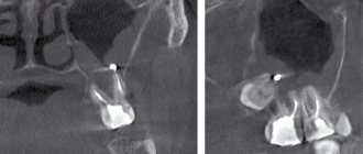
Computed tomography for sinusitis
In some cases, in addition to X-rays, a CT scan of the maxillary sinuses is used. This is a more informative diagnostic method, which has its advantages:
- This is the most reliable way to examine bone structures.
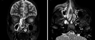
- The result of the study is a three-dimensional image that allows you to view the examined area from different angles and in all planes.
- The procedure takes the least time (when compared with X-rays and MRIs).
- The picture turns out to be as detailed as possible.
- The technique allows you to accurately determine the location and amount of liquid.
When answering the question of what is better to do for sinusitis - CT or MRI, you need to definitely make a choice in favor of computed tomography. This examination can be carried out even in the presence of crowns and implants, which is not possible with magnetic resonance imaging. In addition, MRI has the greatest diagnostic value when examining soft rather than bone tissue.
Is it possible to determine sinusitis without a picture?
If there are significant symptoms, the doctor collects an oral history and conducts an examination. The forehead area and around the nose are palpated for pain. The nasal passages are also examined, the purpose of which is to assess the condition of the mucous membrane.
Based on the manipulations performed, an experienced doctor can diagnose sinusitis without x-rays. But a visual examination does not give a clear picture of the disease, so in order to draw up a competent treatment regimen, it is necessary to take a picture.
X-ray - have been conducting X-ray examinations since 2015. Modern equipment allows you to obtain images of high accuracy and detail. We clearly understand how important diagnosis is for further therapy, therefore we are strict in fulfilling our professional duties.
You can make an appointment with us using the contact phone number listed on the website. Contact us for more detailed advice.
Sign up for a study by phone
+7 (812) 332-52-54
How often should x-rays be taken?
Despite the fact that radiation exposure from X-rays using modern equipment is minimal, it should not exceed certain levels per year. The procedure can be done 3-4 times in a row. Before each new x-ray, the health worker must look at the patient’s card, and if he finds an exceeded radiation dosage there, he will refuse the procedure.
During treatment, it is recommended to conduct two x-ray examinations: before and after therapy or 2 weeks after the start of treatment. If the disease is serious, examinations can be done 3 times a year.
Cost of radiography of the sinuses
At the ENT-Praktika clinic, you can take a 3D image of the maxillary and other sinuses using a Japanese cone-beam computed tomograph (CBCT) of the latest generation from Morita quickly and inexpensively, at a convenient time by appointment. The great advantage of our study is the excellent quality and information content of the three-dimensional image with minimal radiation exposure to the patient in comparison with spiral CT and even conventional radiography. The price of the study is in the price list. In urgent cases, there is no need to make an appointment; the diagnostic procedure will be carried out immediately after the doctor’s examination. Here in the center you can undergo other additional tests and cure any diseases of the ear, nose and throat.
