Upper jaw:
Central incisor - average length 25.0 (22.5 - 27.5 mm) number of canals - one main, in 10% of cases an additional canal.
Lateral incisor - average length 23.0 (21.0 - 25.0 mm) one main canal, in 9% of cases an additional canal.
Fang - average length 27 mm (24.0 - 29.7 mm) one main canal, in 23% of cases an additional one.
First premolar - average length 21 mm (19.0 - 23.0 mm) 85% two canals; palatal, buccal 9% one canal - main.
6% - three canals: palatal, buccal, accessory.
Second premolar: average length 22 mm (20.0 – 24.00 mm)
75% - one main channel
24% - two canals - palatal, buccal
1% - three canals: palatal, buccal, accessory.
First molar : average length 21 mm (18.0 – 26.0)
86% - three canals: palatal, buccal, distal, buccal - medial.
14% - two canals: palatal, buccal
45% - four canals: palatal, buccal - distal, medial - buccal (MB1), medial - buccal (MB2) this is the most common name for the buccal canals - international.
Second molar - average length 20.5 mm (19.0 - 24.0 mm) 1% - one main canal.
4% - two canals - palatal and buccal.
55% - three canals - palatine, buccal - medial, buccal - distal,
40% - palatal, buccal - distal, medial - buccal (MB1), medial - buccal (MB2).
Third molar - the number of root canals varies from 1 to 5.
Treatment of a double-rooted central incisor: tomography capabilities
Before performing endodontic manipulations, the doctor must be fully aware of the anatomical structure of the tooth to be treated. After all, missed canals are the main reason for the failure of endodontic treatment. According to available data, only 0.6-2.0% of maxillary central incisors are characterized by the presence of two roots. Usually the presence of a second root is associated with anomalies in tooth development, such as gemination, fusion, dens invaginatus, or simply with the formation of another additional root.
The outcome of endodontic treatment directly depends on the clinician’s knowledge regarding various forms of variations in the structure of the endodont. Due to the low prevalence of diagnosis of cases of identification of the second canals of the central incisors, clinicians often simply ignore the possibility of their presence, which may result in a compromise of the final treatment result. Diagnostics using radiography taken at certain angles can help verify additional canals and endodontist morphology. For a more accurate diagnosis, it is recommended to use the cone-beam computed tomography method, which allows you to evaluate the structure of the root canals in three dimensions. Comparison of data obtained during studies of extracted teeth using CBCT and two-dimensional radiography methods revealed that CBCT helps to identify a larger number of canals in the root structure.
The purpose of this study is to demonstrate the process of diagnosis and treatment of the maxillary central incisor, in which one canal, present in the orifice, bifurcated along the root, ending in two apical foramina (Vertucci type V). CBCT was used as the main diagnostic method. In addition, this clinical case demonstrates the potential of CBCT as a primary diagnostic approach to help select the most appropriate endodontic treatment method.
Clinical case
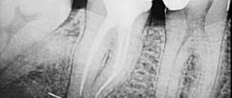
A 72-year-old patient was referred for endodontic treatment of tooth No. 9, in the projection of which it was planned to subsequently fix a fixed orthopedic structure. During the clinical examination, a fracture of the tooth crown and lack of sensitivity to temperature stimuli, percussion or palpation were diagnosed. Based on periapical radiography, the presence of two canals was suspected (Figures 1-2). Based on the available data, the doctor referred the patient for a CBCT procedure to clarify the morphology of the endodontist of the upper central incisor. During the analysis of CBCT data, the patient was confirmed to have 2 canals in the structure of the maxillary central incisor (photo 3). The final diagnosis stated the presence of pulp necrosis without any accompanying periapical changes.
Photo 1. X-ray of the 9th tooth before treatment: unusual anatomy of the root canal system.
Photo 2. X-ray of the 9th tooth before treatment: unusual anatomy of the root canal system.
Photo 3. The axial CBCT section visualizes the presence of two root canals.
Before the start of endodontic intervention, the crown part of the tooth was treated with antibacterial agents through hygienic cleaning, after which the local anesthesia procedure began (2% mepivacaine with adrenaline 1: 100,000). The tooth was isolated using a rubber dam, access to the canals was formed with ultrasonic tips in such a way as to maximize visualization of the mesial canal from the projection of the furcation in the middle third of the root. The length of the root canals was checked using an apex locator and confirmed radiographically (photo 4).
Photo 4. Determination of working length.
The formation of the carpet was ensured by hand files of 10 and 15 sizes, and mechanical processing of the canals was carried out with a ProDesign S rotary file system (25.06 and 25.08, Easy Dental Equipment) and hand files of 30, 35, 40 and 45 sizes. A 2.5% sodium hypochlorite solution was used as the main irrigant. To complete the cleaning of the canals, 5 ml of 17% EDTA (ethylenediaminetetraacetic acid) solution was introduced into the canal space using the passive ultrasonic irrigation (PUI) technique using an E1 ultrasonic tip, the exposure of which was 1 minute. After this, 5 ml of 2.5% sodium hypochlorite was introduced into the endospace. Obturation was performed using a hybrid technique with zinc-oxide-eugenol cement (photo 5-7).
Photo 5. Radiograph taken immediately after endodontic treatment.
Photo 6. Radiograph taken immediately after endodontic treatment.
Photo 7. Radiograph taken immediately after endodontic treatment.
A temporary restoration was provided using composite material, after which the patient was referred for orthopedic rehabilitation. Clinical monitoring of the patient was provided for 1 year (photo 8-10).
Photo 8. X-ray obtained one year after endodontic treatment.
Photo 9. X-ray obtained one year after endodontic treatment.
Photo 10. X-ray obtained one year after endodontic treatment.
Discussion
Several cases of endodontic treatment of maxillary central incisors with two roots and two canals have been reported in the literature, and in most of these, patients also presented with signs of macrodontia, dental fusion, gemination, or dens invaginatus. However, in the analyzed clinical case, the patient had a fracture of the coronal part; therefore, it was impossible to clarify the presence of additional morphological variations in the structure of the crown. The most common cause of the formation of an additional root is developmental disorders of the Hertwig epithelial sheath.
According to the recommendations of the American Association of Endodontists, the use of CBCT to study the anatomy of the root canal system is indicated only in selected cases when conventional intraoral radiographs do not provide sufficient information for proper planning of endodontic procedures. Traditional radiographic research methods with 2D visualization of endodontist structures are quite limited in terms of the ability to detail morphological changes in the root, while CBCT allows you to assess the structure of the tooth in three planes, and, thus, take into account all the anatomical nuances in the process of further iatrogenic interventions. In the described clinical case, CBCT made it possible to verify the presence of a single canal emerging from the projection of the pulp chamber and dividing into two in the middle third, which end in two independent apical foramina. This type of endodontist structure is classified as class V for Vertucci.
Although the clinician may have the appropriate knowledge of intraradicular anatomy, and mechanical and medical treatment of the root canal system, as a rule, creates the appropriate conditions for the restoration of affected periapical tissues, anatomical variations in the structure of the endodontist complicate the process of providing adequate endodontic treatment. Deficiency in root canal treatment is associated with the proliferation of pathogenic microorganisms, which are usually found in pulp and periodontal pathologies.
In the described clinical case, sodium hypochlorite solution was used as the main irrigant due to its high antimicrobial effectiveness. However, the complex anatomical structure of the root canals limits the ability for this solution to penetrate the entire working length. To enhance the antimicrobial effect, a PUI approach was used. Boff et al compared the effectiveness of PUI and traditional irrigation techniques in cleaning the apical portion of root canals using 2.5% sodium hypochlorite and concluded that the apical cleaning of the PUI method showed superior results. That is why we applied the above described approach in our clinical case.
Conclusion
This clinical case describes an approach to the verification of additional canals and subsequent endodontic treatment of maxillary central incisors in the absence of additional morphological variations in the coronal part of the tooth. The results obtained indicate that effective diagnosis, as well as carefully planned treatment, can achieve a successful long-term prognosis for the treatment of maxillary central incisors with an existing additional root canal.
Authors: Natália Gomes de Oliveira, DDS, MSc Casimiro Ricardo Oliveira Passos, DDS Luís Felipe Espíndola-Castro, DDS, MSc Paulo Maurício Reis de Melo Júnior, DDS, MSc, PhD Sandra Maria Alves Sayão Maia, DDS, MSc, PhD Marianne de Vasconcelos Carvalho, DDS, MSc, PhD