1036
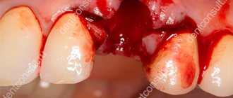
After tooth extraction, a blood clot forms in the alveolar socket, stopping bleeding and protecting the wound from contamination and infection.
The formation of a blood clot is a prerequisite for high-quality and rapid healing of the hole. Its absence is considered a complication or, in any case, a prelude to it.
General overview
Usually, after a tooth is removed, the dental surgeon places a gauze turunda on the hole and asks the patient to squeeze it with his teeth. If everything goes well, after 20-30 minutes the turunda is removed, and the normal healing process continues in the socket, which ends completely after 2-3 weeks.
Sometimes, fortunately, relatively rarely, the primary bleeding does not stop, or it resumes later, thus becoming secondary.
This problem is solved by tamponade of the hole - filling it with iodomorphic bandage, fibrin glue, biological antiseptic tampon (BAT) or other material used to stop bleeding.
The most effective and modern means of stopping bleeding and eliminating other post-extraction complications are hemostatic sponges. These include alvostasis sponge, produced by the Russian company Omega-Dent.
Like other similar materials, alvostaz sponge is intended for filling wounds for preventive and therapeutic purposes. The drug has a complex effect:
- Absorbs exudate.
- Stops bleeding.
- Protects the wound from contamination and bacterial infection.
- Stops inflammation.
- Eliminates swelling.
- Pain relief.
And ultimately it prevents complications and speeds up healing.
Alvostaz sponge is a porous preparation in the form of a cube consisting of collagen impregnated with active and auxiliary medicinal solutions.
The material has an antiseptic, hemostatic (hemostatic), analgesic, and sorbing effect. It is used in dentistry mainly for the prevention and treatment of alveolitis.
If the sponge is installed immediately after tooth extraction, it performs a preventive function - preventing the development of alveolitis. If after continuous primary or secondary bleeding - therapeutic.
The cause of ongoing primary or renewed secondary bleeding may be:
- damage during extraction of large vessels;
- blood clotting disorders due to pathology of the body or the action of anticoagulants;
- destruction of a blood clot due to non-compliance with doctor's recommendations, for example, rinsing the mouth too vigorously with water or an antiseptic solution;
- inflammatory process in the hole;
- highly traumatic tooth extraction due to its characteristics (impacted tooth, third molar, etc.).
The drug is available in 3 forms - with iodoform, with chloramphenicol and neomycin, with chlorhexidine and metronidazole.
In what cases is suturing of the tooth socket performed, and what material is used.
Visit here to learn about the features of suture material for dental surgery.
At this address https://www.vash-dentist.ru/hirurgiya/udalenie-zubov/easy-graft-vosstanovlenie-defektov.html you will find a detailed description of the Easy Graft Classic bone replacement material.
Hemostatic sponge Alvostaz
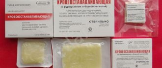
In addition to the jar with medicinal cubes, there is a hemostatic sponge with a similar effect. It looks like a porous plate, dark yellow in color with a faint smell of vinegar. This is a pressed powder of medicines:
- collagen;
- nitrofural;
- 2% substance-solution;
- Boric acid.
The size of the plates is from 50 to 90 mm, in the form of a square. Dry porous elastic structure, perfectly absorbs liquid. Plates are used for open bleeding, as well as internal bleeding, when blood does not flow out. This is typical when organs located in the abdominal cavity are damaged.
The hemostatic sponge does not need to be removed; it dissolves independently in the human body. This will ensure the safety of the surgical procedure. Complete dissolution of the plate occurs after 14-28 days. The medicinal composition of the sponge accelerates wound healing and acts as an antiseptic.
But there are also contraindications to the drug. There should be no individual intolerance to the drugs included in the sponge. The plates should not be used to stop arterial bleeding or purulent wounds.
Hemostatic sponge alvostasis
After removing the sponge from the package, it is applied to the wound and held for a couple of minutes. After soaking in blood, the plate adheres to the body and does not require special fastening with a bandage. To enhance the effect, the plate is moistened with a thrombin solution.
Active components and principle of action
The properties and features of the action of different forms of the drug are determined by the composition of the impregnation:
- Alvostaz Sponge No. 1 (with iodoform) contains collagen, eugenol, calcium phosphate, lidocaine, iodoform, thymol, propolis.
- Alvostaz sponge No. 2 (with chlorhexidine and metronidazole) – collagen, dexamethasone, metronidazole, chlorhexidine.
- Alvostaz sponge No. 3 (with chloramphenicol and neomycin) contains collagen, neomycin sulfate, chloramphenicol, chlorhexidine, dexamethasone.
The individual components of sponge alvostasis have the following effects on the wound:
- Collagen absorbs exudate and blood, creates compression inside the socket that stops bleeding, and serves as a framework (matrix) for newly formed tissues. Having fulfilled its function, depending on its shape, it is resorbed or rejected by the hole.
- Eugenol (“clove oil”) is added as a flavoring agent.
- Iodoform and chlorhexidine have an antiseptic effect.
- Thymol is an antiseptic, anesthetic and preservative.
- Calcium phosphate has a complex effect. For dentists, its osteoregenerative function is most important.
- Lidocaine relieves pain.
- Propolis has antiseptic, antimicrobial, immunomodulatory and antioxidant effects.
- Dexamethasone is a glucocorticosteroid with an anti-inflammatory effect.
- Chloramphenicol and neomycin sulfate are broad-spectrum antibiotics.
- Metronidazole is an antiprotozoal (directed against protozoan parasitic microorganisms) and antimicrobial drug.
The product performs the following actions in relation to the wound:
- It swells from the adsorption of blood and exudate , tightly adhering to the walls of the socket.
- Stops bleeding. For uncomplicated primary bleeding, this takes 3-5 minutes. The duration of elimination in case of complicated primary or secondary depends on the clinical picture.
- Relieves pain for several hours.
- Causes accelerated healing of the hole thanks to the impregnation components.
- It resolves or is pushed out of the socket after 14-21 days.
The principle of action is determined by the action of their active substances. In particular, when reacting with wound exudate, iodoform releases free iodine, activating its antiseptic effect.
The antibiotics contained in the impregnation create a bactericidal and/or bacteriostatic effect.
Help from qualified dentists at the International Center for Health Care
The International Center for Health Protection provides treatment for alveolitis at the prices indicated in the price list. All necessary services are already included in the price. If necessary, we will explain all components of the final amount. Here you can undergo a comprehensive examination, instrumental and laboratory diagnostics, and receive advice from experienced dentists. The quality level of services provided is confirmed by the international TUV certificate.
Treatment prescribed for alveolitis of the tooth socket includes freeing the socket from foreign bodies and pus, antiseptic treatment of tissues, and follow-up to promote complete healing and recovery. Our multidisciplinary medical center has a convenient location in Moscow - it is a 5-minute walk from the Dostoevskaya metro station, a 15-minute walk from the Novoslobodskaya metro station. You can make an appointment by phone.
Release form and absorption time
The material is produced in the form of cubes measuring 1x1x1 cm. One package contains 30 cubes (sponges) placed in a plastic jar.
Resorption of resorbable cubes (alvostasis sponge No. 1) completely ends after 14-21 days. Rejection (pushing out of the hole) of non-resorbable ones (sponges No. 2, 3) - after a few days.
It is unacceptable to forcibly remove (tear off) material from the wound. It must be resorbed or rejected by healed tissue .
The mechanism of development of alveolar process atrophy and the degree of the pathological process.
In this publication, we will tell you which dentist recommendations should be followed without fail after a complex wisdom tooth removal.
Here https://www.vash-dentist.ru/hirurgiya/udalenie-zubov/pezohirurgiya-bez-boli.html everything about the use of piezosurgery in dentistry and its effectiveness.
Solution
Such a diagnosis must be treated in time, then after a few days the person feels better. The process includes:
- removal of remnants of a blood clot, this is done under local anesthesia;
- processing the hole and its walls with a special tool;
- treating the hole with an antiseptic solution so that the infection does not go further;
- prescription of antibiotics;
- the use of drugs that relieve pain;
- rinsing the mouth, for this you should take antiseptic solutions;
- physiotherapeutic procedures that promote rapid wound healing.
In the absence of timely treatment, osteomyelitis of the jaw bone may occur.
Indications and restrictions
Alvostasis sponge is indicated in most cases of maxillofacial surgery. In particular:
- to prevent complications after traumatic tooth extraction (most often during the extraction of wisdom teeth);
- for post-extraction complications - alveolitis, periodontal inflammation, abscissus;
- in the treatment of deep gum pockets (with open curettage);
- for various surgical interventions on the dentofacial apparatus, characterized by a medium or high degree of trauma.
The use of alvostasis sponges does not pose any danger to patients. They are contraindicated only for people with allergies to the components of the impregnation - iodine, eugenol, bee products, lidocaine.
Reasons for the development of the disease
Signs of alveolitis can be detected after poor-quality, incomplete tooth extraction. Most often this happens when the doctor leaves some tissue in the socket. But the pathology can also be caused by the patient’s neglect of the rules of personal hygiene.
The etiology of the lesion includes other causes. The following circumstances can trigger the development of infection:
- chronic inflammation of the gum tissue;
- accumulations of soft or hardened plaque;
- caries on adjacent teeth;
- weakened, decreased immunity, autoimmune disorders;
- insufficient antiseptic treatment;
- fragments of pathological tissue remaining deep in the cavity;
- eating rough, hot, spicy food after tooth extraction.
Application technique
The technique of application depends on the type of operation, and may have its own characteristics depending on it. Let's look at it using the example of tooth extraction. Post-extraction use of the material involves the following procedures:
- Removal of dead, necrotic and infected tissue from the socket.
- Rinse the wound from bone and tissue fragments with saline solution heated to body temperature.
- Drying the cleaned hole with a hardware suction, pipette or gauze swab. Cotton swabs cannot be used due to the possibility of clogging the wound with cotton fibers (lint). Before this, it is necessary to ensure that the hole is isolated from saliva.
- Selecting an alvostasis sponge from a jar and filling the hole with it. This must be done with gloves and sterile tweezers.
- Suturing the wound, if necessary.
- Cover the wound with a sterile dry gauze pad.
In difficult cases, if wound healing is delayed, it can be treated multiple times and the old sponge replaced with a new one. Although in most cases once is enough.
The patient is informed that it is impossible to forcibly tear off the gauze swab; he must wait until it comes off on its own.
In the video, watch the process of using collagen hemostatic material.
Types of alveolitis after tooth extraction
Alveolitis comes in several types:
- Serous
. It is characterized by constant, never-ending aching pain. When trying to eat food, they intensify. Lymph nodes are not enlarged, body temperature does not increase. This form of the disease lasts a week, after which the transition to the next stage is inevitable. - Purulent
. The developing infection affects the patient's condition. Due to intoxication, weakness, lethargy appear, temperature rises, and pain increases. The affected area swells, and the face may also swell. Upon examination, pus and plaque are visible on the hole. The mouth opens with difficulty, which causes an unpleasant odor. - Hypertrophic
. A chronic purulent process, which is characterized by subsidence of symptoms. The lymph nodes are normalized, most of the clinical manifestations disappear, and the temperature drops. The examination can reveal the proliferation of granulations. The gum tissue takes on a bluish tint, and pus is released from the wound.
Analogs
There are a sufficient number of complete or partial analogues of Alvostaz sponge on the pharmaceutical market:
- Alvostasis flagellum . Material from the same company as alvostaz sponge. Similar to it in composition and properties. It is a drug-impregnated collagen tourniquet with a cross-section of 1×1 cm. It has the same indications.
- Iodomorphic bandage. Cotton bandage soaked in iodoform. It has an adsorbent, homeostatic, anesthetic, antiseptic and bactericidal effect.
- Dental micro-tuffer. It consists of a collagen cone impregnated with sanviritrin (a broad-spectrum antibiotic) and lidocaine (an anesthetic). Resorbed in the wound.
- Alvanes sponge. It is lyophilized (cold-dried) collagen impregnated with antiseptic and antibacterial solutions.
Available in 2 forms - with iodoform and chlorhexidine and metronidazole. It has hemostatic, anesthetic (contains lidocaine), antibacterial, anti-inflammatory and astringent effects. Bioactive (stimulates tissue regeneration). It is resorbed in the wound within a few days. - Stimulus-Oss. It is a porous material consisting of hydroxyapatite and various additives. Available in the form of plates 50x50x7 mm and cones with a diameter of 11 mm.
In addition to hemostatic and bactericidal effects, it also has an osteoregenerating effect due to the presence of hydroxyapatite. Therefore, its scope of application is wider than that of alvostasis sponges.Used for any maxillofacial surgery where replacement of missing bone tissue is required. Can also be used after tooth extraction.
Complementary therapy
To speed up the recovery of the inflamed area, especially if necrosis has already developed, additional methods are used. Your doctor may recommend:
- treat the hole using UV radiation / infrared laser;
- use the balneotherapy procedure;
- perform smoothing in case of exposed bone tissue;
- undergo a course of fluctuarization/microwave therapy;
- start taking vitamin complexes.
It is allowed to make baths with herbal decoctions. St. John's wort, chamomile, oak bark, and calendula are suitable for these purposes. Vitamins should be chosen that are aimed specifically at strengthening dental tissues.
These procedures can be continued until pain stops completely. After a week, the walls of the hole heal and begin to become covered with young mucous tissue. However, symptoms of inflammation may still be present. After about 2 weeks, the swelling subsides and the mucous membrane acquires a normal pink color.
Reviews
In most cases, dental surgeons do not see the need to use alvostasis sponges or related materials with every tooth extraction.
The hole heals due to the formation of a blood clot. But if the latter is absent for some reason, the use of collagen sponges and other similar products becomes necessary.
If you have had a tooth removed using alvostasis sponges, tell us about your feelings and results of treatment. You can leave your comment at the bottom of this page.
If you find an error, please select a piece of text and press Ctrl+Enter.
Tags: tooth extraction
Did you like the article? stay tuned
Previous article
Universal dentures - a funny joke or a banal scam?
Next article
Perflex thermoplastics for the manufacture of dentures
Treatment of neuritis
Treatment of neuritis in traumatic injuries must be timely and wait-and-see tactics are unacceptable. For mild damage to the inferior alveolar nerve, decongestant therapy (prednisolone, veroshpirone) is sufficient. In case of moderate severity of damage, drugs that improve the conductivity of the nerve trunk (neuromedin) are added to decongestant therapy. In case of severe damage to the nerve trunk, in the absence of positive dynamics in the restoration of sensitivity within 4 months, the patient should be referred for a consultation with a neurosurgeon in order to resolve the issue of the possibility of restoring the anatomical integrity of the nerve trunk.
Various reflexotherapy methods are widely used in the complex treatment of diseases of the peripheral nervous system. In the complex treatment of diseases of the peripheral nervous system, reflexology methods such as electroacupuncture and transcutaneous electrical neurostimulation are widely used.
Prevention of neuritis
Prevention of neuritis of the inferior alveolar nerve is the correct technique for removing teeth, correct diagnosis and correct reading of the radiograph, and a gentle technique for dislocating the roots of teeth in the lower jaw with an elevator.
Basic Concepts
Plate dentures
Removable structures include plate and clasp dentures.
Plate dentures, in turn, are partial removable and complete removable.
Partial removable laminar dentures are used when one or more teeth are missing.
Complete removable plate dentures are indicated in the absence of teeth in the upper and/or lower jaw.
A partial denture is a partial removable denture consisting of artificial teeth, artificial gums and special fastening elements (hooks, locks).
Why can complications arise after the installation of removable dentures?
- Poor preparation for prosthetics. Before treatment, it is necessary to carry out sanitation of the oral cavity, restoration, and more often, prosthetics of teeth, which will serve as a support for fixing the prosthesis;
- Insufficient oral hygiene. Can lead to inflammation of the gums under the prosthesis, the formation of caries, pulpitis of the teeth that serve as support for the prosthesis;
- Fracture of the prosthesis. As a result of breakage or displacement of parts of the prosthesis, its fixation in the oral cavity is disrupted, discomfort and pain occur when wearing it;
- Inaccurately manufactured prosthesis. The design should be easily fixed and also removed without much effort. There should be no gaps in the oral cavity between the structure and the gum, but the prosthesis should not put pressure or rub soft tissues. In the first case, food debris will accumulate in the gap, in the second, sores will appear on the mucous membrane, and subsequently bedsores;
- Changes in the position of individual teeth and bite, leading to displacement of the prosthesis.
