Molars are divided into canines, incisors, premolars, and molars. The first three types of teeth erupt in place of similar milk teeth. In turn, molars do not have predecessors and appear behind the temporary ones, therefore their second name is accessory.
Each jaw has 16 teeth - four incisors, two canines, four premolars and six molars. In some cases, third molars, better known as wisdom teeth, do not erupt because they do not have buds in the jaw - then instead of 16, a person has 14 teeth above and below. Experts explain this phenomenon by a reduction, or simplification, of the dental system caused by changes in dietary style.
Functional properties of elements
Teeth perform several functions at once. Each element has a distinctive shape according to its purpose. The main role of the units is digestive. The quality of assimilation of useful elements by the body depends on the thoroughness of grinding the products. Only after chewing and moistening with saliva will the food be ready for subsequent processing by the digestive organs.
Chewing one piece lasts on average 10-20 minutes. The force exerted by the teeth on food sometimes reaches 80 kg. Incomplete dentition can lead to disturbances in the digestive system.
Another function of the elements is aesthetic. With malocclusion, a person may develop psychological problems, since the first thing others pay attention to during a conversation is a smile. Teeth also play a role in the formation of syllables. In the absence of one or more elements, the pronunciation of syllables changes: a lisp appears, speech is distorted.
Pathologies
Factors that can trigger gum disease include mechanical damage, poor hygiene, infection, and the development of ulcers or cancer. Characteristic signs of deviation from the norm are loss of sensitivity, the formation of edema, bleeding and abundant accumulation of plaque on the surface. Common diseases include:
- Gingivitis is an inflammatory process that occurs as a result of chronic pathologies, errors in the selection of prosthetic structures, as well as tartar deposits.
- Periodontitis is an inflammation of bone tissue, often developing from a local to an extended form. It becomes the result of infection or food debris getting into the gum pockets, and, in the absence of proper treatment, leads to a weakening of the structure of the dentition.
At the first signs of abnormalities, it is recommended to undergo diagnostics at a dental clinic. Timely identification of the cause of gum disease allows comprehensive treatment to begin and helps to avoid serious consequences.
Tooth anatomy
The structure of all units is the same, but each tooth has a distinctive shape and dimensions, depending on the functions it performs.
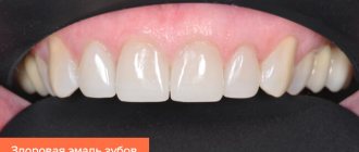
Internal structure of teeth in the photo
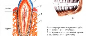
What does a tooth consist of? The top layer (enamel) is predominantly composed of minerals. It protects the inner layers of the tooth from mechanical, thermal and chemical damage. The rest of the solid surface of the elements consists of proteins and liquid involved in metabolic processes. This layer tends to wear out and wear off over time. Typically, caries first affects the surface of the tooth. For this reason, you should carefully monitor the condition of the oral cavity and carry out hygiene procedures in a timely manner.
If you examine a tooth in cross-section, you can also see dentin. It also consists of mineral substances and protects the pulp with the nerve fibers located in it. Many small channels are localized in dentin, with the help of which metabolic processes in bone tissue are carried out. Through these structures impulses are transmitted between nerve endings. For 1 sq. mm of dentin there are about 70,000 microscopic tubules.
The internal cavity of the elements is represented by pulp. It consists of nerve endings and lymph nodes.
The structure of the tooth should be considered depending on its location relative to the gum. The visible part of the element is the crown. The area of contact between the tooth and the gum is called the periodontium. The root is located in a special cavity, which dentists call the alveolus. The root of the tooth ends at an apex, which is supplied with blood vessels. Through these structures, the elements are fed.
No Ads
What other options exist for naming a person’s teeth by numbers?
There are three other systems common among dentists that can be used to designate and record dental units:
- A universal alphanumeric system developed by the American Dental Association.
- Zsigmondy-Palmer system. One of the oldest counting systems (it was created back in 1876), it is often used in their practice by maxillofacial surgeons and orthodontists.
- Haderup system.
All of them are convenient and easy to use in their own way, and in all such systems the arrangement of teeth by numbers in adults and children is indicated differently.
Alphanumeric system
It provides an indication of not only the number of the dental unit, but also its functional purpose. Thus, incisors are designated by the letter I, canines by the letter C, premolars by the letter P and molars by the letter M. As for the numbers, they indicate the serial number of a specific functional unit in the segment (whereas in other systems the functions of the dental unit are not taken into account, only its position is taken into account in a row). For example, if according to the Viola system the wisdom tooth is numbered 8, then according to the alphanumeric system it is also designated as M3, that is, the third molar in the segment. Additionally, digital segment designations are also used, which is why the serial number becomes two-digit. The same 48th tooth mentioned more than once with such a scheme will be designated as M43.
For milk teeth, lowercase (small) Latin letters are used in the recording, and instead of numbers, sometimes letter values from A to K are also indicated, counting dental units clockwise from the upper right incisor.
Zsigmondy-Palmer system
In old, and sometimes in new dental outpatient records, you can see a special plate for recording the condition of dental units, based on the Zsigmondy-Palmer square-numeric system. The order of growth of an adult’s teeth is indicated here by the familiar Arabic numerals from 1 to 8; for children, they use Roman numerals from I to V. The numbers of the jaw segments are not indicated here, the data is simply entered into the corresponding parts of the table diagram. The system is quite convenient and visual, but in an oral conversation it can cause difficulties when indicating a specific dental unit.
Kinds
What are the units? Teeth in the oral cavity are divided into 4 types: incisors, canines, molars and premolars. The human jaw is symmetrical and each part (right and left) has the same number of elements. If this does not happen, then they talk about malocclusion development, which requires immediate correction.
Incisors
Incisors are the elements located in the center of the jaw. There are 8 such units in total - 4 at the bottom and 4 at the top. The peculiarity of the anatomy of the incisors is that they have a flat crown with a sharp cutting surface. There are 3 tubercles on the cuts of the incisors, which wear off over time. The centrally located elements are usually larger than the lateral incisors, since they bear more load when chewing food.
The upper central teeth are most susceptible to damage in the form of cracks in the enamel and chips. It is in these areas that aesthetic defects associated with improper placement of elements in the oral cavity and trema are usually noted.
Crown – part of the tooth protruding above the gum
It should be noted the main functions of the incisors are the primary capture of food, cutting pieces and chewing them. The central elements are the first to begin to chop the food and pass it further. Due to the sharp cutting edge, units and twos crumble and crush food for further normal absorption. The incisors protect the remaining elements from severe damage during chewing loads. In addition, they protect the digestive organs from the development of chronic diseases associated with insufficient absorption of nutrients by the intestines.
On the upper jaw, the units have impressive dimensions - up to 23 mm including the root system. Unit width is from 4 to 6 mm. Thanks to the flat crown, the chewing load is distributed evenly between all areas of the tooth. The thinnest enamel of the incisors is in the area of contact with the gum. It is this part that is most vulnerable to carious processes.
Several features of incisors should be noted:
- rounded root shape;
- the presence of grooves on the surface of the root;
- chisel shape;
- greater curvature of the incisors located on the sides.
Fangs
A person has 2 fangs on each jaw. They resemble a triangle in shape and are easily recognized among other elements of the series. With the help of fangs, a person can bite off pieces of hard foods - apples, carrots, crackers. The elements have only one long root. The upper units are larger than the lower ones. Some patients resort to extension procedures to give greater effect to the canines on the upper jaw.
No ads 2
Premolars
The first premolar is also called a quadruple. The main function of the tooth is transitional and exciting. Due to the wider crown, the elements take part not only in biting off pieces of food, but also in grinding it to the desired state.
On the chewing surface of the element there are several types of chewing tubercles - flat and vestibular. The first tubercle is located closer to the cheek, and the last one is directed towards the oral cavity. In rare cases, three-tubercle or four-tubercle teeth are found. An anomaly is observed if the palatine tubercle passes into the median tubercle.
Due to the many grooves and fissures, pathogenic bacteria can accumulate on the surface of premolar teeth, especially if oral hygiene is insufficient. For this reason, these segments should be given the most attention when brushing your teeth. The second premolar of the upper and lower rows has larger dimensions, but is shaped like a quad.
Molars
Molars have the same structure as the rest of the series. But, despite this, several features can be identified that are characteristic only of sixes and sevens. The main function of molars is to grind and grind food during chewing. They play an important role in the formation of an anatomically correct bite.
Each half of the jaw has 4 molars below and above. Molars have several chewing surfaces. The buccal surface of the tooth is convex. There is a groove on it, which is a place where pathogenic flora accumulates. These areas also need to be given special attention when brushing your teeth, since the salivary glands are located near the buccal surface, which are affected during the development of carious processes.
There are 3 tubercles on the chewing surface: paracone, protocone and metacone. On the buccal side there is also the most important morphological structure of bone tissue - the Carabelli tubercle. The degree of expression of the structure is varied.
In some cases, the Carabelli tubercle may have its own process
The mesial border of the molars has a slightly convex shape. The root system on this side varies in shape among different patients. It can be cylindrical or barrel-shaped. The structure of the distal surface of the molars is similar to the mesial one. The only difference is inaccessibility during hygiene measures. For high-quality cleaning of the last elements, it is better to use floss or irrigator.
Wisdom tooth
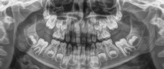
Eight, or wisdom teeth, usually appear on the surface of the gums in adulthood. The shape of the element is similar to 6, but has more powerful roots. The number eight is considered the most problematic tooth: it is difficult to break through to the surface and quickly collapses. Dentists may have difficulty removing figure eights due to their abnormal position in the mouth and crooked roots, which can be diagnosed using x-rays. In such cases, the problem area is immediately extracted, otherwise purulent inflammation of the soft tissues may develop. Another serious complication of improperly positioned wisdom teeth is the destruction of the roots of adjacent units.
No ads 3
Gingival fluid
Gingival fluid is transudate or exudate (more protein compared to transudate) from the connective tissue of the gum and its blood vessels. Normally there is very little of it - 0.5-2 ml; with inflammation this figure increases. The composition of this liquid is very diverse:
- Microorganisms;
- Leukocytes;
- Desquamated epithelium;
- Enzymes;
- Immunoglobulins;
- Other serum and connective tissue components.
It is logical that its composition determines the functions it performs: it cleanses the gingival sulcus, has antimicrobial properties and even improves the attachment of the epithelium to the tooth.
The gingival sulcus (2) is followed by the attachment epithelium (attachment, connective) (3).
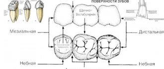
Classification of dental surfaces
To conveniently describe the localization of the pathological process, dentists use a term such as “crown surface”. There are 4 types of such surfaces:
Where is the eye tooth + photo
- Chewable. This part of the tooth faces the elements on the opposite jaw. In canines and incisors, this area is represented by the cutting edge.
- Vestibular. Located in the vestibule of the oral cavity. The vestibular surface of the anterior teeth has an area of contact with the lips. In the molars, this part is in contact with the inner shell of the cheek and is called the buccal surface.
- Lingual or lingual - the area of elements facing the tongue. It is in this area that the largest amount of plaque accumulates due to the difficulty of carrying out hygiene measures. The lingual side of the elements is not visible to others during a conversation, so many patients seek to install corrective systems on the inside of the tooth.
- Approximal. This type of surface is divided into medial and distal. In the first case, the area is located closer to the middle of the dentition, in the second case, closer to the edge of the arch.
Tooth surfaces
Bone tissue
As mentioned above, the jaw bones consist of spongy and cortical bone tissue. Cortical tissue is bone plates, 95% consisting of various mineral salts. These plates are arranged tightly, which is why the cortical bone is hard and durable.
Spongy tissue, on the contrary, is softer, since, for the most part, it consists of soft tissue, has a cellular structure, and the bone plates in it form partitions.
Bone tissue is made up of different types of cells, and each cell performs its own functions. Thus, osteoblasts are responsible for the formation of new tissue, and osteoclasts, on the contrary, for the destruction of old tissue, stimulating the renewal of bone tissue. Osteocytes are the main cells of bone. Interstitial cells are a kind of “lining” of bone.
Features of milk elements
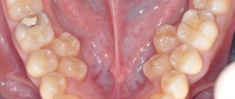
Milk units act as precursors of permanent elements. They usually appear in the first year of a baby's life. Usually the period of eruption of the first incisors is accompanied by painful symptoms for the baby. About 2-3 years may pass between the appearance of the first and the eruption of the last milk tooth. A 3-4 year old child has 20 fully formed baby teeth in his mouth.
The structure of teeth in children
The change of the primary occlusion to a permanent one is carried out from 6-7 years of age. The shape of the permanent elements will differ little from their predecessors. The only difference is the more massive crown part and dense enamel.
In children, the degree of mineralization of enamel and dentin is weaker than in adults. Therefore, their dental care should be carried out with special care. The milk tooth is designed in such a way that most of its volume is occupied by the pulp, so caries in children becomes more complicated faster.
Another difference between milk units and permanent ones is short roots. For this reason, tooth loss in children sometimes occurs unnoticed and painlessly.
Malocclusions
With a correct bite, the teeth of the upper jaw protrude onto the elements of the lower jaw by no more than 1/3. This position will make it easier for the jaws to chop food. Malocclusions are usually associated with the following conditions:
- The upper units overlap the lower ones by more than ½. This type of bite is usually called a deep bite.
- Certain groups of teeth may not fit together at all. In this case, they talk about an open bite.
- Distal bite. What it is? The problem is said to occur when the upper jaw is overdeveloped.
- If the lower jaw is pushed forward, then there is a mesial deviation.
- When the structures are displaced relative to each other, a cross anomaly occurs.
If not treated promptly, malocclusions can cause toothless jaws. For this reason, it is important to contact an orthodontist at the first sign of a problem.
Eruption of canines in infancy
The order of formation of the primary occlusion provides for the appearance of fangs after the incisors and the first chewing units have erupted. The upper elements emerge at the age of 1.2-1.5 years, which is slightly earlier than the period of formation of the lower molars. Late eruption is determined by the deep position of the units in the structure of the jaw, which also determines the characteristic painful sensations that may occur during growth.
To relieve the discomfort experienced by the child, the following techniques are recommended:
- Massage of gum tissue - before the procedure, you need to thoroughly wash your hands, then use your index finger to massage the area above the canine for a couple of minutes. It is recommended to repeat the massage several times a day;
- The use of teethers that provide a cooling effect - a product filled with distilled water does not pose a danger to the baby’s health even if the baby manages to chew the shell;
- Applying anesthetic gels that relieve inflammation within a few minutes;
- For nasal congestion, use children's nasal medications that promote vasoconstriction;
- At elevated temperatures (more than 38 degrees) - take fever medications, available in the form of syrup or suppository.
It is worth emphasizing that the use of medications must be agreed with a doctor, who will determine the appropriate list, as well as the duration of the course.