Disturbance = inflammation
Any disruption of the periodontal ligament and restoration is likely to cause inflammation. Any inflammation in this area can negatively affect the result of the entire work. Understanding the function and location of the ligament increases the likelihood of successful restoration. In particular, factors such as
- position of the gum edge,
- marginal fit,
- contact areas,
- prosthesis shape,
- Iatrogenic damage.
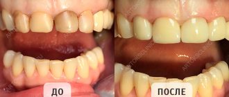
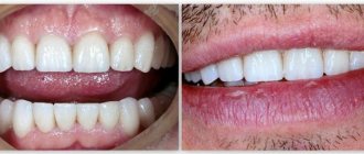
Lack of chewing teeth, difficulty chewing food
Before
Stages
After
Specialists:
Kyalov Grigory Georgievich
Description:
The patient came to the NK clinic on recommendation. Complaints at the time of treatment were the absence of chewing teeth and difficulty chewing food.
After the initial diagnosis, the absence of teeth 17 16 15 14 24 25 26 27 25 26 35 36 37 44 45 46 47 was established. Previously, a bridge prosthesis 34-38 and tooth 48 were installed. Mobility, mobility and pain of the supporting teeth under the bridge prosthesis were noted. The patient was offered a plan for the rapid rehabilitation of the chewing group of teeth using the installation of dental implants in the area of 16 14 24 26 37 34 44 47 teeth and the production of metal-ceramic bridges supported by dental implants. Prosthetics were planned to be carried out with the determination of the optimal position of the lower jaw to create the physiological position of the temporomandibular joint.
Impaired passive eruption
When planning restorations, if the volume/height of the gingival tissue is large, it is necessary to remember the phenomenon of altered passive eruption. Such a violation leads to changes in the connection of the periodontium and restoration, which should be taken into account.
Passive eruption involves displacement of the gingival margin in the apical direction, gradual exposure of the crown and securing the edge in the area of the tooth neck. Violation of this process leads to an expansion of the zone of fixation of the gingival margin, that is, it can “stop” away from the CEJ. According to a study by Garber and Salama (1996), impaired passive eruption occurs in 7-14% of the population, predominantly in patients with thick and flat gingival tissue biotypes.
In 1977, Coslet et al proposed the following classification of passive eruption disorders:
- type 1A – wider band of keratinized tissue, normal bone ridge level;
- type 1B – a wider band of keratinized tissue, a bone ridge at the level of the CEJ;
- type 2A – less wide band of keratinized tissue, normal bone crest level;
- type 2B – a less wide band of keratinized tissue, a bone ridge at the level of the CEJ.
Treatment options depend on the type of passive eruption disorder:
- type 1A – gingivectomy, displacement of the flap in the apical direction;
- type 1B – gingivectomy, bone grafting, flap displacement in the apical direction;
- type 2A – displacement of the flap in the direction of the apex;
- type 2B – bone grafting, displacement of the flap in the apical direction.
If passive eruption disorder remains undiagnosed and is not taken into account when drawing up a treatment plan, the complications mentioned above are possible, that is, periodontal inflammation around the restoration (thick biotype) or gingival recession (thin biotype).
Coronal lengthening
Lengthening the coronal part is a procedure that can minimize the risk of violation of the biological width. When considering the applicability of this procedure to a specific case, one should calculate the feasibility of its implementation in general. Preparation for surgery should include determining the biotype of gingival tissue, assessing its condition, and high-quality x-rays. In some cases, such as severe abrasion/wear of teeth, a sensitivity test should also be performed. A wax up, a wax-up model, is often helpful in discussing the treatment plan with the patient.
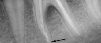
Presence of root caries in teeth and severe dentoalveolar deformations
Before
Stages
After
Specialists:
Kyalov Grigory Georgievich
Description:
The patient came to the clinic on a recommendation from friends who had undergone successful implantological treatment at the NK Clinic.
After an initial consultation, X-ray examination and functional planning, it was proposed to remove teeth 38, 37, 47, 48 and 26. The reason is the presence of root caries in each tooth and severe dentoalveolar deformations, in the presence of which it is impossible to create an effective and correct chewing function. We removed the corresponding teeth and performed simultaneous dental implantation. Three months later, we produced separate ceramic crowns on individual zirconium abutments, supported by dental implants. Thus, we were able to completely restore chewing function.
Biotype of gingival tissue
When lengthening the coronal part, it is important to take into account the biotype of the gum. According to research results, the gingival margin can “grow” along the crown from the level determined during the procedure. This growth is more often observed in patients with a thick biotype of gingival tissue, but there are also individual characteristics. In addition, it is likely that the amount of gingival retraction depends on the position of the flap relative to the alveolar ridge at suturing, as described by Deas (2004). Therefore, if the patient has gum tissue of a thick biotype, it is possible to compensate in advance for the mentioned reverse movement of the edge during the formation of the gum during the operation. A temporary crown should be placed 6-8 weeks after surgery to ensure proper gum formation before the permanent crown is placed.
Medical Internet conferences
Purpose: Selecting the most suitable method for preparing hard dental tissues for permanent restorations in clinical practice.
Objectives: 1) Familiarize yourself with the goals and principles of preparation.
2) Consider methods for preparing tooth surfaces.
3) Consider methods for forming a ledge.
4) Compare preparation methods to determine which is most suitable for use in clinical practice.
1.Purposes of preparation:
1) Preparation of all necrotic and damaged tooth tissues.
2) Maximum preservation of healthy tissues
3) Creation of conditions for retention of a fixed structure
4) Providing functional occlusion
5) Minimizing injuries to the oral mucosa.
Principles:
Preservation of hard tissues. Preparation of a tooth for a crown is accompanied by reduction of a sufficient amount of hard tooth tissue in order to provide space for a sufficient amount of restoration material, especially on the occlusal surface, where tissue reduction can reach 1.5 mm; it is necessary to bevel the functional tubercle (palatal in the upper jaw and buccal in the lower jaw) . It is important to preserve the anatomical shape of the prepared tooth and its group affiliation.
Retention of the restoration is ensured mainly by the shape of the tooth stump, such as: stump height (must be at least 2/3 of the initial crown height), stump area and taper.
The taper of the stump should be on average 6-10*, mainly for the anterior group of teeth. Retention reaches its maximum strength at a taper of 3*. As for the stability of the restoration, the following pattern can be identified: the greater the height of the stump and the smaller the radius, the more stable the restoration will be.
Tooth preparation has a number of contraindications:
1) General: previous myocardial infarction, hypertension, allergies to anesthetics, mental illness, etc.
2) Local contraindications are associated with the possibility of preserving vital dental pulp and are a reason for devitalization.
Preparation principles:
1) Biological aspect - maximum preservation of tooth tissue, elimination of excessive grinding, creation of a supragingival ledge and harmonious occlusion.
2) Aesthetic aspect – to achieve minimal visualization of the metal and maximum thickness of the ceramic coating.
3) Mechanical aspect - creating reliable fixation, stability, eliminating the possibility of deformation of the structure.
2. Preparation methods consist of separate stages, with the help of which in clinical practice the doctor maintains the biological width of the tooth and creates an adequate prosthetic space for restoration.
1) Oblique preparation method according to Martignoni and Schonenberg.
The method involves programming the depth of preparation of the hard tissues of the tooth by means of a saw cut beveled at an angle of 45* to the vertical axis along the cutting/chewing surface; later, the ledge is marked at the same angle, followed by excision of the remaining surfaces.
Pros:
— control of the depth of preparation in the cervical area.
— creation of sufficient occlusal space due to the formation of a beveled surface.
Minuses:
— The line of the ledge on the plaster model can only be determined with sufficient magnification.
— The method requires high manual skills of the doctor and great concentration during preparation.
2) Two-plane preparation method according to Kuwata based on the theory of three planes of the tooth.
The essence of the method is to prepare tissues while maintaining the inclination of the natural planes of the tooth and creating a ledge of 50° to the long axis of the tooth.
Pros:
— Manipulation does not require high manual skills.
— Rational direction of preparation, taking into account the individual topographical characteristics of the tooth.
— Preventive “protection” of the pulp of vital teeth.
Minuses:
— inability to control the depth of preparation.
— Relative consideration of tooth safety zones in terms of its position in the dentition.
3) Method of guiding grooves according to Stein.
The method is based on applying furrows of a certain depth to the prepared surfaces through the use of various calibration burs and further preparing the surfaces to a given depth.
Pros:
— Controlled preparation depth.
— the ability to more accurately take into account safety zones and individual topographical features of teeth.
Minuses:
— The need for accurate calibration of burs and its evaluation with a special measuring tool.
— The position of the tooth in the dentition is not taken into account.
— The impossibility of adequately assessing the depth of reduction of the tooth surface in case of dentition deformations.
4) Gürel's method.
The method is based on the preliminary fixation of a temporary composite restoration on non-prepared teeth, made on the basis of wax modeling. Without removing the composite restoration, we mark the depth of the preparation and then form the core.
Pros:
— Preparation is carried out taking into account the individual characteristics of the tooth.
— Visualization of the thickness of the surface preparation, taking into account the position of the tooth in the dentition.
— Minimization of tooth reduction during preparation.
Minuses:
— Requires additional time and economic costs, equipment and materials.
— Limited use, mainly when using adhesive indirect restorations.
— the difficulty of using the method in the distal parts of the jaw and with pronounced vestibular inclination of the teeth.
5) Method of preparation by tooth segments according to McLean.
The essence of the method is to divide the tooth into segments, the number of which depends on the functional surface of the tooth. First, one half is prepared from the vestibular, oral and occlusal surfaces. Later the second half. Subsequently, the cervical border is formed and finishing is performed.
Pros:
- Ease of manipulation.
— visualization of the thickness of the preparation.
— preparation taking into account the individual topographical characteristics of the tooth.
Minuses:
— relative control of the volume of preparation.
- difficulty of preparation in case of deformation of the dentition.
6) Two-stage preparation according to Massironi.
The essence of this method assumes that at the first stage the tooth is prepared for a certain structure with the location of the border of the closing line above the gum level and the subsequent fixation of provisional crowns. At the second stage, gum retraction is carried out with the final formation of a circular ledge and finishing of the tooth.
Pros:
— Controlled preparation taking into account individual topographic features.
— Protection of the epithelial attachment of the tooth.
Minuses:
— The need for a high level of manual skills, materials, tools, as well as time.
— Difficulty taking into account the peculiarities of the morphometric parameters of the biological zone of the teeth when forming a circular ledge.
3. Formation of a ledge is the most difficult stage in the preparation process, both its localization and choice of shape. The shape of the ledge determines: the amount of restoration material, marginal fit, the possibility of correction and constant care for it.
Principles of ledge formation:
Only with the correct connection and adaptation of the boundaries of the ledge and the orthopedic structure is its durability, functionality and aesthetics possible.
The ledge options are determined by the choice of design:
1) The groove-shaped ledge is used for MC and ceramics.
2) Ledge 120-135* for MK.
3) Shoulder shoulder 90* with an internal right angle for MK and ceramics.
4) Shoulder shoulder 90* with rounded internal corner for ceramics.
Also, the ledge is selected depending on the periodontium and hard tissues of the tooth.
There are two types of ledge arrangement:
1) Subgingival. Indications are aesthetic requirements, carious lesions and hyperesthesia in the area of the necks of teeth, low crown part, subgingival tooth fracture, repeated prosthetics.
2) Supragingival and at the level with the gingival margin. They are used in cases of low lip line, absence of visual necks of teeth in the smile, likelihood of complications from periodontal tissues, high clinical crown, generalized periodontitis of moderate severity, absence of previous sublingual preparation.
In order to minimize complications from periodontal tissues, the preparation of the ledge requires compliance with the biological width.
Biological width is a complex of tissues located above the alveolar ridge, including connective tissue and tooth-attached epithelium, which fill the space between the bottom of the sulcus and the alveolar ridge.
The edge of the subgingival preparation should end in the area of the middle of the periodontal sulcus.
The formation of the final ledge is carried out after installing the retraction thread with an increasing tip at low speeds.
Conclusion: Having considered all the above preparation methods, we can draw the following conclusion: due to the variety of clinical cases, indications and contraindications, it is impossible to choose one best preparation method. You should also take into account the huge variety of materials, manufacturing technologies for fixed dentures and the different technical capabilities of medical institutions. Based on this, in practice it is permissible to use any of the above methods, which is most convenient for the attending physician to use and allows one to obtain the most positive result for the patient’s health.
Gum enlargement after orthodontics
After orthodontic treatment aimed at lengthening the coronal part, enlargement of the gums is often observed, i.e. its edge rises above the desired level. In such cases, you should not immediately take up a scalpel and surgically remodel the gum. The gums will return to the desired shape on their own within 3-6 months after treatment. At these marks it is already possible to assess its condition and decide on the need for surgical correction of the edge.
In the “Orthopedics” department of the Stommarket.ru online store you will find a variety of materials necessary for successful restorations of various levels of complexity:
https://stommarket.ru/catalog/ortopediya/
Nice prices and excellent delivery conditions!