The root system of the tooth and knowledge of its anatomy are an important factor for successful endodontic treatment. The complex anatomy of the canals can significantly complicate treatment.
The main goal of endodontics is complete obturation of the root canal. For successful treatment, it is necessary to clean the canals and tooth cavity from necrotic debris, bacteria and their waste products. We can achieve this through careful mechanical and chemical treatment of the canals.
Persistence of infection due to a missed canal or incomplete treatment of a difficult-to-reach canal can lead to poor results. It is important to study the anatomy of the tooth before treatment to avoid complications.
Anatomy of mandibular molars
In the permanent dentition, the first molars of the mandible have the greatest chewing force and are therefore the most functionally significant. Most mandibular molars are double-rooted with two mesial and one distal canals. But the number of roots and canals may vary.
Additional root
Carabelli was the first to note such a frequent change as an additional root. It is located on the lingual ( radix entomolaris) or buccal ( radix paramolaris ) sides.
Three-rooted mandibular first molars require special attention during endodontics because the accessory root is usually smaller and thinner than the others. Or such a root is partially fused with other roots and is strongly curved.
Identification
To identify additional roots, an x-ray is needed. But sometimes several pictures from different angles are necessary, since one picture may be uninformative.
Rice. 1 - SLOB method allows you to take pictures from different angles.
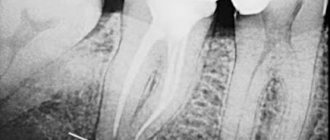
Rice. 2 – On the radiograph we see the accessory root (Radix Entomolaris) of the first molar of the lower jaw.
Rice. 3 – We see the mouth of the additional canal (Radix Entomolaris), located distally lingually. Asymmetry and deviation from the midline are noticeable.
How can root canals cause pain?
- deep caries, which affects the tooth enamel and forms a carious cavity, which becomes the cause of serious dental disease;
- infection entering the pulp can inflame it. As a result, the pulp loses its vitality, so pulp disease must be treated. Inflammation of the pulp leads to inflammation of the soft tissues of the gums - periodontitis. Signs of pulp disease: acute pain, swelling of the gums, the tooth reacts to cold and hot food.
Root curvature
There are 3 types of Radix Entomolaris based on root curvature:
Type 1: Straight root/root canal
Type 2: Curved entrance to the root canal, the rest of the canal is straight.
Type 3: The canal is curved in the upper and middle third. Moreover, the root deviates towards the buccal side. It is also not uncommon for the apical part of Radix Entomolaris to deviate by 90°.
Fig.4
Overlaying roots on a photo
Classification based on the degree of root overlap (distal-lingual and distal-buccal) on x-ray:
Type 1: Slight overlap
Type 2: Moderate overlap
Type 3: Significant overlap
Rice. 5
How to save teeth?
- To keep your teeth healthy, you need to properly organize your diet, including foods rich in proteins, amino acids, vitamins, calcium and phosphorus.
- Carry out oral and dental hygiene regularly and thoroughly. Change your toothbrush every three months, choosing one with bristles that are more suitable for your teeth. Choose a good toothpaste and teeth rinse. You should definitely brush your teeth immediately after sleep and before bed. After each meal, you should rinse your mouth or chew gum.
- If your teeth are healthy, you should visit a dentist once a year, who will conduct a visual examination of your teeth, remove plaque or tartar, and give recommendations on how to care for your teeth.
Be that as it may, if there is pain or discomfort in the teeth, then you need to seek help from a dentist. This will help to eliminate the problem in time and get rid of the causes of pain.
Endodontic treatment of lower molar (Clinical case)
Rice. 6 – By curvature – 1 type, by overlap – 1 type
Rice. 7 - By curvature - type 2, by overlap - type 2
Rice. 8 - By curvature - type 3, by overlap - type 3
Rice. 9 - By curvature - type 3, by overlap - type 3
Complications of endodontic treatment
Accessory roots of mandibular first molars are a common occurrence. In addition, such roots are often very curved. Therefore, to prevent complications of endodontic treatment, it is necessary to find such roots and study their anatomy. In this case, you cannot do without x-rays.
The translation was made specifically for the OHI-S.COM website. Please, when copying material, do not forget to provide a link to the current page.
conclusions
1. According to CBCT data and a dental microscope with a magnification of up to ×40, the anatomy of the root canals of the distal root of the first lower molar directly depends on the patient’s age and changes significantly throughout life.
2. Modern cone-beam computed tomography makes it possible to obtain high-definition images of root canals with a radiation dose in the range of 1.79–3.5 mGy. In turn, visualization of the anatomical and topographical features of the structure of the root canals of teeth even at the stages of treatment planning makes it possible to determine the frequency of visits, the choice of instruments, the technique of filling root canals, as well as reduce the percentage of errors at all stages of treatment and make endodontic treatment more predictable.
The authors declare no conflict of interest.
The authors declare no conflict of interests.
Non-standard anatomy of the mandibular incisor canals in an individual patient
In clinical practice, there is a very significant divergence of tooth root canal systems. Different teeth may have different numbers of root canals, and their anatomy and relationships continue to be the subject of research to this day. This article describes a clinical case of successful endodontic treatment of all four mandibular incisors, with each tooth having four canals of different morphology. X-ray examination revealed the presence of several canals in the central and lateral incisors of the mandible. According to the literature, the presence of several canals in the central and lateral incisors of the mandible is quite rare; The uniqueness of this clinical case lies in the fact that in each of the four incisors two canals of different anatomy and morphology were found.
Introduction
The success of endodontic treatment directly depends on a thorough knowledge of the external and internal anatomy of the tooth. One of the reasons for the negative outcome of endodontic treatment is ignorance of the types of morphological diversity of the tooth root canal system. Mandibular incisors typically have one root and one canal, but anatomical variations occur - two canals with two apical foramina and two canals opening into one apical foramen.
Before commencing endodontic treatment, it is critical to visualize all internal anatomical relationships of the canals. It is mandatory to take and carefully study two or more periapical radiographs. To obtain information about the morphology of the tooth root canal, radiographs must be performed in direct, lateral, and also angular projections. The diagnostic minimum examination of the patient includes:
- Taking two or more radiographs before surgery;
- Assessing the bottom of the pulp chamber with a sharp probe;
- Channeling cracks with an ultrasonic nozzle;
- Staining the bottom of the chamber with a 1% solution of methylene blue;
- Performing a sodium hypochlorite test (“champagne bubble tests”);
- Visualization of the source of bleeding from the canal.
All of these methods are used to identify additional holes in the canal.
Stropko recommends, before beginning a visual inspection of the root canal system, to clean and dry the bottom of the pulp chamber using a 17% aqueous solution of ethylenediaminetetraacetic acid (EDTA), a 95% ethanol solution, and a Stropko irrigator filled with a 27G endodontic needle with lateral perforation.
The use of an intraoperative dental microscope also plays a major role in visualizing the root canals. It saves time and allows the operator to selectively and accurately remove dentin, minimizing the number of errors arising from operating errors.
Rice. 1. X-ray taken before surgery.
Rice. 2. Vertucci classification.
Description of a clinical case
A 45-year-old man was admitted to the clinic with complaints of high tooth wear, which caused the formation of a periapical abscess of both central incisors and irreversible pulpitis of the lateral incisors. X-ray examination revealed the presence of several root canals in each of the four incisors (see Fig. 1). Surgical access was performed, the root canals of the teeth were inspected, after which it turned out that all four teeth had two separate canals - buccal and lingual. According to the Vertucci classification, the canals were classified as type II (31 and 32) and type IV (41 and 42). The working length was determined with an apex locator (Propex, Dentsply) and confirmed by radiographic data. The relationships between the canals within each tooth were determined radiographically (see Fig. 3). [Figure No. 3 is missing from the original document]
Rice. 4. Working length.
Rice. 5. Obturation.
To ensure that no additional root canal was missed, angular radiographs of the teeth were taken. After confirming the presence of two canals in all lower incisors, the canals were prepared using the “Step Back” technique. At each instrument change, 2.5% sodium hypochlorite solution and saline solution (0.9% sodium chloride solution) were used as irrigants. Calcium hydroxide was injected intracanally; the access cavities were temporarily filled with neutral restorative material for 1 week. At the second visit, the cavities were washed and filled (see Fig. 4). After 6 months, a radiograph was taken at the follow-up examination (see Fig. 5).
Rice. 6. After 6 months.
Discussion
The anatomy and morphology of the root canal system is quite complex, and the success of endodontic therapy depends on the timely detection of an additional root or root canal. Canals not detected in time are one of the main reasons for the negative outcome of endodontic therapy. Also, incomplete removal of irritant substances from the pulpous space can lead to a negative outcome of treatment.
In most cases, two root canals merge into one near the apex. In order to be confident in detecting all additional channels, it is necessary to carefully study and interpret radiographic data (which, in turn, must be performed not only in direct, but also in angular projections). If the additional canal ends suddenly, you should think about the presence of two canals in the tooth.
Manual examination of the tooth root canal system using an endodontic probe or guide is also a fairly reliable research method that allows you to determine the configuration of the tooth root canals, in particular the number of holes. Particular care should be taken when accessing the cavity, since it is at this moment that it is easiest to miss the additional canal opening. On the lower front teeth, it makes sense to also examine the buccal and lingual walls of the cavity.
Numerous antibacterial agents are recommended for internal coating. One of the simplest and most accessible methods of cavity disinfection is calcium hydroxide paste, which was also used in the case described above.
This article describes a successful case of root canal treatment for all four mandibular incisors, each with two separate canals, in two teeth (31 and 32) opening into the same apical foramen, and in two teeth (41 and 42) opening into different apical foramina.
Conclusion
The anatomy of the root canal system can be quite complex. There may be differences in canal configuration, number of canals, and presence of narrowing sites. For successful diagnosis and treatment of such canals, it is necessary to carefully study the anatomy and morphology of the tooth root canals before performing surgery, including taking additional angular radiographs. An integrated approach to choosing a method of surgical access will also help in clinical practice.
Authors:
Sudha Mattigatti , Rushikesh Ramesh Mahaparale , Rutuja Vijay Chopade , Vaibhav Garg (Department of Conservative Dentistry and Endodontics, Faculty of Dental Sciences, KIMSDU, Karad, Satara, Maharashtra. India)