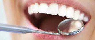
Dental implantation is one of the best methods for restoring lost teeth and allows you to completely restore the anatomical and physiological features of the structure of the dentofacial apparatus. Despite the fact that dental implantation today is performed at a high quality level and is devoid of any disadvantages, sometimes patients still encounter some problems.
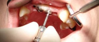
One of them is a change in the color of the gums near the dental implant. The Apex dental clinic uses dental implants from the world's leading manufacturers. They boast of their characteristics and allow you to achieve all your treatment goals.
The clinic’s experienced doctors do not make mistakes in their work, which guarantees impeccable quality of dental services with no negative consequences after the installation of dental implants.
And since doctors at the Apex dental clinic are often asked the question of why the gums near the dental implant turn black, today we will talk about exactly this, paying attention to ways to prevent the development of such a complication. Let's start, perhaps, by identifying the possible causes of darkening of the gums after installation of the implant.
Gingivitis: my gums hurt
The process, which is characterized by gum inflammation, swelling, redness and bleeding, is called gingivitis and is one of the most common periodontal diseases in both children and adults. Only 3% of people can boast of absolutely healthy gums. How to get into such a small percentage of lucky ones? The answer is simple - follow the necessary measures to prevent gum inflammation, regularly visit the doctor and not let even seemingly harmless signs of an incipient disease take their course.
Gingivitis is the last of all diseases in periodontology in which the inflammatory process can still be stopped. Next comes periodontitis, a disease in which inflammation spreads to other periodontal tissues. From this point on, treatment is based only on bringing the disease into remission and attempts to relieve symptoms as much as possible at the time of relapses, as well as in the future when it is necessary to resort to tooth extraction. Therefore, gingivitis in children and adults requires increased attention in order to avoid serious periodontal problems.
Questions for the doctor
Teething
Tell me, my son has erupted one upper incisor, and in place of the second, the gums have slightly swollen and a dark spot has appeared. Do I need to take any measures or just wait until the tooth erupts?
The appearance of an eruption cyst at the site of a future tooth is a common occurrence. And usually such a cyst goes away on its own, without outside intervention. But to monitor the condition of the child’s teeth and gums, it is necessary to show him to a pediatric dentist.
Hematoma on the gum
My daughter has a small black dot under one of her lower incisors. We can see a doctor only in a week. What could this be and does it need to be treated somehow?
If, apart from the black dot on the child’s gum, nothing bothers him, there is no swelling or redness, then most likely this is the result of mechanical damage to the gums from a fall or impact. Such a hematoma usually resolves on its own within a few days, so no additional therapeutic measures are required.
Types of gingivitis
Gingivitis differs in the nature of its course:
- Acute gingivitis is a disease whose symptoms appear suddenly and progress quite quickly.
- Chronic gingivitis is a sluggish process, the symptoms of which increase gradually.
- Aggravated gingivitis (recurrent stage of a chronic process) is an increase in the symptoms of a chronic disease.
- Gingivitis in remission is the moment of complete relief of all symptoms.
The form is:
- catarrhal gingivitis, which is manifested by swelling and redness;
- ulcerative (ulcerative-necrotic) gingivitis, with necrotic (dead) areas of the gums;
- hypertrophic gingivitis, in which there is a significant increase in the volume of gum tissue and its bleeding;
- atrophic gingivitis, on the contrary, is characterized by a decrease in the volume of gingival tissue;
- desquamative (geographic) gingivitis, which is manifested by intense redness and abundant desquamation of the epithelium of the mucous membrane.
According to its distribution in the oral cavity, gingivitis can also be local (affects some areas of the teeth) and generalized (the process affects the gums of the entire jaw or both jaws). And according to severity - mild, moderate and severe gingivitis.
Consequences of cervical caries if left untreated
Advanced caries near the gums is very dangerous, it can progress quite quickly and cause complications such as:
- periodontitis;
- pulpitis;
- inflammation of the gums;
- an abscess or a well-known flux.
In addition, the development of caries in the gums is often associated with pathologies of the thyroid gland and diabetes mellitus. It is necessary to conduct a comprehensive diagnosis to determine the exact cause of the disease. After the therapy prescribed by the endocrinologist, the disease may recede.
Causes of gingivitis
Most often, gingivitis develops as an independent disease, but sometimes the causes of its occurrence are acute and chronic diseases of the gastrointestinal tract, cardiovascular system, hematopoietic organs, infectious diseases, and changes in hormonal levels. In this case, gingivitis is one of the symptoms of the underlying pathology. The causes of gingivitis can be internal or external.
Internal reasons include:
- tooth growth that injures the gums - the eruption of wisdom teeth;
- vitamin deficiency, hypovitaminosis (most often lack of vitamin C and zinc);
- weakened immune system;
- metabolic disease;
- allergic diseases;
- diabetes;
- stress, mental illness;
- anomalies and various deformations of the gums;
- diseases of the gastrointestinal tract.
External reasons are a number of factors:
- physical (injuries, burns);
- chemical (the influence of aggressive substances);
- medical (incorrectly applied fillings, incorrectly installed veneers, traumatic wearing of braces);
- bad habits (smoking, mouth breathing);
- biological (infectious process);
- hygienic (insufficiently thorough hygienic procedures).
Toxins from microorganisms that enter the oral cavity with food and water, as well as those that live there permanently, form dental plaque (plaques) due to insufficient hygiene measures. They are the most common cause of the inflammatory process.
Inflammation can develop differently depending on the cause. Chronic catarrhal gingivitis occurs most often due to unsatisfactory hygiene measures or as a result of gum injury or burns. Hypertrophic gingivitis is caused by crowded teeth, incorrectly installed fillings or dental crowns, as well as changes in hormonal levels, for example, during pregnancy (pregnant gingivitis) or puberty (adolescent or juvenile gingivitis). Necrotizing ulcerative gingivitis (Vincent gingivitis) is usually caused by an infectious process. It occurs due to the activation of two microorganisms (Vincent spirochete and spindle bacillus) against a background of weakened immunity, hypothermia, stress or malnutrition.
Forms of gingivitis and symptoms
Signs of gingivitis directly depend on the nature of the disease and its form. Let's look at each form of gingivitis separately. So, complaints and visual inspection.
Catarrhal gingivitis
This form of the disease usually occurs without obvious pain. Its immediate symptom is bleeding gums when brushing teeth, eating solid foods and other mechanical effects on the dental system.
Ulcerative-necrotizing gingivitis
This is one of the most unpleasant forms of gingivitis, which is characterized by a feeling of itching of the gingival papillae, severe pain, copious flow of saliva, fever, inflammation of the lymph nodes and the formation of necrotic areas of the gums.
Hypertrophic gingivitis
Patients suffering from this form of gingivitis complain of severe pain, constant bleeding of the gums and a significant increase in the volume of the gums, which can partially cover the crowns of the teeth from the outside (not from the tongue). At the same time, the patient’s gum remains quite hard and under it, on the teeth, tartar forms, which creates favorable conditions for the proliferation of microorganisms. With hypertrophic gingivitis, teeth may move slightly.
Atrophic gingivitis
The last and most advanced stage of gingivitis, often leading to periodontitis, is atrophic gingivitis. With it, the gum tissue becomes thinner, decreases in size, the necks of the teeth, and sometimes their roots, are exposed. Teeth become more sensitive to temperature changes (cold or hot drinks, frosty air), to sour or sweet foods, to the mechanical impact of a toothbrush.
Desquamative (geographic) gingivitis
The symptoms of this form of gingivitis differ from others by pronounced red spots on the gums, desquamation of the upper layer of the epithelium, the appearance of blisters on the gums and the formation of mouth ulcers and erosions.
Professional help
Treatment begins with diagnosis and determining the causes of the pathology. After eliminating the causes that provoke the darkening of the enamel, appropriate treatment is prescribed.
The method of treating cervical caries depends on the stage at which the pathology was noticed:
- At the dark spot stage, remineralizing therapy is used: the tooth is cleaned of soft plaque and hard dental deposits, and the affected area is covered with an application containing fluoride.
- In case of superficial and medium caries, the affected area is prepared, after which the tooth is filled.
- At the stage of deep caries, as a rule, the pulp is removed and the canals are cleaned. After this, the tooth is filled.
Diagnostic tests
- Schiller-Pisarev test
This test is based on determining the level of glycogen in the gum. Its amount increases significantly during inflammation, while healthy gums do not contain glycogen. Lubricating the inflamed gums with Schiller-Pisarev solution gives a color change reaction from light brown to brown. This research method is used to make diagnoses of both periodontitis and gingivitis.
- Assessment of oral hygiene level
A solution (2 g of potassium iodide, 1 g of crystalline iodine, 40 ml of distilled water) is applied to the outer surface of the six lower front teeth.
The assessment is carried out using a five-point system and each tooth is assessed separately:
- 5 points – complete staining of the entire tooth surface;
- 4 points – staining of ¾ of the tooth surface;
- 3 points – staining of half the tooth surface;
- 2 points - staining of a quarter of the tooth surface;
- 1 point - absence of any staining of the tooth surface.
Then the scores of all examined teeth are summed up and divided by their number (usually the test is carried out on six teeth). This is how the hygiene index is obtained.
As a result, the quality of hygiene is assessed:
- 1.1-1.5 points – good hygiene index;
- 1.6—2.0—satisfactory hygiene index;
- 2.1—2.5—unsatisfactory hygiene index;
- 2.6—3.4—poor hygiene index;
- 3.5-5.0 - very poor hygiene index.
- Vacuum test according to Kulazhenko
Using a Kulazhenko vacuum apparatus, it is possible to determine the time of hematoma formation when a vacuum is applied to the gum area. Typically, the test is carried out in the incisor area by placing a tube of the device on the gum. The formation of a hematoma in 60 seconds indicates the normal condition of the gums, while the appearance of a hematoma in 29-30 seconds signals an inflammatory process.
- Oxygen tension in gum tissue
The sensor of the device is applied to the gum, and the device records the level of tissue hypoxia. Reduced oxygen tension indicates a prolonged inflammatory process.
Features of the disease
This type of caries is especially dangerous and unpleasant, as it has some peculiarities in its course and location. Its manifestations are associated with the following factors:
- Carious lesions are localized in the weakest area near the neck of the tooth (this is the part covered by the gum). In this zone, the enamel is also weakly mineralized, and this factor enhances the development of caries.
- Circular distribution. Cervical caries affects the tooth in a circle, covering increasingly larger areas. As a result, part of it may break off, since the integrity will be broken in many places.
- Damage to the front teeth. An unpleasant feature of gingival caries is its frequent location on the incisors. A person experiences psychological discomfort when he has to talk, smile, laugh.
Under the influence of the bacteria Streptococcus Mutans, which causes the development of the disease, the enamel and dentin underneath are actively destroyed. Inflammation can spread to the pulp and ultimately lead to pulpitis or periodontitis.
Differential diagnosis of gingivitis
It is based on the complaints presented to the patient, a visual examination of the patient, the results of functional tests and laboratory tests. The goal of differential diagnosis is to distinguish gingivitis from other periodontal diseases, such as periodontitis and periodontal disease.
The main feature that distinguishes gingivitis from other periodontal diseases is that the inflammatory process affects only the gum tissue, the remaining structures (muscle ligaments that hold the tooth in the jaw and bone tissue) remain unchanged.
Along with this symptom, gingivitis is not characterized by periodontal pockets, exposure of the necks of teeth, or their mobility. And the x-ray shows no signs of bone resorption.
Identifying gingivitis in a timely manner, determining its form and prescribing the correct treatment is the task of a periodontist. But not to forget about prevention and regularly visit the dental clinic is the maximum program for the patient. This is the only way to avoid a more serious periodontal disease – periodontitis.
Prevention of subgingival caries
The development of the disease is provoked by many external and internal factors. And even if caries has never bothered you, preventive measures will help prevent its occurrence and development. Here are some simple rules doctors recommend following:
- Brush your teeth with toothpastes high in minerals.
- After each meal, use a mouthwash and dental floss - it will help effectively remove pieces of food stuck between your teeth. Oral irrigators can also be used.
- Half an hour after eating, brush your teeth if possible. This should be done twice a day for a couple of minutes.
- Include in your mandatory daily diet foods rich in fluoride, calcium and other beneficial microelements to strengthen enamel, eat more solid vegetables and fruits.
- Avoid excessive consumption of sweets, flour and sour foods.
- Periodically massage the gums: it improves blood circulation in the gum tissues, protects against the formation of deep periodontal pockets and the accumulation of deposits in them, which reduces the risk of caries formation at the neck of the tooth.
If the enamel is thin and susceptible to the rapid formation of tartar on it, regular remotherapy helps a lot - this is the name for applying fluoridating compounds to the teeth. The procedure reduces the risk of enamel destruction and improves the condition of hard tissues. To further strengthen the enamel, you can periodically take special vitamin complexes containing useful minerals. In addition, it is necessary to visit the dentist’s office at least once every six months for a dental examination. The doctor will make sure that everything is fine with them, or will prescribe additional diagnostics using x-rays. Once every 6-9 months, it is recommended to carry out professional enamel cleaning using ultrasound or other methods. During the procedure, plaque and tartar are removed from the surface of the teeth. It polishes and becomes smoother, and less deposits accumulate on the enamel. During cleaning, the doctor can also clean periodontal pockets if plaque and deposits accumulate under the edge of the gums.
People who are at risk for endocrine diseases need to be especially attentive to pain in their teeth. With them, teeth are often affected by cervical caries. Treatment must be comprehensive and systemic to prevent caries from damaging the tooth root. Gingival caries is a dangerous pathological process if it is started, but it can be successfully treated in the early stages.
Modern dentistry eliminates caries of various types, but it also happens that if the stage of the disease is too deep, some teeth cannot be saved. We have to remove them and resort to various methods of restoring the dentition. You just need to be attentive to your oral health, taking simple preventive measures, so as not to have to deal with expensive treatment later. With regular dental examinations, you will be able to detect the problem in time and solve it.