Home / Articles / Teeth falling out - what to do
The absence of a tooth, especially the front one, always provokes a lot of problems. The main one is a decrease in a person’s self-esteem, because in the absence of a beautiful smile, everyone will avoid communication. At the same time, tooth loss can be accompanied by numerous physiological problems. The contours of the face may noticeably change, and insufficient chewing of food can even provoke disturbances in the functioning of the stomach, although even this is not the worst consequence.
Sign up for a free consultation >>
Why do teeth fall out
If your teeth begin to fall out, think about the reason for their loss. There are several common reasons why teeth fall out in humans:
Caries
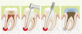
Caries disease is accompanied by a violation of the mineralization of teeth, which ultimately destroys bone tissue in the oral cavity. Inside the tooth, the cavity gradually grows and becomes empty, which leads to acute sensitivity and food particles getting inside the teeth. Ignoring the symptoms of caries and unwillingness to consult a professional dentist ultimately leads to the fact that the tooth completely ceases to perform its function. The only solution in this case is to pull out the tooth.
It is possible to avoid caries by brushing your teeth properly and using toothpaste rich in fluoride. If even small holes appear in the teeth, it is recommended to immediately fill them in a specialized clinic.
Periodontitis
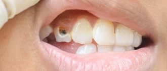
The first symptoms of the disease are bleeding in the gums. Next, an unpleasant odor appears in the oral cavity and a purulent process begins when pressure is applied to the gums. After some time, the affected tissues become completely incapacitated. Therefore, if your tooth falls out with blood, often the reason lies precisely in periodontitis.
Prevention of the disease in this case is quite simple: monitor the health of your gums and always visit the dentist in a timely manner, without waiting until the pathology becomes incurable. After each meal, try to brush your teeth for three minutes using special dental floss and toothpaste.
Chronic diseases
The reason why teeth fall out without pain and blood, and sometimes with them, can also be inflammatory processes as a result of chronic diseases. Common causes are hypertension, thyroid problems, diabetes, and respiratory problems. Often, aggressive methods of treating serious diseases, in particular chemotherapy, can provoke dental problems.
Negligent oral care, improper diet
It's no secret that citrus drinks can deplete enamel, which causes erosion. Gradually, this stimulates the development of caries and leads to the final loss of the tooth. A similar effect can occur if you carelessly eat nuts, crackers and other hard items. Clefts appear in the teeth, they become loose and subsequently fall out. Try to avoid eating excessively hot and cold foods.
Tobacco products, drugs
About a third of patients who systematically lose teeth are people with bad habits, in particular, heavy smokers. It is reliably known that drugs and tobacco not only worsen the color of teeth, but can also be the reason why molars fall out.
Recommendations for everyone
Are there any general recommendations that are effective in all cases?
Yes, I have. The first and most important thing is not to panic. Usually, tooth loss is accompanied by panic and surprise. Especially if an unpleasant event occurred during a meal or during another event, when nothing foreshadowed trouble.
When panicking, a person makes mistakes that will still have to be corrected later. Therefore, the first and most important recommendation is not to panic.
The second recommendation is to trust dentists. It is better not to self-medicate, but to turn to specialists. Let them examine, make a diagnosis, and do everything necessary to keep your mouth healthy. After all, health is not something that can be entrusted to amateurs, not professionals.
Otherwise, healthy teeth and may none of the readers ever need dental services.
Why teeth fall out in adults and children.
Tooth loss is a bad sign that most often causes anxiety in people. The problem is, of course, serious, but it also has various solutions.
Tooth loss in children
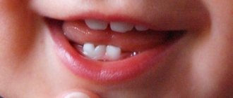
When (at what age) do baby teeth fall out? Typically, the first milk teeth fall out in children aged 6-7 years, but deviations from the timing are possible based on the individual characteristics of the body. Although baby teeth erupt at the age of 6 months, their formation begins in the uterine period during the mother’s pregnancy. With the birth of a child, the formation of the rudiments of permanent teeth begins, which is why it is important to monitor the health of the milk teeth, because infection with caries can easily harm the rudiments of permanent teeth.
Do teeth fall out on their own?
Yes, they fall out if they are dairy. In this case, changing teeth can be accompanied in children by acute painful sensations, swelling of the gums and sensitivity of the enamel, and in some cases even itching. If you experience these symptoms, it is important to seek the advice of a professional dentist. At the same time, while waiting to see which tooth falls out first, you should not forget about the child’s adequate nutrition so that the body and teeth in particular continue to receive all the necessary vitamins and microelements.
When a baby tooth falls out, the wound may continue to bleed for 5-10 minutes. Simply give your child a sterile gauze or cotton swab to bite on the area where the baby tooth has fallen out. If bleeding continues for a long time, be sure to take your baby to the pediatrician to check blood clotting. The help of a dentist may also be needed when baby teeth complicate the eruption of permanent teeth. In this case, the teeth are forcibly removed.
Preservation of teeth with root fractures
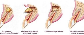
Primary root fractures (PRF), traditionally referred to as “vertical root fractures,” are defined as “a complete or incomplete fracture that occurs in the root region of a tooth at any level, and is usually in a buccolingual direction.” It is logical that this definition of PPC is based on its anatomical location.
Since almost all teeth with PCD are pre-treated endodontically, it is reasonable to assume that there is some connection between the characteristics of endodontic intervention and the development of PCD. Based on the nature of the occurrence of PPC, the following definition was proposed: “a complication of endodontic treatment, which is characterized by complete or incomplete fracture of the dental tissues, begins in the root structure at different levels, and usually spreads in the bucco-lingual direction.” It is fundamentally important to understand that although PPC is a complication of endodontic treatment of a tooth, this does not indicate that PPC is directly related to any type of medical error during the procedure. And although treatment errors can lead to complications in the form of a fracture, not all root fractures are caused by procedural errors by the dentist, and some of the fractures develop even after completely adequate root canal treatment. Considering that PPC develops both among teeth after high-quality endodontic intervention, and in the structure of teeth after poor-quality canal treatment, we can conclude that the expanded definition of PPC includes both purely clinical and medical-legal aspects of assessing the quality of endodontic interventions.
The prevalence of PPC reaches 11-30%. A recent study by Yoshino et al in 2015 found that high prevalence of PPC was the leading cause of removal of endodontically treated teeth. In an analysis of 736 teeth from 24 different clinics, the authors were able to determine that 626 of them were extracted within 6 months after treatment, while 233 teeth were extracted as a result of the diagnosis of PPC (31.7%). Anatomically, RPCs often develop lateral to the root canal wall toward the outer root surface. Incomplete fractures extend only on one side of the root surface, while complete fractures extend in both directions, affecting two surfaces. Occasionally this leads to the root splitting into separate segments.
Timely diagnosis and proper treatment of such complications are crucial to prevent loss of alveolar bone, which can complicate further manipulations, including the possibility of dental implantation. However, the clinical and radiological diagnosis of PPC is quite difficult, and in some cases, the correct diagnosis can only be made through direct visualization of the root after surgical separation of the flap (Figure 1).
Photo 1. Diagnosis of tooth root fracture during direct imaging after flap separation.
Traditionally, teeth with PCD were considered hopeless from the point of view of functional prognosis. Possible treatment approaches for PPC have included tooth replantation after bonding the two fractured segments, but such approaches are not sufficiently evidence-based to be implemented in daily clinical practice. Therefore, the remaining treatment alternative was extraction.
For many years, there has been an ongoing debate among clinicians about whether it is worth trying to preserve a compromised tooth as much as possible, or whether it is better to remove it and immediately proceed to the dental implantation stage. In recent years, given the level of prevalence of peri-implantation complications, it has been suggested that the treatment of one’s own teeth, even those compromised by a fracture, is simpler and more predictable compared to the treatment of peri-implantitis, the problem of which continues to progressively increase in modern dental practice.
In recent years, a number of new algorithms have also been proposed, including promising alternatives for the treatment of teeth with PCD, which until now have traditionally been referred to extraction. These unique single-unit treatments for teeth with root fractures are in the early stages of development and refinement, and data on them are mainly based on single publications. However, modern endodontics offers a number of technical means such as devices for magnification and lighting, as well as a set of modern materials such as bioceramic cements, which expand the possibilities for the restoration of teeth with root fractures with the correct selection of appropriate clinical cases with a clear understanding of the difference between which tooth can still be saved and which can no longer be saved. This article will discuss modern possibilities for treating teeth with PPC.
The importance of choosing a clinical case for treatment
Modern endodontics provides many treatment options that can save compromised teeth. However, PPC still poses a diagnostic and therapeutic challenge for dentists, as root fracture is the reason why most endodontically treated teeth have to be removed. In addition, with the development of modern endodontic treatment options, new dilemmas have emerged regarding the functional prognosis of treated teeth. The topic of discussion remains the decision about the possible preservation of a compromised tooth with PPC or its extraction with restoration of the edentulous area using a dental implant. Traditionally, it was believed that the sooner a tooth with a root fracture is removed, the better, because the presence of one in the jaw provokes the development of inflammation, which, spreading to the periodontal area, provokes the formation of a destructive bone defect. Under such conditions, bone loss, in turn, compromises the prognosis of the future implant, which can be installed in place of the problematic tooth. However, each individual clinical case should be assessed objectively, since some data have shown that with an adequate selection of a suitable clinical situation, teeth affected by a root fracture with a limited distribution do not provoke the development of inflammation in the surrounding periodontium over a fairly long period of time. The selection of clinical cases of PPC suitable for conservative treatment should be based on evidence-based protocol data, taking into account the completeness of the diagnosis and available therapeutic options.
The dentist must also take into account the aesthetic needs of the patient, the duration of functioning of the treated tooth and the costs required for its restoration. When making decisions about possible conservative treatment of teeth with PCD, one should take into account not only endodontic aspects, but also periodontal, prosthetic and aesthetic components of the patient’s rehabilitation. Many factors, such as the location of the tooth, the presence of predisposing periodontal disease, the type of coronal restoration and the possibility of using modern endodontic materials and instruments, must be taken into account by the doctor when assessing the risks and benefits of conservative treatment of teeth with a root fracture.
In cases of development of PKD in multi-rooted teeth, there may be no need to preserve the root with a fracture, since it can simply be amputated. With single-rooted teeth, the situation is different, and the functional prognosis of such teeth depends on the adequate choice of treatment method. Predisposition to periodontal pathology negatively affects the prognosis of teeth with PCD. Thus, during the initial diagnosis of teeth with root fractures, it is necessary to carefully assess the presence and severity of possible adjacent periodontal lesions.
The inability to preserve a tooth (even if one with a root fracture) in the dentition significantly reduces the patient’s aesthetic profile. And although in most clinical cases implants take root absolutely without problems and serve successfully for many years, the long-term prognosis of such implants must also necessarily include the parameters of the achieved aesthetic rehabilitation. After all, if disintegration of the implant occurs in the frontal area, then the aesthetic profile of not only the immediate area of implantation is disrupted, but also the entire aesthetic profile, which is subsequently quite difficult to correct.
It should not be forgotten that in cases of preservation of teeth with PCD, gum recession and alveolar loss of surrounding bone tissue often develop in the area. Especially often, such symptoms develop in patients with a thin biotype of periodontal tissue. Thus, a thorough assessment of aesthetic and periodontal parameters should be a mandatory component of planning a comprehensive treatment algorithm for patients with PPC.
The long-term prognosis of the functioning of treated teeth with PCD is also influenced by prosthetic parameters: for example, the crown-to-root ratio, the amount of vital tooth structure and the presence/absence of tooth ferrule. In fact, adequate crown restoration is another of the most important factors affecting the survival of a tooth after treatment. In addition, due to the absence of pathognomonic radiographic signs or clinical symptoms of PPC, the prognosis of the future functioning of the compromised unit of the dentition can only be assessed after completion of the restoration of its coronal part with an appropriate superstructure.
Treatment Alternatives
If preserving a tooth with PCD is the initial goal of complex treatment of the patient, to achieve this goal, the doctor can use a number of surgical and conservative approaches, including root amputation or hemisection, apicoectomy with resection of the fractured root coronally relative to the fracture line, performing a replantation procedure, flap separation and restoration areas. Despite the variability of approaches over the past decades, there has been a change in the trend towards the subsequent effect after treatment: if previously the majority of treated teeth with PCD eventually had to be removed, now their survival rate has increased throughout the entire monitoring period.
Root amputation or hemisection
When diagnosing PPC in a multi-rooted tooth structure, the most logical treatment option is surgical amputation of the fractured root. About a hundred years ago in 1884, Farrar proposed a surgical approach that involved resection of one of the roots of the problem tooth and filling the endospace of the remaining one. At the same time, Farrar performed root resection at different levels in different clinical cases, thus sometimes leaving only a short “snip” of hard tissue in the gums (photo 2).
Photo 2. Root amputation procedure during endodontic surgery.
a. The patient had a fistula in the area of the upper molar. After diagnosis, a diagnosis of chronic apical abscess was made. b. During the surgical procedure, the mesiobuccal root was removed. c. Two years after the operation, the tooth showed no symptoms, and the presence of replacement hard tissue was noted in the amputation area.
In their study, Anitha and Rao (2015) reported that the root resection procedure to remove the cracked root fragment is completely predictive of the relative prognosis of the remaining tooth. But again, to achieve successful results of the intervention, you need to carefully approach the selection of a clinical case, taking into account the condition of the periodontium and the design of the future prosthetic structure. Depending on the specific clinical conditions, the resection procedure has several subtypes and modifications, for example, the resection can be extended to include part of the crown, and in some cases it is carried out together with orthodontic extrusion of the remaining part of the tooth. Root amputation, or hemisection, is more indicated for upper or lower molars in cases where only one of the roots is broken. Taking into account the condition of the periodontium and the location of the fracture line, resection can be performed at different levels, and sometimes it is possible to preserve even the coronal part of the root, which is subsequently filled. In cases of fused roots, amputation is not recommended. It should also be noted that although root amputation can be performed in mandibular molars, hemisection or resection is a more predictable procedure for these teeth. The large length of the root and the short distance between the teeth in the area of their furcation negatively affect the surgical prognosis of resection, since in such cases the implementation of this procedure is purely technically complicated.
Cementation of the root fracture area with further replantation
Many case reports have reported attempts to cement the root fracture site followed by tooth replantation, which have shown fairly variable results with long-term monitoring. Hayashi et al in 2004 described a case of treatment of PPC by restoring the fracture site with a composite bonded to the tooth wall, after which the latter was replanted back into the socket. In 18 of the 26 cases analyzed, the teeth continued to function successfully for another 4-76 months. At the same time, the authors noted that the treatment of teeth with a fracture that extended more than two-thirds of the length from the neck of the tooth to the apex area, as well as the restoration of distal teeth, is less successful compared to other clinical cases.
Nizam et al 2021 evaluated the results of 21 replanted teeth that were preextracted and restored in the PPC area with resin cement. After replantation, all teeth were splinted to adjacent teeth. The authors reported that out of 21 treated teeth, 2 were extracted within the first month after treatment. The values of periodontal indices (plaque index, gingival index, probing depth and clinical attachment level), as well as periapical index (PAI) in the area of the treated teeth 12 months after surgery were significantly lower compared to the initial situation. Taking into account the data obtained, the authors summarized that performing adhesive cementation and deliberate replantation of teeth with PPC can be relatively effective only in cases of treating single-rooted maxillary teeth.
Kawai and Masaka 2002 proposed a modified method for replanting teeth with PCD after restoring the fracture site by rotating the tooth 180 degrees from its original position. In this way, it is possible to reposition the fracture line below the level of the bone covering and periodontal ligaments, minimizing the risk of complications. Other authors (Hadrossek and Dammaschke 2014) have presented cases of retrograde root filling with fracture using calcium silicate cement (biodentine).
Although intentional tooth replantation may be considered a predictable treatment option in some clinical cases, restoring the fracture line with any material is itself a controversial therapeutic approach. Indeed, for such treatment, the tooth must first be extracted, without provoking any segmentation of it into parts during removal. The aspect of the development of resorption after replantation also remains problematic. Thus, a contraindication to replantation is a condition of the teeth in which they cannot be safely removed, or their periodontal status is compromised. Also, such manipulation is not recommended for patients with severe somatic diseases.
Cementation of root fracture after flap separation
More than two decades ago, Selden (1996) described the treatment of teeth with incomplete PCDs using glass ionomer cement and bone graft. Ultimately, however, the author reported that all six cases treated in this manner ultimately failed. At this time, using the capabilities of lighting and magnification, the doctor has a wide range of methods for working with the root system of the tooth, which allows achieving high accuracy of iatrogenic interventions. Modern endodontic materials, such as mineral trioxide aggregate (MTA), have been repeatedly used by doctors to restore root fracture sites. The protocol for this treatment involves creating a groove along the fracture line, which is subsequently sealed with MTA. Taschieri et al in 2010 analyzed the results of treatment of maxillary anterior teeth that were diagnosed with a root fracture. Teeth with deep periodontal pockets and radiographic evidence of periapical lesions were excluded from the study. During treatment, the flap was separated; a groove was formed along the fracture line with an ultrasonic instrument, which was then filled with MTA. The existing bone defect was filled with calcium sulfate material. After 33 months, 5 of the 7 initially treated teeth showed successful results, while the remaining 2 had to be removed during monitoring. Floratos and Kratchman in 2012 tried to treat 4 distal teeth with a similar intervention. After separation of the flap, the authors performed an oblique resection of the root, and its residual part was filled with MTA. The osteotomy area was covered with a collagen membrane, and the treated teeth were observed for 8-24 months. The results obtained were classified as successful (Floratos and Kratchman 2012). In a recent study, Taschieri (2016) reported a case of treatment of a maxillary left central incisor in which an incomplete root fracture was diagnosed. After separation of the flap, the doctor confirmed the presence of a fracture. The broken part of the root was removed and the defect area was filled with platelet-rich plasma. After 24 months, the tooth remained asymptomatic. The authors suggested that a similar surgical approach could be successfully used in the treatment of endodontic-periodontal lesions associated with incomplete PPC. Although most reports show promising results, flap separation itself carries the risk of possible gingival scar formation and the potential risk of further recession. Also, such manipulation can provoke excessive bone loss in the surgical area. Given such complications, before carrying out such manipulations, the potential risks should be carefully assessed, especially if the intervention is carried out in an aesthetically significant area.
conclusions
In order to decide what to do with a tooth that has a root fracture, you need to take into account a number of specific factors that affect the success of a possible treatment algorithm. The choice, in fact, is simple - the tooth can either be removed and the edentulous area can be restored with an implant, or the tooth can be tried to be saved. Despite the fact that extraction of teeth with root fracture remains the method of therapeutic choice, new endodontic instruments and materials allow the doctor to compete for the prognosis of the problematic unit of the dentition. To formulate any evidence-based conclusion about the algorithm for choosing an appropriate method of treating teeth with PPC, a number of additional studies are required that will make it possible to unambiguously assess the prognosis of the functioning of restored teeth in the long term.
Authors: Eyal Rosen, Ilan Beitlitum, Igor Tsesis
What to do if teeth fall out
Many patients are interested in the question of what to do if a tooth falls out at home. Of course, in a number of such cases no additional measures are required - for example, if a tooth falls out in the side of the jaw, which is not visible when communicating with other people. However, the need to restore a lost tooth may not only become necessary if the tooth falls out from the front. Since the load on the jaw is unevenly distributed, this can ultimately lead to jawbone atrophy. The only way out of the current conditions is to install a dental implant.
If a patient has lost a front tooth, or if it was deliberately removed, it is possible to restore a beautiful smile with the help of a bridge. A similar solution is appropriate if the permanent lower tooth falls out, which is subject to the greatest load when eating food. The use of a bridge is also appropriate if false teeth, rather than permanent ones, have fallen out.
Classification
Based on the identified clinical picture and x-ray examination, several types of pathology were identified .
Type I
The first type is characterized by supernumerary fusion, in which the second tooth is smaller . It is distinguished by its spiky shape and bumpy surface.
Most often, supernumerary specimens act as the second part.
Type II
This type is different in that fusion occurs only in the supragingival region, involving the enamel and a minimal layer of dentin.
III type
of fusion occurs at the root level in the area of the cementum . As a rule, in this case, the teeth are connected with deep damage to the dentin.
Moreover, both parts have the same shape and, practically, the same dimensions. The only difference can be in the length of the cutting part. The complementary part is already the main one.
IV type
The fourth stage is characterized by the fusion of dental tissues throughout the entire height of the tooth . Both the coronal and root parts are involved in the joining process.
In this case, the pulp chambers can be separated or merge into one cavity.
Why does a filling fall out?
Filling loss is an equally common problem with the human oral cavity. This also creates an uncomfortable feeling, especially when eating: the chewing process becomes more difficult, and pain symptoms begin. If a filling has fallen out of your tooth, be sure to seek help from a professional dentist in your city.
There are a wide variety of reasons why a filling may suddenly fall out of a tooth. The most common among them is the use of low-quality materials in the filling, as well as improper shrinkage of the filling inside the tooth . This can cause a small hole to form between the tooth and the filling, which becomes clogged with food and becomes a breeding ground for bacteria. Often this situation is complicated by the appearance of secondary caries.
Often the loss of a filling is associated with the expiration of its service life . On average, based on installation technology and manufacturing material, a filling lasts about 7 years. Regular visits to the dental clinic will help determine when the need to replace the filling arises.
Other reasons why fillings may fall out are a lot of pressure on the tooth (in particular, due to strong impacts or cracking of a nut), as well as failure to maintain proper oral hygiene.
Lack of prevention?
There is a sign: a lost tooth attracts wealth.
This concept probably dates back to childhood, when parents gave chocolates or something tasty for a lost baby tooth. Unfortunately, usually if teeth fall out on their own, this probably indicates poor oral care. This could be expressed, for example, like this:
- Brushing your teeth not every day
.
Dental statistics are terrifying: almost half of the country's population neglects daily brushing or brushes their teeth once a day. In the long term, this can lead to serious oral diseases. In extreme cases, it will be necessary to remove damaged teeth that cannot be restored, but infect neighboring ones. - Lack of
mouth rinsing after meals. The idea of the need to rinse your mouth every day after eating is still not widespread among Russians, and this is causing an increase in dental diseases. - Lack of calcium-rich foods. Calcium is the main source of strong and healthy teeth. She cannot be neglected. Teeth often become loose and fall out precisely because of a lack of calcium in the body.
What to do in these cases? First of all, resume daily oral hygiene and switch to a healthy diet. Sometimes, if the disease is not too advanced, this can help. However, you shouldn’t count on this too much and if your teeth continue to bother you, you should consult a doctor.
What to do if a filling falls out
Losing a filling is a common problem, but does not have any negative consequences other than the pain associated with the reopened nerve. If a temporary filling falls out at the most inopportune moment, try to contact your dentist as soon as possible. Given that the tooth cavity is opening up again, delaying the process of installing another filling can lead to aggravation of the problem, especially if food particles get into the tooth cavity.
Before starting treatment for a temporary filling that falls out, dentists determine the reason for the loss. Based on specific conditions, a certain material is selected, and treatment occurs using a certain technology. During the treatment, the remains of the previous filling are eliminated, the oral cavity is thoroughly processed, and then the damaged tooth is restored.
Until the dentist restores the lost filling and numbs the pain, the only possible measure the patient can take is not to place excessive stress on the damaged tooth. It is advisable to chew food on the other side of the jaw, and when brushing your teeth, treat the damaged area more carefully. It is also recommended to rinse your mouth more often to protect the damaged tooth from possible penetration of food debris with microbes and bacteria into it.
Statistics confirm that about 39% of people who complain about dental problems try to avoid visiting doctors. Fear of instruments and drills is quite justified, as is fear of possible discomfort when treating teeth and gums. However, it is precisely those patients who undergo systematic examination by dentists who avoid the biggest troubles throughout their lives. These include tooth loss.
Sign up for a free consultation >>
View prices
Making an appointment with a dentist
Physical effects
It happens that after a fight, a tooth falls out by the root. If this happens, you should not only consult a doctor, but also use the previous recommendations. After all, if a tooth falls out due to a blow, it means that it didn’t hold on well before.
Other influences also affect teeth. For example:
- Smoking
. If a smoker notices the appearance of serious problems with his teeth, this is a reason to seriously think about his health and quit smoking. Cigarettes have an extremely harmful effect on teeth and, sometimes, it is enough to get rid of the bad habit for oral problems to disappear. - Coffee
. Another food enemy of dental health. Excessive coffee consumption not only removes the natural whiteness of teeth, but can lead to other, more serious diseases. The fact is that coffee flushes calcium from the body, and it, in turn, directly affects the health and strength of teeth. Do you drink a lot of coffee? Increase your diet with foods high in calcium. - Quality of water and food
.
Remember how, as a child, you were warned not to drink tap water? Bad water, rich in harmful substances, can sometimes corrode the tooth, and in the case of a genetic predisposition, even lead to tooth loss.
What to do in these and other similar cases?
First of all, limit the harmful effects on the teeth. Stop smoking, drinking too much coffee and start boiling your drinking water. Such reasons are easily removed, since the presence or absence of harmful factors depends solely on the individual person.