All tissues of the oral cavity, both teeth and gums, are interconnected and naturally have a complex structure. Gums, just like teeth, require careful and thorough care. Periodontology is a separate science in dentistry that deals with the study of gum disease. Inflammation from the teeth can spread to periodontal tissue, and vice versa. Therefore, it is so important to maintain careful hygiene of the entire oral cavity and promptly treat any dental diseases.
Gum structure
The periodontium consists of a complex of tissues that form the entire periodontal space.
- Periodontium is a complex of fibers that hold the dental unit in the socket. The periodontium is located between the cementum and the alveolar wall. Nerve fibers, lymphatic vessels, veins and arteries are also located here, which together are responsible for the proper metabolism of the tooth.
- The gums are the outer part of the entire complex. The gums are the first to bear the brunt of harmful microorganisms that enter the oral cavity.
- The alveolar process is a bone plate that has a spongy structure and serves as a bed for the dental unit.
- Cement is the outer covering and protection of the tooth root.
- Enamel covers the crown of the tooth and is the hardest element of the entire complex.
- The pulp is the main source of metabolism in the tooth. Consists of blood vessels and nerve endings.
- Dentin is a substance located around the pulp and consisting mainly of mineral components.
Anatomical and physiological features of periodontium
Basic concepts and provisions of the topic:
Periodontium is a complex of tissues that have genetic and functional similarities: dental tissue, periodontium (ligamentous apparatus of the tooth), alveolar bone and gums.
Dental cement is acellular and cellular (in the area of the apex and bifurcation); under pathological conditions it can undergo resorption. Cellular cement contains cells (cementocytes and cementoblasts) and calcified intercellular substance.
Periodontium is the ligamentous apparatus of the tooth. It consists of bundles of collagen fibers that combine bone and dental cement (Sharpey's fibers), and immature elastic fibers. They go in different directions and perform a supporting function. Between the bundles of fibers there are spaces filled with loose fibrous connective tissue containing blood vessels and nerve fibers; epithelial remains (islands) of Malasse, which are involved in the development of cysts, are also located here.
Periodontal cells are represented by:
— cementoblasts necessary for cement formation;
— osteoblasts located in the lacunae of bone tissue;
- fibroblasts oriented along the collagen
fibers;
- poorly differentiated progenitor cells.
On the surface of the tooth root and bone there are odontoclasts (cementoclasts) and osteoclasts that destroy these tissues; The interstitial periodontal tissue contains macrophages, mast cells, and leukocytes that perform protective functions.
The bony wall of the dental alveolus (the alveolar bone itself) is a thin plate of bone that surrounds the root of the tooth. It consists of lamellar bone tissue. Alveolar bone includes:
1 - compact bone that lines the tooth socket and covers the septa of the alveolar bone, which have a different structure: in the area of the front teeth - pointed; premolars – rounded, dome-shaped; molars - a type of truncated pyramid;
2 – spongy bone, which is formed by anastomosing trabeculae; between them there are bone marrow spaces filled with bone marrow.
The cortical plate of the alveoli has its own characteristics: elements of the periodontal ligament (Sharpey's fibers), Volkmann's canals are embedded into it, through which vessels and nerves penetrate into the periodontium. The presence of such structures during the development of acute inflammation in the periodontium can contribute to the spread of the process into the bone, and lymphatic capillaries and venules associated with the general blood flow can become the basis for the generalization of the process up to the development of sepsis and bacterial endocarditis.
The gingival mucosa is part of the oral mucosa that covers the alveolar processes of the jaws. There are three zones that differ in structure: attached, free gums and sulcular gums.
The attached gum is relatively inactive, since it does not have a submucosal layer and is tightly fused with the periosteum by connective tissue fibers.
The free part of the gum is not connected to the surface of the tooth and does not have a strong attachment to the periosteum by connective tissue fibers. Free and attached gums are formed by stratified squamous keratinizing epithelium and the lamina propria of the mucous membrane, consisting of loose connective tissue with a rich network of microvessels.
Periodontal functions
In a healthy state, the periodontium performs a number of functions assigned to it:
- Support. The main function is due to which the tooth is held between bone plates.
- Shock-absorbing function. Correctly distributes pressure over the entire dentition.
- Trophic. A function responsible for nutrition and ensuring metabolism of the tissue complex.
- A protective function that helps create a barrier against the effects of bacteria.
- Reflex – affects the correct distribution of the chewing load.
- The plastic function is responsible for the elasticity of periodontal tissues.
General factors:
These are diseases of the body that contribute to the development of periodontal diseases. These primarily include:
- atherosclerotic vascular lesions (as a result of poor nutrition, increased cholesterol levels or hereditary predisposition);
- deficiency of vitamins C, B, A, E (poor nutrition, spring-autumn vitamin deficiencies);
- decreased body resistance to infections (most often respiratory);
- endocrine diseases (diabetes mellitus, parafunctions of the thyroid and parathyroid glands);
- diseases of the gastrointestinal tract (peptic ulcer, gastroesophageal reflux disease);
- blood diseases (anemia, pathology of the blood coagulation system).
Periodontal diseases
Reasons why gum disease occurs:
- soft and hard plaque on teeth;
- anomalies in the location of dental units;
- poor-quality prosthetics or treatment;
- genetic predisposition;
- reduced immunity;
- diseases of internal organs;
- hormonal imbalances in the body;
- constant stress;
- various bad habits;
- Irregular oral care.
In contrast to the large number of causes influencing the development of periodontal diseases, there are not so many diseases themselves:
- Gingivitis is the initial stage of gum inflammation.
- Periodontitis is an inflammatory process in the gums, gradually spreading to the alveolar processes of the jaw.
- Periodontal disease is a fairly severe form of the disease, characterized by exposure of the roots of the teeth.
- Periodontoma is the formation of tumors in soft tissues.
Structure and functions of periodontium
The periodontium is a complex morphofunctional complex of tissues that surrounds and holds the tooth in the bone. All elements that make up the periodontium (gingiva, periodontium, alveolar bone tissue and cementum) are closely related in development and structure, which ensures the performance of various and very complex functions - barrier, trophic, plastic, support-retaining, etc. At the same time, each individual the element has its own characteristics.
The gum, over a considerable length, tightly fuses with the periosteum of the alveolar process of the jaw. The multilayered squamous epithelium covering the alveolar part of the gum (on the side of the oral cavity) contains cells that, under normal conditions, become keratinized, which provides a protective function in response to chemical, mechanical and other irritants.
The structure of the main (intercellular) substance of the gums is also aimed at performing a barrier function, increased regeneration, and maintaining homeostasis. The protection of the gums from various irritating factors, including microbial ones, is provided by the hyaluronic acid - hyaluronidase system. With an increase in the activity of hyaluronidase of microbial origin, the permeability of the basic substance of connective tissue is sharply disrupted and conditions are created for the development of inflammatory changes.
Fibrous structures with a predominance of collagen fibers ensure normal gum density. Cellular elements, and above all fibroblasts, carry out collagen formation and collagen renewal. A variety of cells (micro- and macrophages, plasma cells, mast cells, etc.) provide a protective function (phagocytosis, pinocytosis, antibody formation).
In elucidating the etiology and pathogenesis, as well as in determining ways to prevent inflammatory periodontal diseases, the concept of epithelial attachment to the gingival sulcus becomes important. It is these sections of the periodontium that are a barrier to various irritants, primarily of microbial origin, and it is in these areas that the pathological process of inflammatory genesis begins.
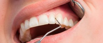
The gingival sulcus refers to the gap-like space between the surface of the tooth and the adjacent gum. The gingival groove and epithelial attachment perform a protective function for the periodontium.
The epithelium of this section never keratinizes and consists of several layers of cells located parallel to the surface of the tooth and quickly renewing themselves (every 4-8 days). The epithelial attachment is not adjacent to the tooth surface, but fuses tightly with it, and as long as this barrier is intact, the underlying periodontal tissues are not infected.
Gingival fluid plays an important role in the protective function of the periodontium. It contains enzymes involved in carbohydrate, protein and other types of metabolism. Normally, the activity of some enzymes in gingival fluid is 8-10 times higher than those in blood serum. The proteins contained in the gingival fluid, including immunoglobulins, have the same properties as plasma proteins.
Leukocytes are constantly found in the gingival fluid, the number of which increases significantly during inflammation, which is the body’s protective reaction in response to damage to the oral mucosa, in particular, periodontal disease.
Treatment
Treatment of major periodontal diseases is as follows:
- removing all plaque and then polishing the teeth;
- treatment of existing carious formations;
- carrying out high-quality prosthetics, if necessary;
- splinting of the dentition (also carried out if necessary);
- treatment of existing common diseases;
- taking vitamins or medications;
- Regular cleaning of the oral cavity not only at home, but also in the dental office.
In the most difficult situations, in addition to the listed treatment, surgical intervention may be necessary.
Local factors:
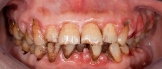
Local factors include, first of all, dental plaque, which is a consequence of improper and untimely oral hygiene. It is a conglomerate of food debris and the bacteria that live in them.
Figure 3. Plaque under a microscope
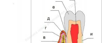
At first, the plaque is soft and can be easily removed with a toothbrush, but over time, bacteria firmly bind to the tooth enamel and the plaque gradually turns into tartar (1), which constantly injures the gums (2) and is a constant source of infection of the tissues surrounding it ( 3-6).
Figure 4. Local causes of periodontal disease development
In addition to dental plaque, local factors also include overload of periodontal tissues, insufficient chewing of food, and underload of certain groups of teeth.
CLASSIFICATION
Periodontal diseases include:
- Gingivitis, or inflammation of the gums. The main symptom that may indicate that you have gingivitis is bleeding gums when brushing your teeth. Gingivitis can be catarrhal - that is, expressed only in redness of the gums, hypertrophic - gum growth is observed, ulcerative - ulceration occurs.
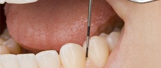
Periodontitis
- inflammatory disease of periodontal tissue. In addition to bleeding, periodontitis causes tooth mobility and pain. But the most important symptom of periodontitis is the presence of a so-called periodontal pocket between the root of the tooth and the gum, from which purulent discharge often comes. The process can be localized - within a group of teeth, or generalized - spread to the entire dentition.
Periodontal disease
– non-inflammatory periodontal disease. A characteristic feature is that with this form there are no inflammatory phenomena and periodontal pockets.
The teeth are well fixed, but their necks are exposed. If you do not engage in active treatment
, you can lose the affected teeth very quickly.