Dental hyperplasia is an excessive increase in the structural components of dental tissues due to their excessive formation. The exact causes of such defects, including the most pronounced one, enamel hyperplasia , have not yet been established, although it is assumed that they lie in the area of impaired cell differentiation during the development of tooth germs and the growth of dental tissues. Provoking factors can be various effects on the reproduction and differentiation of dental tissues: metabolic disorders, violations of correlations in the process of growth and reproduction of tissue components, etc. Based on this, it is clear that for the identification and treatment of dental tissue hyperplasia, it is very important timely preventive examinations and sanitation of the oral cavity.
Dental hyperplasia can be hereditary (for example, focal enamel hyperplasia) or acquired (for example, hypercementosis - excessive formation of cement in the roots, observed against the background of certain pathologies). It is estimated that this defect of varying severity is observed on average in 1.5% of the population.
Dental hypoplasia
It affects the enamel of the teeth and, as a result, underdevelopment of the tooth or its tissues. It is a fairly common dental disease and is observed in approximately 30% of patients. A weak degree of enamel underdevelopment is manifested by a change in color in the form of pronounced pigmentation of white or yellow shades without any unpleasant sensations. Small single spots may not be treated, but if the spots are visible to the naked eye, for example during communication, treatment is mandatory!, and as soon as possible, since inaction can cause the most irreversible consequences. Such as:
- Increased tooth wear
- Destruction of tooth tissue
- Complete loss of affected teeth
- Development of malocclusions
A popular and effective treatment method is dental restoration, and in case of serious changes, orthopedic treatment is indicated. In some cases, tooth enamel may be prepared to smooth the surface of the teeth.
Erosion of dental tissue
A non-carious lesion that is difficult to diagnose in the early stages and seriously worsens the appearance of the frontal teeth. It develops gradually; in the early stages, negative changes are almost invisible, so the pathology is often diagnosed when destructive changes become obvious: the tooth darkens and begins to hurt. If erosion is not treated, then there is a threat of penetration of pathogenic microorganisms, including those that cause caries, into the deep layers of dentin, which is fraught with the development of pulpitis, and subsequently periodontitis.
Causes of tooth erosion:
- mechanical damage (scratches, abrasions) resulting from the use of toothbrushes with hard bristles and pastes or powders containing coarse abrasives and operating on the principle of sandpaper;
- changes in the structure of the enamel and the appearance of microcracks on it under the influence of acid contained in food (marinades based on vinegar and lemon juice, fruits, carbonated drinks, freshly squeezed juices);
- malfunctions of the thyroid gland and digestive system, leading to metabolic disorders, including loss of calcium contained in enamel and dentin.
Symptoms of enamel erosion
The first sign of erosion is the appearance of spots on the enamel that lack their natural shine. The formations are non-carious in nature and are clearly visible, since due to their porous structure, pigments contained in food easily penetrate into them. As the pathology develops, the spots transform into round or oval-shaped depressions with a smooth bottom. Loss of tissue leads to increased sensitivity of teeth, which begin to respond with pain to thermal, chemical and mechanical stimuli. In addition, due to the loss of enamel and dentin, the teeth are deformed and look worn at the base.
Treatment of tooth enamel erosion
Erosion is a lesion diagnosed by a dentist during a visual examination of the oral cavity. In the early stages of the disease, therapy is carried out aimed at stopping the destruction of dental tissue. To do this, applications of preparations containing phosphorus and calcium are made, followed by treatment of the affected area with a fluorine-containing composition. In case of significant tissue destruction, the defects are filled, thereby restoring the integrity and aesthetics of the teeth. However, even in this case, remineralizing procedures cannot be avoided - they are done to stop the erosive process and prevent the filling from falling out. If the teeth are so damaged that it is impossible to restore them using artistic restoration techniques, prosthetics are performed with crowns or bridge structures. Erosion can be prevented to some extent by using a soft brush and low-abrasive cleaning pastes without bleaching agents for hygiene procedures, always rinsing your mouth after sour and sweet foods, and also limiting the duration of eating sour and sweet foods to 5 minutes.
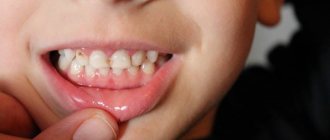
Dental hyperplasia
Manifests itself in the form of excessive formations of hard dental tissues. In 70% of cases the disease is acquired, in the rest it is hereditary. An example of dentin and enamel hyperplasia are oval-shaped formations, measuring from 0.5 to 3.5 mm diagonally, consisting of either enamel or dentin covered with enamel. There are cases in which the cavities of such formations were filled with pulp. They can be located almost anywhere on the tooth. This dental disease occurs in 2-3% of the population. Identifying this disease and 3-5 visits to the dentist will allow you to get rid of it for life, .. or for a long period of time, if after treatment you stop visiting the dentist again.
Treatment of hyperplasia
Therapeutic treatment, in which grinding of enamel drops and filling of pinpoint manifestations of the disease are prescribed. In children, treatment of hyperplasia is carried out using a restorative method.
To restore teeth, photopolymers are used, which completely restore the structure and color of the enamel.
Veneers are installed or artificial crowns are made. Of all types of hyperplasia, only pearl drops on the neck of the tooth can be cured, which over time can cause inflammation of the gums. During the treatment process, excess enamel is removed from the tooth using diamond grinding, and a course of phosphorus-containing medications is prescribed.
Dental fluorosis
Dental disease that occurs when drinking water and food with a high fluoride content, and in large cities and during the usual inhalation of combustion products (exhaust gases from cars, factories, etc.). It is one of the most common non-carious diseases affecting tooth enamel. One of the reasons for the appearance is the entry of fluoride elements into the blood and manifests itself in the form of stains on the tooth, of different sizes, shapes and colors. The disease usually destroys the incisors. When treating the disease, professional whitening is used, or restoration of the damaged part of the tooth with a composite material or an onlay (orthopedic design).
Fluorosis
A non-carious lesion that develops as a result of large doses of fluoride compounds entering the body. As a rule, we are talking about a cumulative effect - a microelement enters the body over a long time before causing pathological changes in hard tissues, not only tooth enamel and dentin, but also bones. Even slightly exceeding the level of fluoride in drinking water can be fatal for people with calcium deficiency, especially for children during the formation of the skeleton and molars (baby teeth are rarely affected by fluorosis). There are several forms of the disease, differing in the shape of the spots and the degree of tooth decay. Thus, streaked, spotted and chalky-speckled forms are characterized by the appearance of white stripes, yellowish, brown and dark brown spots on the enamel, and erosive and destructive forms are accompanied by deep lesions of hard tissues, leading to tooth destruction.
Causes of fluorosis:
- consumption of water containing fluoride more than 1.5 mg (6.0 mg is a critical indicator leading to pathological changes) The optimal concentration of fluoride, which has a pronounced preventive effect on teeth: 0.6 - 1.5 mg/l.;
- work in production in conditions of excess fluoride in the air;
- lack of calcium with excess fluoride.
Symptoms of fluorosis
Teeth become brittle, their enamel wears off, and dark brown (brown) stains appear, which over time turn into erosions. The non-carious nature of the disease is confirmed by the fact that other hard tissues of the body also suffer from it, as evidenced by bone pain and decreased joint mobility. The development of fluorosis is often indicated by muscle weakness, as well as disturbances in the functioning of the liver and autonomic nervous system.
Treatment of dental fluorosis
First of all, you need to eliminate the source of excess fluoride intake - change the water and working conditions, and then begin to put your teeth in order. Treatment is determined by the form of fluorosis - sometimes, in order to return the teeth to an aesthetic appearance (in some cases after grinding off the enamel), it is enough to carry out bleaching and remineralization with preparations enriched with phosphorus and calcium, and if the process has gone deep, the teeth are restored with crowns or veneers. By the way, residents of Russia more often suffer from a lack of fluoride in water than from its excess. But it is better to find out exactly the microelement content in the water you drink, and not to use fluoride-containing toothpastes and mouth rinses if there is enough fluoride in it.
In tap water in St. Petersburg, the fluorine content is 0.15 - 0.20 mg/l of water, which is significantly lower than the optimal level.
Pathological abrasion of teeth
Characterized by severe loss of hard tissue on all teeth. Traditionally, three types of abrasion are distinguished: horizontal, vertical (extremely rare) and mixed. In this case, tissue loss occurs either in the horizontal plane, then the cutting edges of the teeth, cusps and chewing ones are erased. There are several subtypes of increased abrasion and they depend on the depth of the tissues involved in the process and the diminishing tissues. Often this disease is promoted by: traumatic occlusion, bruxism and frequent chewing of very hard foods. Minor abrasion of two or more teeth is a consequence of increased, systematic load on the teeth. If you have teeth worn down by time? If yes, then you definitely need two dentists, an orthopedist and an orthodontist, since it is impossible to get rid of tooth wear without a dentist. I would like to note that solving this disease is not the cheapest pleasure, which means installing braces at a young age will allow you to maintain healthy teeth and a very significant budget.
Complications
Hyperplasia is dangerous because without treatment it provokes inflammatory processes in the periodontium: gingivitis and periodontitis. This leads to loosening of teeth and their loss.
Due to the fact that it is impossible to clean teeth well, caries often develops. Enamel demineralization occurs. Metabolic processes are disrupted.
Due to increased sensitivity, patients try not to chew on the side where the gum has grown. The chewing load is distributed unevenly, and the risk of losing teeth increases.
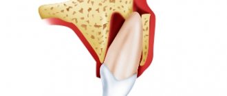
Wedge-shaped tooth defect
Non-carious lesion of the hard tissues of teeth, manifested in the form of a formation in the area of the neck of the tooth. Non-carious lesion of teeth, observed in the form of a wedge-shaped defect. The diagnosis is observed in the front teeth (smile area). Outwardly it appears in the shape of a “ladder” (hence the “wedge-shaped defect”). Symptoms of the disease manifest themselves in the form of a medium defect in the enamel and up to the complete chipping of part of the tooth. The painful anomaly most often appears in people aged 35 to 65 years. Treatment of this pathology is possible, but it is necessary to identify the nature of the disease accompanying the appearance of such a lesion.
- Is there a risk of tooth fracture?
- Is there a risk of developing pulpitis?
- Is the patient embarrassed by his appearance?
- Is there sensitivity?
- Is the patient concerned about further progression of the lesion?
Only by collecting all the answers to the questions can the orthopedic dentist make a final diagnosis and offer several methods of dental treatment. But in any case, you need dental intervention. Inaction with such a tooth defect can lead not only to its loss, but also to a number of symptoms not related to teeth, for example, insomnia.
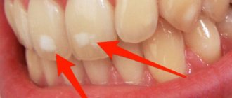
Characteristic symptoms
The main symptoms of the pathology include a change in the color of the enamel: white spots appear on the outer surface. Such changes do not cause unpleasant feelings. In addition, the surface of the teeth in the affected areas is smooth and non-pigmented.
More severe forms of hypoplasia are characterized by the presence of pronounced depressions. At the initial stages, the lesions have a natural shade, but over time they become pigmented. Sometimes on such teeth you can notice deep grooves located horizontally or vertically. Despite the hyperpigmentation of certain areas, the integrity of the enamel is not compromised.
Aplasia (complete absence of enamel) is characterized by pain upon contact with any irritant. In addition, aplasia is associated with underdevelopment of dentin. This leads to the fact that the teeth gradually begin to change shape.
Increased tooth sensitivity
In dental language - Hyperesthesia. If you feel a sharp pain lasting 5-20 seconds, then be sure that this diagnosis has reached your teeth. The disease manifests itself in the form of sensitivity of one or more teeth, or is generalized, affecting almost all teeth.) There are several types of hypersensitivity
- reaction to hot and cold.
- reaction to sugar, salt and acids.
- reaction to everything, including tactile stimulation.
Hyperesthesia almost always leads to many other dental diseases. Sensitivity between teeth is a symptom of the development of dental caries. There are many reasons for sensitivity, but the main ones are:
- Carious process
- Tooth wear
- Enamel chipping
- Wedge-shaped defect
- Hypoplasia
- Erosion of tooth enamel
Note that this disease is a direct consequence of loss of tooth integrity. Treatment of the disease depends on the causes of its occurrence (caries - filling; chipping of enamel - tooth restoration, etc.)
Diagnosis and treatment of gingival hyperplasia
The main task facing a physician diagnosing a pathology is not to confuse it with other diseases. For this reason, in addition to external examination, X-ray examination and histological analysis are necessary.
Treatment for gum hyperplasia today is carried out exclusively through surgical intervention. It is performed by removing excess gum tissue. In parallel with this, complete sanitation of the oral cavity is carried out.
If the pathology occurs after taking special medications, then they are changed to other medications. In this case, refusal of the drug leads to the return of the gums to a physiological state. If this does not happen, the excess tissue is removed surgically. If a person returns to taking medications that caused gum hyperplasia, then a relapse of the disease is inevitable.
Tooth injury
Enamel damage
.
Enamel is the only tissue of ectodermal origin with an acellular structure, without blood vessels and nerves. According to modern concepts, tooth enamel is the strongest tooth tissue. Although the proportions of the enamel are quite small. According to research, the volume of enamel in chewing teeth is 2.3–3.5 mm, and in baby teeth – no more than 1 mm. There are several types of tooth enamel structure. Type 1
is defined as having a uniform coating that is an organic coating of enamel.
The organic matter has a dense structure with poorly defined enamel prisms; this structure corresponds to areas of non-crystalline enamel. Type 2
is characterized by zones in which the organic coating only partially covers the underlying inorganic component, resulting in the formation of scalloped tissue.
The contours of the enameled prism can be observed on the surface. Type 3
is characterized by the absence of an organic coating on the enamel surface and clearly defined contours of the enamel prism.
Type 4
is characterized by a homogeneous organic coating on the enamel surface with faintly defined contours of the enamel prism, which give the design a wave shape.
Different types of enamel structure determine the unequal resistance of the surface layer of enamel to various external factors, leading your teeth to injury, including the development of dental caries. Enamel decay
is extremely common.
Most cracks are not very noticeable and can easily be missed during a routine dental examination. Most cracks are vertical, with the exception of the mandibular incisors. Such changes in the enamel structure are the basis for subsequent tooth destruction. Dental intervention is necessary in order to subsequently avoid problems with dental health, otherwise the development of caries is guaranteed. Dentin destruction.
Dentin is one of the strongest biological materials in the human body.
In humans, teeth come into contact almost 5,000 times a day during normal use. Despite the fact that we use force while chewing, healthy teeth do not break, since the structure of the teeth is quite rigid because the base is dentin. Dentin
is a component of a special component in the bone, which consists of mineral particles, collagen and water. Despite the fact that enamel and dentin are composed of the same mineral (carbonated hydroxyapatite), dentin is a complex microcomposite material. When the integrity of dentin is violated, many clinical situations arise that can destroy your tooth. -Tooth fracture A tooth fracture is a rupture or crack in the dentin of a tooth. The outer shell of the tooth protects the pulp, which contains nerves and blood vessels. A fracture is a break in the chewing surface of a tooth. Typically, the tooth cracks from the chewing surface downwards and in some cases reaches the root of the tooth. A vertical fracture also occurs, when cracks begin at the root and move toward the chewing surface. The main cause is chewing hard food or accidentally biting off a hard object. But the occurrence of injury associated with external threats, including a car accident or sporting event, is no exception. Tooth fractures are more common in older people because teeth wear down over time. Teeth with dental caries or decay are especially susceptible to fracture. We would like to note that not all tooth fractures cause painful symptoms; often the fracture causes irritation or infection in the pulp, which is not accompanied by toothache. Main symptoms of a tooth fracture:
- Pain when chewing food.
- Chew food on one side only to avoid discomfort.
- Acute pain when biting into dense food. -Pain when interacting with cold or hot food and air.
- Occasional pain lasting 3 to 10 seconds (even when swallowing saliva).
Vertical root fractures
may not be noticed until the infection affects the bone or gum.
If the tooth itself is visually intact, the fracture may not be visible to the naked eye. That’s why modern dental clinics are equipped with a special dental microscope. Although this is not the only way to detect a fracture, other options include staining (a solution is applied to the tooth to help see the crack), transillumination (passing light through the tooth), periodontal probing (using special instruments to determine the extent of the crack), bite testing (you will be asked to bite down on a special object to find the specific tooth causing the problem), x-ray (to look for certain defects, as not all fractures can be seen on x-ray. Early diagnosis can help save the tooth before the fracture progresses). Teeth do not heal on their own.
The goal of treatment is to protect the tooth and the inside of the pulp. Talk to your dentist and choose a treatment plan that suits your cost and treatment time. Treatment of the tooth will depend on the severity of the damage. Tooth restoration options may include:
- A crown is a dental covering. However, a temporary crown will be installed first to make sure that it solves the problem. A permanent crown is installed only after wearing a temporary onlay for a short time (this allows not only to properly restore the tooth, but also to avoid problems with its further use).
- A veneer or Lumineer is a thin covering that is placed over a tooth if there is a small chip on the surface; in some cases, dentists resort to restoration with a composite material. However, if the fracture has affected the root of the tooth, you cannot do without root canal treatment. Root canal infection is a common clinical situation in cases of severe pulp damage. The root canal is depulped and filled with filling material. Tooth extraction is used as a last resort only in emergency and very difficult situations when saving the tooth is not possible.
Pigmentation of teeth
A non-carious pathological condition in which teeth become brown, yellow, gray, pink, black, red or another unusual color or shade. In most cases, pigmentation develops against the background of demineralization of the enamel, accompanied by hypoplasia and hyperesthesia. Pigmentation occurs under the influence of general (diseases, medications) and local (smoking, food colorings) factors and occurs in 85% of people, more often in men than in women. Treatment of this non-carious dental disease involves regular professional cleaning and whitening, but often the only way to restore the aesthetics of the dentition is prosthetics.
Causes of tooth pigmentation:
- demineralization of enamel;
- chips and other injuries to hard dental tissues;
- poor oral hygiene;
- treatment using iodoform and resorcinol-formalin, as well as “silvering” of teeth;
- rupture of the neurovascular bundle (pulp);
- smoking;
- taking certain medications and foods with added food coloring;
- hemolytic disease;
- uroporphyria;
- Excessive production of pigment in the body.
Pigmentation, like many other non-carious lesions, develops not only after, but also before teething. Thus, pigmentation in a child is often a consequence of the mother taking tetracycline antibiotics during pregnancy, which, penetrating the placenta, are deposited in the bones and tooth buds. Since the composition of dentin and enamel remains virtually unchanged over time, one cannot expect that yellow-brown pigmentation will disappear with age.
Symptoms of tooth pigmentation
The natural color of enamel is milky white, all other options indicate pathological pigmentation. The color of teeth affected by non-carious diseases varies. Thus, with a hemolytic disease accompanied by bilirubin deposition, the teeth are painted in various shades of gray, blue, green and brown, while “tetracycline” pigmentation is yellow-gray. Red staining is characteristic of uroporphyria, and gray staining is characteristic of pulp necrosis. In cases of exogenous pigmentation (associated with external factors), enamel and dentin are colored dark brown by nicotine, and pink by resorcinol-formaldehyde paste (not currently used in dentistry).
Treatment of tooth pigmentation
If, based on the patient’s complaints, clinical examination data and the results of additional studies (radiography and thermography), pathological pigmentation of the teeth is diagnosed, then treatment begins immediately. The therapy consists of sanitation of the oral cavity, including thorough removal of dental plaque (professional cleaning with ultrasound and using the Air Flow air-abrasive system). In case of deep pigmentation, intracanal and external bleaching is carried out using special preparations. However, not all types of pigmentation are eliminated by bleaching. Thus, the procedure will not eliminate tetracycline discoloration and color changes that appear as a result of oxidation of metal pins installed in the channels and silver amalgams. In these cases, pigmented teeth are covered with crowns and veneers (if their condition allows this). When treating non-carious pigmentation in children, consultations with a pediatrician and a genetic specialist are often required.
What is tooth sensitivity?
In other words, dentin hypersensitivity. If cold, heat, sugar, salty, sour or just cool air have a painful reaction on your teeth, then you have all the symptoms of tooth sensitivity. The symptoms are very common and it is estimated that a large proportion of the population experiences this sensation. Why does tooth sensitivity (dentin hypersensitivity) occur? Tooth sensitivity is usually caused by dentin in areas of the roots exposed due to gum extraction or periodontal disease. Receding gums are very common, and by age 65, four-fifths of people will have gum recession.
Mechanism of development of hyperplasia
The mechanism of development of the hypertrophic process remains not fully understood. However, scientists have discovered that the pathogenesis of the disease is influenced by impaired calcium transport. This leads to increased production of collagenase, which causes the gums to grow. Hyperplasia develops gradually, so patients do not pay attention to it right away.
Stages of the disease:
- First: the tissue covers a third of the tooth, there are no signs of an inflammatory process.
- Second: covers half of the tooth, bleeding may occur, especially with mechanical impact.
- Third: overlap from two-thirds to the entire crown, sometimes even the cutting edge and chewing surface are covered with soft tissue, granulations are visible over the entire surface.
Why does tooth sensitivity (dentin hypersensitivity) occur?
When the root of a tooth becomes exposed, it does not have a layer of enamel like the crowns of your teeth. The roots have a very vulnerable coating - cementum, which, after the loss of the top layer, leaves the dentin exposed. Excessive brushing with highly abrasive dental paste causes abrasion of the surface of the tooth enamel and exposes dentin. A very acidic diet with lots of acidic fruits and pickles can also cause tooth decay and dissolve the surface of the tooth, exposing dentin. It is important to visit your Doctor periodically if you have any symptoms of sensitivity so that he can examine your mouth and identify any symptoms associated with dentin sensitivity and suggest you the best treatment plan. When teeth react painfully, brushing them can be painful, and if you don't brush your teeth because of pain, it can lead to the destruction of enamel and gum disease. Reactions to heat, cold, sugar, sour and salty foods and drinks are also a clear sign of tooth decay, or a sign of a broken tooth, in which case your dentist will treat you comprehensively. Some of the most common causes of sensitivity are:
- Gum recession due to age or improper brushing of teeth
- Acidic drinks (such as soda), which cause enamel erosion and dentin attack
- Brushing with very low quality toothpaste, improper brushing, and brushing more than three times a day will result in loss of enamel.
- Gum disease that can lead to gum recession
- A broken or fractured tooth can expose dentin
Additionally, some dental procedures may cause sensitivity. Procedures such as teeth whitening, professional teeth cleaning, braces or fillings are known to cause sensitivity during or after the procedure.
Treatment tactics
Hypoplasia of the enamel of primary or permanent teeth is irreversible. Therefore, all therapeutic measures are aimed at protecting the altered areas of the dentition and restoring the enamel coating. Mild pathology does not require special treatment, but only requires constant monitoring. In most cases, a person does not experience pain: non-carious changes in dental tissues do not interfere with everyday life.
For severe forms of hypoplasia, for example, deep lesions of the enamel or extensive spots, special therapy is carried out. Without timely treatment, there is a risk of developing various complications:
- pulpitis, periodontitis;
- malocclusion;
- pathological abrasion;
- increased sensitivity of teeth.
Hypoplasia can also lead to dentin destruction and complete tooth loss. There are several methods for eliminating the manifestations of hypoplasia. Therapeutic tactics are selected by the dentist depending on the severity of the pathology. The doctor takes into account the condition of the patient’s dental tissues.
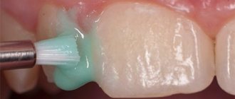
Remineralization
Remineralizing therapy involves saturating tooth enamel with fluoride and calcium. Artificial mineralization is carried out using dental gels, pastes, varnishes and other means. Treatment can be done in a clinic or at home.
Remineralization in a clinical setting consists of several stages:
- Professional oral hygiene.
- Application of restorative gel.
- Coating with a fluorine-containing compound using a tray or brush.
The dentist selects the frequency of procedures and the type of drug individually. In addition, your doctor will often prescribe oral vitamins and minerals as additional support for your body.
Whitening
Bleaching is carried out after professional hygiene and remineralizing therapy. This method is effective if the defects are located in the surface layers of the enamel or slight clouding of the enamel layer is observed. Chemical bleaching gives the most pronounced results.
In cases of severe damage to the enamel layer and numerous foci of hypoplasia, chemical bleaching with solutions of carbamide peroxide and hydrogen peroxide is contraindicated.
Filling and prosthetics
Filling is used for pronounced erosive depressions, as well as mixed forms of hypoplasia, when the integrity of the dental units is compromised. Composite materials are used to restore teeth. In some cases, the vestibular surface is covered with veneers. This technique helps to give teeth an aesthetic appearance and prevent their destruction.
Prosthetics are used for severe damage to the enamel of primary or permanent teeth. Crowns help maintain the health and aesthetics of your teeth. Installing crowns for hypoplasia in childhood contributes to the formation of a correct bite and the development of normal diction.
Depending on the extent of the damage, the condition of the soft and hard tissues, it may be necessary to remove the affected tooth followed by implantation.
Can you prevent tooth sensitivity on your own?
Of course, yes, if you keep your mouth clean, observing all oral hygiene standards, which will help stop gum loss and, as a result, periodontal disease. Thorough flossing and flossing as recommended by our dentist can help reduce the chance of avoiding this disease. A diet without consuming acidic foods also helps stop the development of this symptom. Ignoring the disease can lead to other health problems, leaving you vulnerable to tooth decay and gum disease.
What it is
Hyperplasia is a random rapid increase in gum tissue cells. Hyperplasia is not a tumor; it is benign changes in cell tissues, in which the cells themselves continue to perform their functions without changes. With hyperplasia, unlike oncological diseases, cells do not grow into surrounding tissues and do not metastasize. Many doctors are inclined to believe that it is not always a pathology, but only an experienced doctor can decide on a treatment method.
Gingival hyperplasia is manifested by excessive growth of tissue that can partially or completely cover the entire crown of the tooth. With this disease, only the gum tissue grows; the cheek and tongue tissues are not affected by changes. Hyperplasia is not an inflammatory process, so there is no drug treatment for it. In addition, it is necessary to correctly carry out differential diagnosis of the disease, being able to distinguish it from cysts and other tumor neoplasms. And only an experienced dentist can do this. Therefore, if you find that your gums are growing by leaps and bounds, consult a doctor immediately.
Gum hyperplasia is also accompanied by increased blood supply to the gums, so when brushing your teeth, the gums often bleed. Often patients mistake this disease for gingivitis or periodontitis and self-medicate, which does not bring any effect. In order not to waste time and not to advance the disease to such a stage when the tooth is completely under the gum, you must immediately consult a doctor.
What to do if you have sensitive teeth?
See your dentist first. The doctor will offer you all treatment options. It is also important to tell your Doctor if the cause is not dentin (root) hypersensitivity, the tooth may be reacting due to a more serious problem. To treat increased reactivity to stimuli, your Doctor will definitely recommend using a low abrasion dental paste designed for such clinical cases - desensitizing toothpaste. These toothpastes make your teeth less sensitive if you brush them twice a day, and also contain fluoride to protect your teeth from decay. In addition, your dentist may prescribe a fluoride gel, fluoride rinse, or a high-consistency fluoride toothpaste that is specially formulated to make your teeth less sensitive and provides additional protection against tooth decay. These procedures are carried out at home and are inexpensive. Other procedures for sensitive teeth that your dentist or hygienist may suggest in your dental office.
Non-carious lesions of teeth
In addition to caries, the most common dental disease, there is another diverse pathology of hard tissues - non-carious lesions of teeth.
Non-carious lesions of dental tissue occur without tissue softening and without the participation of microorganisms. These processes are based on a violation of the mineralization of hard dental tissues under the influence of external or internal factors. These pathologies occur in approximately 15% of patients, but no more than 5% seek dental care for them, since non-carious dental lesions, as a rule, do not cause pain or other subjective sensations, and often only worsen the appearance. However, if left untreated, a number of complications may occur, in particular early tooth loss.
Hypoplasia of teeth and enamel is most often the underdevelopment of enamel or tooth tissue in the tooth buds under the influence of disturbances in mineral and protein metabolism in the body of the fetus in the womb or in the body of a child. The enamel immediately after eruption has a chalky tint, and then becomes loose and is quickly lost. Hypoplasia is irreversible. Hypoplasia of permanent teeth occurs during the period of their mineralization under the influence of certain diseases (acute infectious diseases, measles, scarlet fever, rickets, diseases of the gastrointestinal tract, dystrophy, brain disorders) at the age of 6 months to 1.5 years. Hypoplasia can appear in the following forms: discoloration (white or yellowish spots on the teeth with a shiny surface and painless upon probing), wavy enamel, absence of enamel in a certain area. Spots with hypoplasia have pronounced symmetry and are characterized by stability (they do not change their shape and color). When the child’s body weakens, caries may occur at the site of the defects. Treatment of dental hypoplasia consists of filling the affected areas of the teeth with composite materials, using sodium fluoride paste, and coating the teeth with fluoride-containing varnish. It is also necessary to normalize general metabolism.
Enamel hyperplasia – “enamel drops” (pearls). These drops are usually located in the cervical area of the crowns of both permanent and baby teeth and have a diameter of 2-5 mm.
Dental fluorosis (endemic fluorosis) is a disease observed in individuals (mainly children) who live for a long time in areas with high fluoride content in water and soil (more than 1 mg/l). Fluorosis is a peculiar form of hypoplasia. Depending on the concentration of fluoride ions, stains that appear on the surface of teeth during fluorosis can have different colors - from white to brown and even black. The more fluoride in drinking water, the more often fluorosis occurs and the less often caries occurs. A predisposing factor to the development of fluorosis is a decrease in the body's reactivity (infectious diseases, endocrine disorders). Fluorosis spots are stationary, dense, with a shiny surface, painless and smooth upon probing. With fluorosis (as opposed to hypoplasia), caries almost never occurs, due to the fact that fluorapatite is deposited in the spots (hence their high microhardness and resistance to acids). Treatment of fluorosis. In the early stages (change in enamel color), bleaching is recommended, followed by remineralizing therapy; calcium and phosphorus supplements are prescribed. It is necessary to increase children's intake of proteins, milk, fruits, vegetables, and limit the consumption of fatty foods. In winter, fish oil, multivitamins, and ultraviolet irradiation are prescribed. It is important to maintain the correct daily routine and conduct general hardening of the body. For complex forms of fluorosis, methods of cosmetic dental restoration or covering teeth with artificial crowns are effective.
Marble disease is a congenital familial osteosclerosis (an abnormal increase in bone density). In this case, the bones of the entire skeleton are affected. After teething, teeth have a chalky tint, and then the enamel becomes loose and is quickly lost. The process can develop into a malignant form.
Wedge-shaped defects are a type of damage to dental tissues located near the walls of the teeth, on the cheek and labial surfaces. The defect has the shape of a wedge with the base towards the neck of the tooth and the apex towards the cutting edge or chewing surface. A wedge-shaped defect, as a rule, does not bother the patient much: pain occurs rarely (only briefly from thermal and chemical irritants), the tooth cavity is not affected and is not opened, the defects slowly deepen, and softening is not determined (this distinguishes the defect from caries). The causes of the wedge-shaped defect have not been fully established. There is a point of view that it occurs under the influence of mechanical factors (for example, a toothbrush). It is sometimes believed that since a wedge-shaped defect begins after exposure of the tooth wall, it is one of the manifestations of periodontal disease. There is evidence of the role of endocrine disorders, diseases of the central nervous system and gastrointestinal tract in the occurrence of a wedge-shaped defect. Treatment of a wedge-shaped defect is aimed at strengthening the hard tissues of teeth through the use of remineralizing therapy (applications of calcium, phosphorus, fluorine, use of fluoride varnish, fluorine gel, etc.). In advanced cases, filling with composite materials or orthopedic treatment (artificial crowns) is recommended.
Increased abrasion of dental tissues . The wear of tooth enamel is a completely natural process that occurs in all people by the age of 45-50. However, in a certain group of people, pathological abrasion occurs already at a young age. Not only the enamel is erased, but also almost completely the crowns of the teeth (most often the front teeth). The causes of the disease are: the habit of grinding teeth during sleep, disorders of the parathyroid glands, almost complete loss of chewing teeth (which increases the load on the front teeth). Treatment – orthopedic or orthodontic (in order to properly distribute the load on the teeth).
Erosion of hard dental tissues is the progressive loss of enamel and dentin on the vestibular surface of tooth crowns. Areas of erosion have an irregular rounded shape. The causes of tooth erosion are considered to be the mechanical effects of toothbrushes, endocrine disorders (in particular, increased function of the thyroid gland - thyrotoxicosis). It occurs mainly in middle-aged people and lasts 10-15 years. There are different degrees and stages of the process. Treatment is aimed at additional mineralization of hard dental tissues. Erosion is filled with composite materials and artificial crowns are made. Calcium and phosphorus preparations, multivitamins with microelements are prescribed internally.
Necrosis of hard dental tissues initially manifests itself in the loss of enamel shine, then chalky spots appear, gradually turning into dark brown. Softening appears in the center of the spots, the enamel becomes brittle and easily chips. Necrosis differs from erosion by softening in the center of the spot. Teeth are extremely sensitive to any irritants. The causes of necrosis are endocrine pathologies (hyperthyroidism, dysfunction of the gonads), pregnancy, diseases of the central nervous system, chronic intoxication of the body, hereditary factors. Treatment is aimed at eliminating tooth sensitivity, remineralization, and normalizing endocrine disorders.
Dental injuries. Acute dental injuries occur when a tooth is exposed to traumatic factors - impact, increased load during chewing, constant pressure from improperly made orthopedic structures. Injuries are more common on children's baby teeth (luxation, tooth fracture, crown fracture). Acute dental injuries include: tooth bruise, tooth dislocation (complete and incomplete), fracture of part or the entire crown, combined injury, tooth germ injury. If a tooth is bruised, it is necessary to rest it, eliminate contact with opposing teeth by grinding off the cutting edge, and exclude solid foods from the diet. It is necessary to monitor the condition of the pulp, and in case of irreversible changes, remove it and fill the canal. Tooth dislocation with healthy gum tissue rarely occurs. However, if there is periodontal pathology, in particular, bone tissue resorption, tooth dislocation can easily occur even when chewing hard food. In case of incomplete dislocation of a tooth, it is necessary to create rest for which the tooth is splinted. If the pulp ruptures, it is removed and the canal is filled. If part of the crown breaks off, it is restored using composite materials. Sometimes special pins are used.
Bibliography:
- Lutskaya I.K., Nichiporovich G.S. Frequency of enamel and dentin cracks in permanent teeth. Dental Journal 2006, number 2. Page. 87-91.
- Nichiporovich G.S. Modern ideas about the increased sensitivity of hard dental tissues. Dental Journal, 2006, number 2. Page. 92-95.
- Leus P.A. Non-carious diseases of hard dental tissues. Educational and methodological manual. Minsk, BSMU-2008, 56 pages.
- Borovsky E.V., Leus P.A. Erosion of hard dental tissues. Dentistry 1971. T. 50 No. 3.p. 1-5.
- Fedorov Yu.F. Clinic and treatment of hyperesthesia of hard dental tissues. - M 1970, 140 pp.
- Leus P.A., Kozel O.A. Disturbances in the development of tooth enamel. Educational and methodological manual. Minsk, BSMU-2004-29 pages.
Author: Shulyak Alesya Vadimovna
Sensitivity due to gum disease, what is it?
Gum disease
is an inflammation of the gums that can progress to affect the bone that surrounds and supports your teeth, which also causes sensitivity, which we often confuse with tooth sensitivity. It is caused by bacteria in plaque, which always forms on tooth enamel. If plaque is not removed by daily brushing and flossing, thick plaque will form and bacteria will then infect the entire mouth. This can lead to weakening of the gums and, as a result, teeth falling out or being removed by the dentist.
Stages of gum disease:
- Gingivitis: This is the earliest stage of gum disease and is caused by the formation of plaque on the gums. If daily brushing is not carried out promptly or does not meet all hygiene standards, plaque produces toxins (poisons) that can irritate the gum tissue, causing gingivitis. You may notice some bleeding during dental hygiene. If gum problems are detected early, the damage may be localized because the bone and connective tissue that holds the teeth in place are not yet affected.
- Periodontitis: At this stage, the bone and fibers are irreversibly damaged. Your gums may begin to form a pocket under the gum that traps food and plaque. Proper dental treatment and improved home care usually help prevent the disease from developing. Advanced periodontitis: In this final stage of gum disease, the fibers and bone that support the teeth are destroyed, which can cause the teeth to shift or become loose. This can affect your bite, and if aggressive treatment cannot save them, your teeth may need to be removed.
Types of gum hyperplasia
Periodontal tissues can grow in different ways, involving different areas of the gums. There are several types of hyperplasia:
- localized: the growth of new cells occurs in a limited area, next to one tooth or group of teeth (only on one or both sides);
- generalized: the gums grow over the entire surface on one or both jaws;
- papillary: new cells are formed only in the area of the gingival papilla, the pathology does not affect other gingival tissues;
- marginal: the gingival papilla and the area of the gingival margin grow;
- diffuse: growth of both marginal and attached gums occurs (an increase in periodontal tissue to the entire height from the gingival margin to the mucogingival border).
Hyperplasia may be limited. The increase in tissue in this case is similar to a tumor, strictly isolated, and has a wide base or pedicle. It looks like a growth or bump.
How do you know if you have gum disease?
Gum disease is most common in adults. If detected in the early stages, the problem can be corrected with minimal consequences, please contact your dentist if you notice any of the following symptoms:
- Gums that are red, swollen, or swollen or tender
- Gums that bleed when brushing or flossing
- Teeth that look longer because your gums have receded
- Gums that have separated or pulled away from your teeth, creating a pocket
- Changes in the way your teeth fit together when you cleave them together
- Pus between teeth and gums
- Constant bad breath
- How is gum disease treated?
When you detect the first symptoms of gum disease and proper oral hygiene, you will rid your teeth of plaque and avoid the development of the disease. Professional cleaning with a Doctor is the only way to remove plaque that has accumulated and turned into tartar. Your Doctor will clean your teeth and remove tartar from above and below the gum line.
Causes of gum hyperplasia
Regardless of the fact that this pathology is registered very often, the reasons leading to its progression have not yet been fully determined.
Often, gum hyperplasia is formed as a result of frequent consumption of the following drugs: cyclosporine (used to prevent rejection of transplanted organs), phenytoin (to avoid epileptic seizures). As a rule, the risk of this disease is highest in 10% of patients whose body receives a medication that blocks calcium channels: verapamil, felodipine, diltiazem, nifedipine.
If we talk about the factors that precede gum hyperplasia, then among them we can highlight:
- Certain diseases of the blood system (such as leukemia),
- Pregnancy period
- Hereditary predisposition
- Puberty period
- Multiple layers of tartar,
- Certain malocclusions.
Gum hyperplasia is often recorded in adults. If we are talking about children, then in them it can only be a consequence of genetic problems. Drug-induced hyperplasia is registered regardless of a person’s age.
Varieties
Depending on the prevalence, generalized and focal hyperplasia . If overgrown gums cover a large surface of several or all teeth, then this type of disease is called generalized. If it grows only near one tooth, then this type is called focal. It should be noted that generalized hyperplasia is much more common than focal hyperplasia.
Depending on the causes of hyperplasia, it can be medicinal or fibrous.
- Drug-induced hyperplasia occurs as a result of taking medications, the side effect of which is the extensive proliferation of gingival tissue.
- The fibrous form is rare and is a hereditary disease. The first symptoms appear in infancy during teething. In the future, the disease progresses, the gums gradually grow, become thicker, and over time, pockets begin to form, which are similar in appearance to the manifestation of periodontitis.
Structure of tooth enamel
Tooth enamel is the main protector of teeth and completely covers their crown part. The average thickness of the tissue is about two millimeters: in the chewing area the layer is slightly thinner, and on the sides of the teeth, on the contrary, it is thicker. The color of tooth enamel depends on its density and the quality of dentin, as well as on some individual characteristics of the body, but this type of tissue is essentially transparent.
The composition of tooth enamel includes, for the most part, inorganic substances (more than 95%). The percentage of water content is very small (approximately 3%): this is partly why such high strength is achieved. Inorganic materials primarily refer to hydroxyapatite crystals (a mineral containing fluorine, magnesium, carbon and other elements). The presence of hydroxyapatite crystals also explains the fact that the enamel is vulnerable to the effects of an acidic environment, i.e. The consumption of foods that increase the acidity of saliva is contraindicated. This is especially true for calcium ions, on which the integrity and density of the enamel largely depends.
Treatment
If the doctor has diagnosed gum hyperplasia, treatment can only be surgical. It is impossible to get rid of this disease on your own, with the only exception being mild forms of drug-induced hyperplasia. In this case, sometimes it is enough to simply cancel the inappropriate drug, and over time the gums gradually restore their previous appearance. In more complex cases, the overgrown gum should be surgically removed after stopping the drug; it will not grow in the future.
When treating fibrous hyperplasia, only surgical removal of overgrown gum tissue is possible. Treatment should begin with an accurate diagnosis. This can only be done by an experienced dentist, using x-rays, as well as histological analysis, which allows differentiating the disease from tumor neoplasms.
Surgical treatment of gum hyperplasia is performed under local anesthesia. The doctor removes hypertrophied tissue and disinfects the oral cavity.
Removal of overgrown hypertrophied gums is carried out using both a scalpel and a laser. The use of a surgical laser instead of a traditional scalpel is less traumatic and allows for faster wound healing.
Unfortunately, sometimes there is a need for repeated surgical interventions. This does not depend on the doctor’s qualifications, since with fibrous hyperplasia the gums grow constantly and over time can again cover the crowns of the teeth. In this case, it will not be possible to avoid repeated surgery.