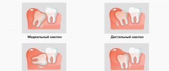
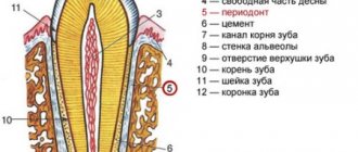
What is bifurcation
The bifurcation of a tooth is the area on the tooth itself where the branching of its roots begins (you can see what it looks like in the photo below). The term "bifurcation" means "branching into two", i.e. for two roots. But some teeth have not two, but three roots - in this situation the term “trifurcation” would be more correct, but doctors often still use the word “bifurcation” as a more common concept. Moreover, bifurcation does not occur on any tooth, but on multi-rooted ones - molars have two or more roots (“sixes”, “sevens” and “eights”).
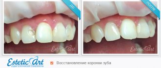
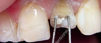
Removing a broken instrument from the root apex area
| After poorly performed endodontic treatment, it is necessary to remove the broken instrument from the area of the apex of the root canal of tooth 15. The tooth hurts when biting. Chronic periodontitis develops, which can lead to serious complications. | A root apex resection operation was performed. A control photograph was taken after 3 months. The inflammatory process has been stopped. The tooth is saved! |
In case of removal of the root apex, it is necessary to perform retrograde canal filling of the remaining part of the root. Such operations make it possible not to involve the crown part of the tooth; in this case, we can save not only the tooth, but also the expensive fixed prosthesis installed on it, for example, a metal-ceramic bridge.
Is it necessary to treat bifurcation?
In fact, when a dentist talks about tooth bifurcation, he means not only the area of root branching itself, but also the pathological processes occurring here. But there should be a clarification here. For example, perforation in the bifurcation area - i.e. the formation of a hole that violates the integrity of the tooth. This pathology not only causes complications (pulpitis, inflammation of the bone socket, etc.), but often calls into question the very existence of the tooth. Therefore, treatment of perforation or other pathologies in the area of root branching should begin as early as possible.
On a note! If there is perforation in the bifurcation area, the tooth is quite difficult to treat and will not last more than 3-5 years, after which it will have to be removed. Such a tooth is not suitable for a prosthesis, since it can no longer withstand increased chewing loads.
Cost of tooth-preserving operations in Moscow
| Name of service | Price |
| Removal of a dystopic tooth | 7000 rub. |
| Removal of an impacted tooth | 13500 rub. |
| Applying and removing sutures | included in deletion |
| Anesthesia, sedation | 12,000 rub./hour |
It is impossible to talk about the cost of the operation with certainty, since it depends on many components. These include:
- Type of surgical intervention;
- Consumables and use of medications, in particular anesthesia;
- Qualification and experience of the doctor;
- Modern equipment;
- The need for additional consultations;
- Additional examinations if necessary and other individual factors.
Carrying out tooth-preserving surgery in Moscow, in any case, will be cheaper than tooth extraction followed by prosthetics. The main thing is to choose a qualified specialist who will perform the treatment correctly and avoid complications.
Example: The cost of tooth-preserving surgery to remove a cyst is 18,000 rubles, there is no way to indicate the cost of bone materials, since often infection does not allow the simultaneous installation of a sterile regeneration stimulator at the time of surgery, and the second stage is carried out in a defect that has already actually healed, so the cost of CT no more than 10,000 rubles.
Sincerely, Levin D.V., chief physician
Why problems arise
Involvement of the furcation area in the pathological process occurs in two cases:
- infection penetrates from the outside (through the gum or bone): normally, the furcation area is in close contact with the periodontium and the bone walls of the tooth socket. But if the bone begins to sag (which happens with periodontitis, periodontal disease), then the furcation area becomes accessible to microbes from the external environment. Also, inflammation of the bone (for example, when an infection enters it through the bloodstream) leads to the “melting” of dental tissue. Over time, the root cement is destroyed, a hole appears in the tooth, and microbes penetrate into the pulp,
- from inside the tooth: for example, with pulpitis or perforation of the bottom of the pulp chamber (during drilling or passage of root canals), which is located just above the furcation area. In this case, microbes from the oral cavity penetrate into the bone socket, causing its inflammation.
Retrograde filling
This atypical filling of teeth from the side of the tooth root is not available in all cases in the treatment of cysts and cystogranulomas, but it is effective under certain conditions. The attending physician will determine when such manipulation is required. Previously, if a cyst was detected and in similar situations, the tooth was immediately removed, but now a retrograde filling method is used to sterilize the area where the cyst was removed and the tooth lasts for a very long time. The essence of the surgical intervention is that access to the affected area is through the alveolar process of the jaw, usually through the fistulous tract. The operation allows you to save teeth that cannot be treated conservatively for a number of reasons:
- Curvature of channels;
- Lack of channel patency;
- The presence of fragments of a surgical instrument in the canal;
- The presence of metal-ceramic crowns, as well as structures with core inlays or pins.
Using retrograde filling without filling the canal, the affected apical surface is hermetically sealed, the cyst is removed and the spread of the pathological process to surrounding tissues and healthy teeth is prevented.
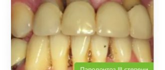
What symptoms should you pay attention to?
In many people, pathology in the area of branching of dental roots is asymptomatic. But some patients note the following signs:
- pain when biting and pressing,
- darkening of the enamel,
- bad breath,
- unsteadiness of an artificial crown or bridge,
- discharge of blood or ichor from under the gums.
Read on the topic: why a tooth hurts “without a nerve” - and how to remove the pain, advice from a dentist.
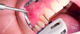
How is diagnostics carried out?
Diagnosis of the pathological process in the area of bifurcation of tooth roots includes several studies. An X-ray examination is required - computed tomography, which shows the smallest changes, is more informative here. The dentist also probes the gum pocket (normally, the gums should not move away from the tooth by more than 3 mm).
It is important to know! If necessary, the artificial crown is removed or the old filling is removed and the dental cavity is examined for the presence of perforations. It is optimal if the doctor uses a microscope during such an examination - after all, with multiple magnification, you can see the smallest defects.
If pathology in the bifurcation area occurs during treatment, the dentist should pay attention to the appearance of blood in the cavity. In this case, we can speak with 99% certainty about perforation, which needs urgent restoration.
Types of tooth-preserving operations
There are many types of tooth-preserving operations, each of which has its own advantages and disadvantages, and is also performed for certain dental diseases. Modern dentistry distinguishes the following methods of tooth preservation:
Excision and correction of the apical portion of the root
The operation is carried out in cases where there is any pathological focus in one of the areas in contact with the root. In order to eliminate the problem, a small incision is made through which the cyst and inflammation are removed and the damaged root is partially removed. The cavity formed as a result of surgery is filled with natural, synthetic or osteoplastic material. This promotes rapid tissue healing and preservation of the full functioning of the natural tooth. The most common reasons for resection of the root apex are the presence of cysts and granulomas, accompanied by inflammation and not amenable to conservative treatment methods.
Complete root amputation
Human teeth do not always have only one root, which makes tooth-preserving surgery such as complete amputation of a dead root possible. It is carried out on those teeth where there are two or three roots, while tissues not involved in the pathological process remain in place. The dentist’s task is to rid the patient not of the tooth, but only of the diseased area of the multi-rooted tooth.
No changes in the functioning of the tooth occur, since the mechanical function of the tooth does not change, the remaining roots completely compensate for the loss of one area and distribute the load among themselves.
Hemisection of tooth root
This surgical intervention involves the removal of one of the affected tooth roots with partial amputation of the crown, usually performed on molars or premolars. During the surgical procedure, the area involved in the pathological process is carefully cut out and removed, after which the resulting space is filled with osteoplastic material. Hemisection of the tooth root is a sparing corono-radicular separation and gives very good results.
Increasing the length of the coronal area of the tooth
This type of surgical intervention is required in cases where the remaining tooth tissue is too weakened, it is necessary to improve the appearance of the tooth, and also when the natural tooth is too short or is quickly worn away. As a rule, such a problem affects several areas of the dentition at once and is caused by an incorrect bite or excessive load on the tissue. There is also a congenital predisposition to increased tooth wear, which also requires lengthening of the crown part. The procedure is non-traumatic, very fast and cosmetically always very successful.
Manipulations in pathological processes in the periodontium
Periodontal soft tissue diseases pose a great danger of losing the patient's entire dentition. In addition, their progression is accompanied by the addition of secondary infections, which aggravates the risk of loss of natural teeth. Treatment of such diseases must be timely and as radical as possible with the removal of all compromised tissue.
The use of tooth-preserving surgery for periodontal diseases is performed not only on damaged teeth, but also to strengthen healthy teeth. This method of treatment is almost the only solution for periodontal damage, which involves preserving teeth. They try to treat diseases conservatively, but very often the desired effect is not achieved. During the surgical procedure, infected soft tissues and formations in them are removed. Microsurgical access is carried out through the mucous membrane of the gums.
There are two main types of operations for periodontal pathologies:
- Flap tooth-saving operations. This surgical intervention is the main solution to the problem of periodontal pathologies. There are several of its varieties: reconstruction and correction of bone pockets, gingivoplasty, the use of the method of directed regeneration for periodontal tissues, the use of an allogeneic graft to strengthen teeth. Thanks to the operation, the infectious process is transferred to the stage of remission, and subsequently, the teeth become less mobile, signs of bacterial damage to the oral cavity disappear (unpleasant odor, discharge of pus, and so on).
- Curettage. This operation involves the delicate removal of affected areas from periodontal pockets without incisions. If the gums around the crown are pulled back, the curettage is called open curettage. It allows you to better eliminate infected areas and achieve a positive treatment result. Bone material is often introduced into the surgical area, but sometimes this is impossible due to severe infection, then the resulting cavities are gradually filled with dense scar connective tissue, which is not sensitive to external irritants, and itself strengthens the tooth. If the empty spaces are too large, they are filled with osteoplastic material in a second stage after several months in clean conditions. Closed curettage allows you to deal with small foci of the infectious process (up to 5 mm) in a less traumatic way.
Modern methods of treatment
When pathology is detected in the area of root bifurcation, a logical question arises: is it worth saving the tooth? Only a dentist can give an accurate answer after a thorough diagnosis, because... There are many determining factors here. It is important to correctly predict the “functionality” of the tooth if treatment is chosen - so that severe inflammation of the gums and bones does not begin (which, by the way, can affect the roots of neighboring teeth).
Therapeutic treatment
Conservative treatment of the bifurcation area is possible if the hole size does not exceed 1 mm, and is most often carried out using the following technologies:
- closing the perforation with special filling materials: these can be glass ionomer cements that absorb moisture - since it is not possible to properly dry the work area. Suitable brands for root canal filling are ProRoot MTA, MTA-Angelus,
- closing the perforation using calcium preparations: they restore the structure of damaged tissues (which mainly consist of calcium),
- reinforcement with metal threads and filling of the hole in the bifurcation area.
In all of the above cases, after restoration of the hole in the bifurcation area, the tooth cavity is closed with a temporary filling, and after 5-7 days the patient comes for a control x-ray to assess the quality of the perforation closure. There may be several such visits. After achieving a positive result (the perforation is completely closed, there is no inflammation near the roots), a permanent filling is installed.
On a note! If the pathology began from the outside, then treating the tooth alone is not enough - you also need to cure all the tissues that surround it (this is called “periodontal”). Treatment of periodontitis is carried out comprehensively - i.e. conservatively and surgically. First, tartar and granulation tissue are removed using curettage, and then regeneration drugs are introduced.
Surgery
If the perforation cannot be closed, or the hole is 1-1.5 mm or more in size, then surgical intervention is indicated1:
- cystectomy: cutting off the root of a tooth is called
- hemisection: in this case, the root is removed along with the adjacent part of the crown,
- complete removal followed by prosthetics.
“During the treatment of pulpitis in the clinic, they “punched” a hole to the bone with a drill. Of course, I didn’t feel it, but the doctor’s face even changed a little. I thought that I would have to remove it, but the filling seemed to fit normally. Although the dentist immediately said that it would not last long, and in 3 years it would have to be removed. But I don’t feel any particular problems yet.”
Andrey, 34 years old, Kirov, review from forum.stom.ru
When the cause of the pathology is inflammation of the bone tissue or periosteum, removal is often required (even along with a section of the bone). The bone volume is replenished with a special graft, and after some time a prosthesis is installed.
Recovery period after surgery
Modern approaches to surgical intervention in dentistry can eliminate almost all risks and adverse outcomes. The rehabilitation period passes quite quickly and ends with a full restoration of the functioning of the dentofacial apparatus.
In the first days after surgery, the following may persist: pain of moderate intensity, swelling, and the release of pinkish saliva. Gradually, clinical manifestations should disappear, completely stopping to bother the patient within a week after surgery.
!Important: If in the postoperative period you notice any questionable symptoms, you should immediately contact +74957752001 for consultation and determine the causes of atypical manifestations; at night and on Sunday you will have access to a 24-hour support telephone number, it will be included in the package with medications and a brochure with postoperative recommendations.
Basic recommendations for the postoperative period:
- Careful oral hygiene without using hard toothbrushes during the first week after surgery;
- Periodic use of antiseptic solutions to disinfect the oral cavity;
- Using healing ointments and taking antibiotics (the doctor can individually prescribe different medications that must be taken strictly according to the instructions);
- Eating only well-chopped food at room temperature without chemical irritants for three days;
- Avoid intense physical activity for a week after surgery.
The patient must strictly adhere to the dentist's instructions, as they are all aimed at minimizing the risk of infection and the development of other complications. Correct implementation of the recommendations contributes to the speedy healing of postoperative injuries and a return to a normal lifestyle.