Osteomyelitis of the jaw is an inflammatory and infectious process with purulent contents that covers the structural elements of the jaw bone. Gradually, the disease provokes bone decomposition, that is, osteonecrosis gradually progresses. The pathology requires urgent professional help to avoid the spread of infection to other organs and systems of the human body. Osteomyelitis of the lower jaw is diagnosed twice as often as the same disease, but affecting the upper jaw system.
Classification of osteomyelitis of the jaw
The inflammatory process requires urgent and competent treatment; for this, the doctor prescribes medications and performs surgery to clear the tissue of necrosis.
Lack of therapy leads to severe complications, for example, the development of meningitis. According to the course, the pathology is acute, subacute and chronic. Depending on the cause of osteomyelitis, the following types are distinguished:
- traumatic;
- hematogenous;
- ray;
- odontogenic;
- after tooth extraction.
The traumatic type occurs as a result of injury, for example, after a broken jaw or injury. If bacteria enter the affected area, an inflammatory process begins. In the hematogenous form, pathogenic organisms enter the jaw tissue through the bloodstream from other foci of inflammation.
The radiation type is a complication of radiation therapy for oncological disorders and is quite rare. Most often, odontogenic osteomyelitis occurs, associated with complications of caries.
Under the influence of the inflammatory process, destruction of bone tissue inevitably occurs, that is, destruction. According to the degree of bone damage, the following types of pathology are divided:
- Destructive is characterized by the formation of sequesters, areas of dead tissue.
- Destructive-productive. Small sequesters and foci of osteosclerosis are formed, and the periosteum thickens.
- Hyperplastic. Occurs in children 7-12 years old, accompanied by necrosis, vascular thrombosis, and facial deformation.
In the chronic destructive form, the jaw bone becomes thinner, resulting in pathological fractures.
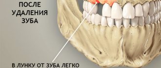
Injuries during tooth extraction
They are considered a complication, most often appear on the upper jaw, the highest risk is when removing premolars and molars. If removal is difficult, there is a risk of perforation of the floor of the maxillary sinus. It becomes damaged, and a hole appears in the bony septum between the nasal and oral cavities. This may be due to the anatomical features of the jaw structure (there is no bone septum or the roots of the teeth being removed are located close to the bottom of the sinus). Another possible cause is chronic inflammation in the area of the tooth root, due to which the bone septum becomes thinner and destroyed. Such an injury is eliminated immediately. If this is not done, liquid may enter the nose when swallowed or chewed. If such a symptom appears, you should contact your dentist so as not to provoke inflammation.
Causes and provoking factors
In 80% of cases, odontogenic osteomyelitis occurs, and its main cause is advanced pathologies:
- carious damage to crowns;
- pulp inflammation;
- periodontitis;
- alveolitis;
- cyst and granuloma of the tooth;
- pericoronitis;
- boils and carbuncles;
- purulent diseases of ENT organs;
- diphtheria, scarlet fever, umbilical sepsis in newborns.
The infection enters the tooth root and then into the bone tissue, thus forming osteomyelitis.
The cause of traumatic osteomyelitis is often gunshot and stab wounds, as well as damage to the nasal mucosa. In such cases, bacteria enter the bones from the external environment and cause inflammation.
Doctors identify a number of factors that contribute to the development of inflammation:
- weakened immunity, poor lifestyle;
- blood pathologies;
- diabetes;
- rheumatic pathologies;
- polyarthritis;
- severe pathologies of the kidneys and liver.
Those patients who refuse timely visits to the dentist are more susceptible to osteomyelitis.
Desensitization therapy
The treatment complex is supplemented with antihistamines:
- diphenhydramine;
- chloropyramine;
- fenkarol;
- diazolin.
Medicines help eliminate intoxication, activate the anti-inflammatory functions of antibiotics, minimize the permeability of the vascular wall, and fight swelling. To increase the effect, drugs containing calcium and glucose are prescribed in combination.
This treatment protocol is used for patients with diabetes, since this approach minimizes the negative impact of concomitant disorders in the foci of infection and facilitates the recovery process.
General restorative therapy
As part of a general strengthening course, the patient is prescribed vitamin complexes (groups B, C), adaptogenic drugs, and stimulants.
In severe forms of the disease, extracorporeal detoxification is performed. For this purpose, hemosorption, plasmapherosis or lymphosorption are prescribed. Courses of autohemotherapy (administration of one’s own venous blood intramuscularly) have a positive effect.
Physiotherapy
Such treatment is allowed only after acute inflammatory symptoms have reduced, so manipulations are prescribed a few days after the start of therapy. The methods may be as follows:
- hardware treatment with UV rays;
- electrophoresis using anti-inflammatory and antibacterial drugs;
- massage;
- oxygenation;
- laser irradiation of the site of the extracted tooth.
Regardless of the course of the disease, the treatment protocol is determined only by the doctor, taking into account the physiological characteristics of the body.
Symptoms of osteomyelitis
Acute pathology manifests itself sharply and is accompanied by the following symptoms:
- pain in the affected area;
- mobility of the diseased tooth, swelling of the gums;
- purulent discharge from the gums;
- numbness of the lip;
- putrid odor from the mouth;
- temperature rise to 38-39°C;
- enlarged lymph nodes;
- headaches, weakness, nausea;
- chills, lack of sleep.
The main symptom is severe pain that radiates to the ear, eye and temple. If the process has spread to soft tissues, the face swells and becomes asymmetrical, the skin feels hot to the touch, the patient cannot open his mouth and it hurts to swallow.
In the subacute form, the symptoms are less pronounced, so the patient’s well-being improves. There is no visible swelling of the face, pain becomes tolerable, but tooth mobility increases.
The chronic stage can occur due to improper treatment of acute osteomyelitis of the jaw; sometimes a primary chronic process also occurs. With this pathology, the symptoms are mild, but a hidden course can lead to tooth loss and pathological fractures of the jaw.
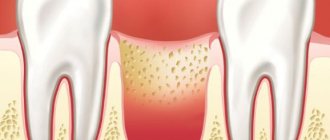
What is periosteum
In everything you need to start small, so first let’s figure out what the periosteum of the tooth is. This is the soft connective tissue that covers the jawbone and also serves as the link that connects the tooth to all the surrounding muscles and ligaments. This tissue is permeated with blood vessels and nerve fibers; it is responsible for blood supply and also performs a protective function for bone.
Like all components of our body, the periosteum can be subject to various inflammatory processes. And the presence of these inflammatory processes in this area cannot and will not even be ignored for a long time (thanks to the striking symptoms).
So, if the periosteum is inflamed, this means that there is a disease in the oral cavity with an associated infection, which necessarily requires treatment.
On a note! You will be interested to know that dentists scientifically call inflammation of the periosteum “periostitis”, and ordinary people call it “flux”. This terminology greatly facilitates understanding among ordinary patients, and now you know what we are talking about.
Diagnosis and treatment of inflammation
Acute osteomyelitis of the jaw is diagnosed by a dentist based on the clinical picture and the collected medical history. Additionally, laboratory tests and x-rays of the jaw are prescribed. In case of chronic pathology, the image will show signs of osteoporosis and sequestration.
Treatment begins with removal of the focus of necrosis, the affected tooth or abscess is removed, the wound is washed with antiseptics and antibiotics, and drainage is installed. If the teeth are hypermobile, therapeutic splinting is performed.
Additionally, anti-inflammatory drugs and antibiotics are prescribed. After stopping the acute process, physiotherapy is indicated to speed up tissue recovery.
In case of chronic pathology, an operation may be required, during which the doctor will remove the affected areas of bone tissue and fill the voids with osteoconductive material. If there is a risk of a pathological fracture, therapeutic splinting is used, which helps to avoid injury.
Conservative therapy
This includes the following activities:
- general strengthening;
- anti-inflammatory;
- physiotherapy;
- desensitizing.
Anti-inflammatory treatments
Therapy consists of treatment with antibiotics. Antimicrobial drugs should be started as early as possible (for example, gentomycin, ciprofloxacin, lincomycin, etc.). They are absorbed by bone tissue and do not cause allergic reactions.
Patients with severe forms of the disease are simultaneously prescribed several groups of antibiotics, taking into account compatibility and side symptoms.
Due to the fact that osteomyelitis is provoked by aerobic and anaerobic pathogenic microorganisms, it is effective to include antibiotics of the metronidazole group in combination with sulfonamides in the treatment protocol. In some cases, patients are prescribed intravenous administration of enzymes. Competent selection of drugs determines the result of drug treatment, and also helps to minimize the risk of complications and adverse reactions.
Osteomyelitis in mild forms can be cured in 7-10 days. Severe forms require a longer course until the symptoms of inflammation are completely relieved.
Complications of the inflammatory process
Osteomyelitis of the jaw requires urgent treatment, otherwise there is a risk of developing a number of complications:
- abscess and phlegmon of jaw tissue;
- meningitis;
- meningoencephalitis;
- brain abscess;
- lung abscess and sepsis;
- pathological bone fractures;
- formation of a false joint;
- impaired mobility of the masticatory muscles;
- damage to the heart and kidneys.
Many of these pathologies lead to death or disability, and pronounced cosmetic defects.
Treatment of osteomyelitis of the jawbone
The standard treatment program for acute osteomyelitis of the jaw includes:
- surgical intervention (removal of infected teeth, dissection of the periosteum, primary treatment of lesions in the bone, periosteum and adjacent soft tissues, opening, drainage and sanitation of foci of infection);
- surgical removal of sequestra (rejected jaw bone tissue);
- anti-inflammatory therapy;
- detoxification measures;
- taking general restoratives;
- symptomatic treatment.
In this case, the scope of surgical intervention, the nature of drug therapy and other treatment methods are determined by the characteristics of pathogenesis, the general condition of the patient and clinical symptoms.
Prevention and prognosis
The prognosis for osteomyelitis is favorable if the patient consults a doctor in a timely manner and undergoes adequate therapy. The occurrence of pathology can be prevented by observing the following preventive measures:
- treatment of caries, pulpitis or gum inflammation should occur in a timely manner;
- Instant sanitation of inflammatory and infectious foci in the body should be carried out;
- you should strengthen your immune system, eat right and lead a healthy lifestyle;
- It is recommended to avoid any injuries to the jaw.
Dentists recommend brushing your teeth daily, morning and evening, for at least 2 minutes, and visiting the dentist annually for a preventive examination and professional oral hygiene.
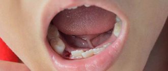
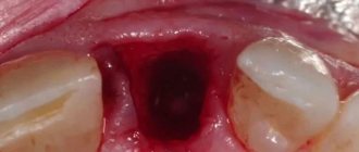
Treatment of alveolar osteitis
Prophylactic antibiotics reduce the risk of alveolar osteitis, as well as relieve pain and reduce the risk of soft tissue infections.
If we are talking about treatment, it is usually symptomatic (for example, taking painkillers), and the dentist also removes debris from the nest by irrigation with saline solution or local anesthesia. Medicinal solutions can be placed in the exposed socket, but their use is limited due to severe pain.
People who experience alveolar osteitis often seek help from a doctor several times after surgery.
Moscow metro station Zvezdnaya, Danube Avenue, 23
Sources
- Lukyanenko V.I. Osteomyelitis of the jaws.
- Modern features of clinical manifestations of odontogenic and traumatic osteomyelitis of the lower jaw. Fomichev E.V.
Attention!
This article is posted for informational purposes only and under no circumstances constitutes scientific material or medical advice and should not serve as a substitute for an in-person consultation with a professional physician.
For diagnostics, diagnosis and treatment, contact qualified doctors! Number of reads: 2371 Date of publication: 04/29/2019
Dentists - search service and appointment with dentists in Moscow
General information
Osteomyelitis of the mandible is more common, more severe and has more complications. With chronic osteomyelitis of the lower jaw, the bone is affected deeper, which can lead to jaw fractures. Inflammation affects a dangerous area - the jaw processes. Acute osteomyelitis of the lower jaw easily turns into subacute and then chronic.
Osteomyelitis of the upper jaw develops more rapidly and can lead to inflammation of the maxillary sinuses. However, since the bone tissue here is less dense, abscesses and phlegmons develop less often with osteomyelitis of the upper jaw, and the disease itself is easier.
The entry point for infection is a diseased tooth, and the causative agents of inflammation are Staphylococcus aureus and Staphylococcus alba, pneumococci, Escherichia coli and typhoid bacilli. This microflora is usually located in areas of chronic infection of the tissues surrounding the tooth.
Inflammation in osteomyelitis does not occur suddenly; initially, a chronic infection develops in the gum for a long time - for example, in periodontitis or periodontitis. Then, if the outflow of waste products from the infection is disrupted, the microflora from the lesion spreads to the surrounding tissue.
Osteomyelitis of other parts of the skeletal system is caused by only one type of pathogen - staphylococcus (streptococcus), which penetrates the tissue through the bloodstream. Since osteomyelitis of the jaw can be caused by different pathogens, the course of the disease has much more distinctive features than with ordinary osteomyelitis.
What happens if you don't get treatment?
If you have inflammation of the periosteum of the tooth, and you do not want to carry out treatment and do not rush to see a doctor, then everything can be complicated by necrosis of the affected tissue and the spread of infection to neighboring organs: palate, throat, larynx, maxillary sinuses, ears, eyes, brain. Blood poisoning and sepsis may also occur, and large fistulas will appear. Some patients with complications cannot avoid osteomyelitis or phlegmon. And in most cases, you also have to say goodbye to the affected tooth, which could become the culprit for the spread of infection.
Such consequences require urgent surgical intervention, and without timely surgery, everything can end in death. Therefore, remember that the disease is not to be trifled with.