X-ray diagnostics during dental implantation is required to identify diseases and contraindications. Using images, the implantologist assesses the condition of the bone tissue, and based on this, selects the appropriate method for installing implants. After the operation, X-ray diagnostics determine whether the structure is installed correctly and whether treatment adjustments are required. For a complete picture, several images are taken using different methods - a targeted image, an orthopantomogram, a computed tomography. Our clinic has its own certified radiology department.
The importance of X-ray diagnostics before implantation
The images allow you to identify obstacles to the installation of implants. They identify problems that can complicate the operation.
Using images, the implantologist determines:
- distance to the mandibular nerve and maxillary sinuses;
- density, volume of bone tissue to select the shape and size of the implant;
- the condition of adjacent teeth to prevent infection from entering the surgical area.
Lack of diagnosis can lead to:
- damage to the nerve and sinuses;
- unstable fixation of the artificial root;
- infection of the operation area;
- reducing the service life of the implant and replacing it with a new one;
- increased sensitivity of the gums due to incorrectly selected orthopedic design.
X-ray diagnostics makes it possible to exclude complications and install implants according to the correctly selected protocol in accordance with the patient’s clinical situation.
Why is a CT scan prescribed before dental implantation?
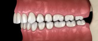
Installing implants to replace a lost tooth or group of teeth is not as complicated as planning an operation. The dentist must take into account many different factors: the thickness of the alveolar ridge, the proximity of the nasal sinus or infraorbital foramen, the location and inclination of implantation of the artificial root, and other features. This cannot be done without special equipment.
When performing surgery using conventional x-rays, the risk of errors is very high. Bone deficiency will not allow the implant to anchor well, so it may fall out. If a pin is installed too small, the chewing load will be too strong, which will ultimately lead to peri-implantitis. If you make a mistake with the location, then during the operation the bottom of the maxillary sinus, nerves, sinuses may be damaged, and the artificial tooth itself will cause a pathological change in the bite.
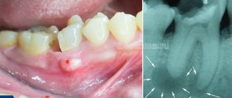
Sight shot
A 2D image makes it possible to analyze the condition of 1-3 teeth. For this purpose, a digital radiovisiograph is used.
Allows you to identify inflammatory processes in neighboring teeth, monitor the condition of sealed canals, diagnose damage, and detect pathologies. It can be periapical (study of the tissues around the tooth) and interproximal (analysis of the crown).
Advantages:
- clear image of 1-3 teeth with image magnification;
- safety for the patient, several tests can be done in a row without harm to health.
Flaws:
- 2D image giving only one perspective;
- small visible range.
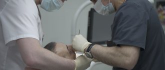
In our Center, the procedure is performed in a separate X-ray room. The patient is put on a protective apron, the position of the head is fixed, and the radiologist directs a beam of rays to the area being examined.
The procedure takes a few seconds, is painless, and does not cause discomfort.
Orthopantomogram or OPTG is an important component of preparation for dental implantation
Article navigation
- Orthopantomogram - the essence of the study
- Indications for OPTG
- Advantages and disadvantages
- CT or panoramic image - differences
- What types of photographs are there and what is better to choose?
- Preparing for the examination
- How is the procedure performed?
- Are there any restrictions
- Acceptability during pregnancy
- Potential dangers and possible complications
- How to decrypt a photo
- How often can OPTG be performed?
- Cost of dental examination
Question for a specialist
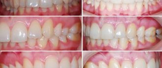
An orthopantomogram is a separate type of x-ray examination that allows you to obtain a panoramic image of the teeth - more precisely, the entire dental system of the patient. It reveals detailed information not only about the condition of the teeth of both jaws, but also about the maxillary sinuses and other important anatomical structures. This is a modern, affordable and effective way to conduct a diagnostic examination, making it possible to identify all problems at once. True, the detail of OPTG is always slightly lower than that of targeted images and computed tomography results. But today we will take a closer look at the orthopantomogram, find out what it is, how it is done and what it shows. Well, let’s answer the most important question – is it needed before implantation.
Panoramic photograph of teeth OPTG
An orthopantomogram gives an idea of the general condition of the dental system - it shows the external and internal condition of the dentition and bone tissue. Performed on an orthopantomograph, a flat two-dimensional panoramic image is obtained - the device combines several images taken in different planes into one. Therefore, OPTG does not accurately convey the image.
The procedure defines:
- ratio of jaw, sinuses;
- location of nerves;
- hidden carious formations;
- damage to seals;
- dental diseases (cyst, periodontal pockets).
Pros:
- full picture of the jaw;
- consideration of a local problem;
- the price is lower than CT.
Minuses:
- two-dimensional images;
- likelihood of misstatement;
- does not determine the angle, thickness of the alveolar processes, structure, position of the jaw.
Before the examination, you need to remove metal jewelry and accessories. To perform OPTG, the patient bites on a special plate to keep the jaws motionless. The orthopantomograph rotates around the patient's head. The procedure lasts from 8 to 20 seconds and does not cause discomfort.
Do implants interfere with CT scanning?
Sometimes dental restoration is required for patients who already have some orthopedic structures installed, for example, crowns, pins, etc. Therefore, the question naturally arises: is it possible to do a CT scan of the jaw for implantation in this case, and whether existing artificial teeth will interfere with obtaining high-quality images.
The first thing you should pay attention to is the material used to make the non-removable orthopedic structure. If it is metallic, then there is a possibility that the CT image may show optical distortions. Although this is not very significant, it can still affect the information content of the final data.
If the structures are made of medical metal, then their presence in the patient’s mouth will not affect the result of a jaw examination with a tomograph. But, before the procedure, it is necessary to inform the radiologist about where and what artificial teeth are already available in the row.
If ceramic implants are installed in the jaw, a tomograph examination is carried out without restrictions and the risk of obtaining low-quality images of the teeth.
When CT scanning teeth for implantation, the qualifications of the radiologist and the technical equipment of the clinic are of great importance. That is why we recruit only experienced specialized specialists to the staff of the Smile Factor clinics and purchase cutting-edge equipment. This allows us to provide high-quality dental services with a guarantee of results for many years.
Computed tomography CT
CT scan is performed on a 3D tomograph. The device's detector generates two-dimensional images, which are combined into a three-dimensional 3D image in a special computer program. The method is more informative than 2D images and allows you to obtain a three-dimensional image of the jaw in high resolution.
CT diagnostics are carried out at the preparatory stage of implantation to identify contraindications and after installation of implants to prevent complications. It is applied for:
- identifying pathologies of the maxillary sinuses;
- determining the condition of bone tissue;
- assessing the location of the mandibular nerve;
- analysis of temporomandibular joints;
- detection of impacted teeth that may interfere with the installation of implants;
- diagnostics of fractures, neoplasms on the jaw.
Advantages:
- accuracy, information content;
- clarity of the picture.
Flaws:
- high price compared to other types of x-ray diagnostics.
The procedure is performed while standing, the patient bites the plate of the device and remains motionless for up to 40 seconds until the device rotates around the head.
Initial examination
Before dental implantation, you need to visit the dentist's office for an initial consultation. During this procedure, the doctor will carefully examine the oral cavity, assess its condition and give a referral for x-ray diagnostics and tests. Already at this stage, the dentist can:
- determine indications and contraindications for implantation;
- inform the patient about the features of the upcoming operation.
In addition, during the initial examination, the patient must fill out a questionnaire (survey card), in which the presence of bad habits, chronic diseases and surgical interventions should be noted.
| We strongly recommend that you fill out the survey card honestly. Then our doctors will know better how your body will behave during implantation. |
Safety
According to the established SanPiN requirements, the annual exposure rate should not exceed 1000 microsieverts. Modern X-ray equipment is safe - the table below shows the radiation exposure during one image and the permissible number of procedures per year.
| Radiation exposure per 1 procedure | Safe number of procedures per year | |
| Sight shot | 1-3 µSv | 500 |
| OPTG | 13-17 µSv | 80 |
| CT | 50-60 µSv | 20 |
| Type of diagnosis | Radiation exposure per 1 procedure | Safe number of procedures per year |
| Sight shot | 1-3 µSv | 500 |
| OPTG | 13-17 µSv | 80 |
| CT | 50-60 µSv | 20 |
X-ray diagnostics are not recommended during the first and last trimester of pregnancy.
Contraindications for CT examination
Computed tomography is not recommended more than twice a year. If the procedure was recently performed in another clinic, the patient can show the doctor the finished CT result of the teeth. In some cases it is contraindicated.
Restrictions on CT scanning:
- period of pregnancy, breastfeeding, as the body is exposed to x-rays;
- claustrophobia - the patient is in a confined space, so panic may occur;
- high motor activity of children under 14 years of age due to the need to remain completely still during scanning.
During breastfeeding, you can temporarily transfer the baby to artificial feeding. The milk needs to be expressed and discarded. Breastfeeding is resumed the next day.
Our clinic uses a safe device that meets international standards
The digital X-ray system with a minimum level of radiation SIEMENS SIRONA GALILEOS allows you to perform the most accurate studies for patients. The device is intended for the study of bone tissue, tooth roots, temporomandibular joint, nasopharynx, and upper spine. An analysis of local problems and the condition of a large volume of jaw tissue is carried out.
Levin Dmitry Valerievich
Chief physician, Ph.D.
Orthopantomogram - the essence of the study
An orthopantomogram is a complete panoramic x-ray of all teeth at once, which allows you to get a general picture of the condition of the dentofacial apparatus. Today, this technique is actively used in cosmetology and various fields of dentistry, including dental implantation and maxillofacial surgery. The procedure is carried out using a special device - an orthopantomograph or computed tomograph (this is more powerful and modern equipment that is installed in clinics that also offer computed tomography services).
Thus, the snapshot allows you to examine the status of the following elements:
- jaws and bite,
- teeth, their shape, position, roots, as well as the rudiments of future teeth, including eights, impacted elements, bone tissue,
- gingival tissue, vascular system, dental canals - examination allows us to identify possible pathological conditions, such as periodontitis or the presence of neoplasms,
- soft fabrics,
- fillings, crowns, dental bridges.
An orthopantomogram (OPTG) is a mandatory procedure in preparation for dental implantation. The examination allows you to identify hidden pathologies, eliminate possible contraindications to treatment and avoid many problems even at the preparatory stage. The diagnosis itself lasts about a minute. After this, the received data is loaded into a computer, where further image processing is carried out.
Important! In addition to OPTG of the dentition, computed tomography is often also prescribed, which allows you to obtain not just a flat, but a three-dimensional image - on it, of course, all elements can be viewed in 3D. This is especially important when all teeth are implanted, immediate loading methods are used, or complex osteoplastic surgery is required.
DENTAL PROSTHESIS WITH 4 OSSTEM IMPLANTS - RUB 170,000!
Implantation even with bone tissue atrophy. Work guarantee! Save RUR 20,000.
on promotion >> Free consultation with an implantologist +7 (495) 215-52-31 or write to us
Price
Diagnostics in our Center is included in the cost of implantation consultation:
- drawing up a treatment plan, CT scan with examination on a computer screen without decoding and recording on DVD - 3000 rubles. ;
- drawing up a treatment plan, CT scan with interpretation, recording on DVD - 5200 rubles.
When installing implants in our Center, consultation and x-ray examination are included in the package, diagnostics after surgery is carried out free of charge. Funds spent before the start of treatment are credited to the patient’s account in the form of an advance.
You can view pricing at our clinic here.
A few words about the shortcomings
3D implantation has a huge number of advantages, but it also has disadvantages. In particular, no software will help an unprofessional doctor who does not thoroughly know the anatomy of the jaw system and does not have computer modeling skills. In addition, this approach requires the presence of special equipment in the clinic - a scanner for performing computed tomography (although the best option would be to contact specialized medical centers to conduct a more in-depth diagnosis on a tomograph with better image detail), as well as computer software itself - a program for planning the process implantation
Three-dimensional dental implantation allows you to obtain an almost accurate prediction of the treatment outcome. Naturally, a computer cannot completely replace a person, but advanced high technologies significantly simplify and speed up the treatment process and, of course, make it better. Naturally, provided that the work is carried out by a professional doctor with high qualifications and extensive experience.
Is it possible for pregnant women to have x-rays and anesthesia?
When treating pregnant women, only local anesthesia is allowed. Modern drugs for such anesthesia are effective and safe for health. But during pregnancy, the choice of anesthetic and calculation of its dose is performed individually for each woman, depending on her age and stage of pregnancy. The issue of the admissibility of radiography for diagnostic purposes in pregnant women is still debated in the medical community. The latest generation of X-ray machines have a minimal radiation dose, but gynecologists still advise refraining from such examination at least in the first trimester. Computer diagnostics for pregnant women is allowed at any stage.
Please note: the “Smile” chain of clinics is equipped with innovative radiographic and computer equipment for high-precision diagnostics. The minimal dose of radiation allows even pregnant women to be examined.
Orthopantomogram in pediatric dentistry
Due to reduced radiation exposure, OPTG can also be used for diagnosing children. Most often, an image is needed if there is a suspicion of malocclusion. You can use an orthopantomogram from the age of 5-6 without risk to the child’s health.
Pediatric diagnostics has its own characteristics. During an orthopantomogram, the exposure mode is used. It allows you to change the trajectory of the sensor and the irradiation area. This is necessary due to the small size of a child's jaw. Thanks to exposure during the examination, the child does not receive unnecessary stress.
The picture clearly shows:
- The rudiments of permanent teeth.
- The location of the teeth, where they are crowded.
- Absence of tooth buds (one of the symptoms of adentia).
In pediatric orthodontics, a panoramic photograph is used to monitor the development of the jaws and the process of teething.
What types of photographs are there and what is better to choose?
As for the classification of OPTG, there are 2 types of panoramic X-rays of the jaw - film and digital orthopantomogram. Sometimes you can also hear about three-dimensional, but in fact this type of OPTG does not exist - all X-ray results here are only two-dimensional, “flat”.
- 1. film – this type of OPTG appeared first. Initially, models of devices were used that involved the use of a special film. It was placed on one side of the patient's head, while the X-ray tube was on the other side. After the procedure, the image was transferred to film, after which it was processed and developed,
- 2. digital – a modern version of the examination involves recording images on a medium in digital format. The obtained data is loaded into a special computer program, and the doctor can carefully study the already processed image from the computer screen.
What is better to choose – digital or film orthopantomogram? Of course, it is better to give preference to the digital option. It is more modern, accurate, and the image can be enlarged many times on a computer to examine the pathological area in detail.
How to minimize risks
What should an expectant mother do so as not to worry about the baby’s health?
- Not all clinics, especially municipal ones, have visiographs. It is better to inquire about this in advance.
- Be sure to use a protective apron and collar.
- You need to inform your doctor about the exact timing of your pregnancy. In case of a delay, even if the test shows a negative result, you must inform the specialist. In unclear situations, diagnosis (and treatment) are carried out as if pregnancy was definitely established.
- Severe pain, swelling, inflammation are good reasons not to delay a visit to the dentist, even in the first trimester. Unless absolutely necessary, the doctor will never prescribe dental x-rays during pregnancy and will try to do everything to postpone treatment to the 2nd, safe trimester or to the postpartum period. However, some acute conditions may require emergency intervention, such as purulent pulpitis or acute periodontitis. In this case, the mother needs to remember that a rapidly developing infection in the oral cavity will cause much more serious damage to the baby than 1-2 pictures on a visiograph.
- If there are concerns that the X-ray has somehow damaged the baby, you should consult with a geneticist and do an ultrasound. This will reassure those mothers who may have taken the photo without knowing that they were expecting a baby.
Since dental treatment during pregnancy is still associated with certain difficulties, the best thing an expectant mother can do is to go to the dentist and have all her teeth treated at least a month before the date of planned conception. And already in pregnancy, mommy should brush her teeth thoroughly and eat foods high in calcium.
Why prepare?
The most unpleasant thing that can happen to a patient who decides to undergo dental implantation is the rejection of the rod, the destruction of bone tissue to a state where subsequent implantation has too little chance of success. It turns out that effort and money are wasted, and the choice is limited to removable dentures or bridges. To prevent this from happening, it is important to do everything possible to prevent the chance of developing reimlantitis or other complications.
Preparing for the examination
The procedure does not require any specific preparation; it can be carried out directly on the day of contacting the dentist or diagnostic center. However, before sending a person for OPTG, the specialist is obliged to conduct a visual examination of the oral cavity and collect a complete anamnesis, that is, take into account all complaints and possible pathological phenomena in the maxillofacial area. After the initial diagnosis, all that remains is to settle some formalities: the doctor will ask you to sign a consent to conduct the examination.
How dangerous is x-ray?
In many cases, dental treatment is simply unthinkable without X-ray examination. When you can't do without it:
- when filling thin and curved tooth canals;
- to detect hidden carious cavities, caries under fillings;
- when the question arises whether wisdom teeth need to be removed (for example, if they are incorrectly positioned);
- to diagnose a tooth root fracture, cyst, determine the degree of inflammation of the tissues around the diseased tooth;
- when the issue of removing or restoring a tooth is being decided.
In the first three months, it is not advisable to take dental x-rays for pregnant women, since the fetus has not yet formed. Radiation can have an adverse effect on dividing cells, cause chromosome abnormalities and provoke intrauterine developmental defects. By the second trimester, at 16–20 weeks, the fetus is already fully formed - a tiny dose of radiation will not harm the baby in any way, so pictures can be taken without fear.
Dental X-rays during pregnancy are much safer than examinations of the pelvis or chest, because the mother’s belly is reliably protected by a lead apron, and dental examinations are always carried out using special devices with reduced radiation exposure. This applies to both old models and modern visiographs.
Indications for OPTG
The diagnostic technique under consideration is widely used in various fields of dentistry. Before moving on to the question of how and where a panoramic photograph of teeth is taken, it is worth understanding in which cases such a procedure is indicated. Among the mandatory indications for an orthopantomogram, experts identify the following cases:
- removal of teeth and roots in order to prevent possible complications,
- treatment of periodontal tissue diseases,
- diagnosis of jaw tumors,
- diagnosis of periostitis,
- orthodontic treatment – before installing plates or braces,
- diagnoses of unknown etiology,
- prosthetics, implantation.
Are there any restrictions
This examination option is considered safe and harmless, however, there are some contraindications. The procedure is not performed if the patient has malignant neoplasms in the head and neck area, as well as blood cancers - leukemia, lymphoma and others. In the first case, under the influence of radiation, the tumor can begin to grow and mutate, which will lead to even more severe complications. In the second case, even a slight effect of radiation on the bone marrow, a hematopoietic organ, can provoke and accelerate the development of the disease, including fatal consequences.
Also, performing OPTG is not always possible in a small child or in a person with serious mental disorders - when the patient is unable to remain still for even 1-2 minutes.